Abstract
[Purpose] The study analyzed the electroencephalographic (EEG) data of the central cortical areas, during execution of the motor gestures of feeding, activation of the system of mirror neurons, and imagery between a right hemiparetic volunteer (RHV) and a healthy volunteer (HV). [Subjects and Methods] The volunteers’ EEG data were recorded with their eyes open for 4 minutes while they performed five experimental tasks. [Results] The alpha band, absolute power value of HV was lower than that of RHV. In the beta band, during the practice condition, there was an increase in the magnitude of the absolute power value of HV at T3, possibly because T3 is representative of secondary motor areas that work with cortical neurons related to planning and organizing sequence of movements performed by the hands. The gamma band is related to the state of preparation for movement and memory. The results of this study indicate that there was increased activation of the gamma frequency band of HV. [Conclusion] The findings of this study have revealed the changes in pattern characteristics of each band which may be associated with the brain injury of the hemiparetic patient.
Key words: Electroencephalography, Hemiparetic, Rehabilitation
INTRODUCTION
The increased life expectancy of Brazilians, due to scientific and technical advances has contributed to the changing epidemiological profile. In this sense, the model of health care focused on infectious diseases has been gradually replaced by a model directed at chronic diseases. Stroke is classified as chronic degenerative disease and is characterized by an abrupt episode of vascular origin, which results in focal or generalized disturbance of brain function that persists for more than 24 hours1). After a stroke, an individual may present neurological alterations related to motor functions, speech and cognition, as well as impairment of independence in daily activities of life2).
Motor disorder may present in the form of hemiplegia/hemiparesis. Hemiplegia is characterized by an abnormal physical state of the hemibody, accompanied by total loss of strength, alteration of the senses, and mental, language and perceptual disorders, while hemiparesis results in partial loss of strength and the other symptoms mentioned above, with hemiplegia/hemiparesis3) being the most commonly found symptom in patients after stroke4).
A functional recovery strategy for limitations resulting from motor impairment, the use of motor imagery (MI) stands out especially with regard to daily activities. MI is defined as the mental process dynamic in which the subject simulates a motor task that occurs without the movement of the segments associated with this task, and can it be presented in two modalities: visual and kinesthetic. These differents modalities of MI activate distinct brain regions, and these activities can be investigated through electroencephalograms (EEG)5).
Besides MI, another therapeutic strategy is the mechanism of mirror neurons. These are visualmotor neurons that are triggered both when an individual performs a certain gesture as when volunteer observes another individual performing the same movement. The presence of mirror neurons in humans has been demonstrated in studies of the reactivity of EEG brain rhythms during the observation of movements6). Furthermore, the utility of EEG as a tool for the analysis of brain function can be observed in protocols that use actual execution of motor tasks, like MI7).
The present study used electroencephalography to compare the values of absolute power of the alpha, beta and gamma frequency bands in the central cortical areas during performance of the motor gestures of feeding, and the drive system of mirror neurons and imagery between a right hemiparetic volunteer (RHV) and a healthy volunteer (HV).
SUBJECTS AND METHODS
This study was conducted at the Laboratory of Brain Mapping and Functionality (LAMCEF) at the Federal University of Piauí (UFPI) and presents the report of two cases, one healthy volunteer and one right hemiparetic; both were male and right-handed. To check the handedness, individuals answered the Edinburgh Inventory. Then, they were evaluated using to the Modified Ashworth Scale to identify hypertonia’ degrees. The mini-mental state examination (MMSE) was conducted as a cognitive screening instrument. In order to verify the overall functionality of the subjects, the Barthel Index was assessed as well. The ability of imagining a motor gesture in the first or third person was investigated through the scale of motor imagery (The Kinesthetic and Visual Imagery Questionnaire; KVIQ). The subjects were duly informed about the objectives, the experimental procedure involved, and the methodology of this study, and voluntarily signed a consent form. The study was approved by the Ethics Committee on Human Research of UFPI, under registration number 246.283/2013.
The EEG signal was captured using a BrainNet BNT-EEG (EMSA). Electrodes were placed according to the 10-20 international system, and the reference electrodes were placed on the earlobes (bi-atrial). The EEG signals were recorded in a prepared soundproof room. The EEG signal frequencies fluctuated between 0.01 and 50 Hz. To ensure a more judicious selection, parts contaminated by muscle artifacts were removed using an automatic rejection algorithm to reject values exceeding a threshold (100 microvolts).
The first EEG measure was performed with the subject at rest for 4 minutes with his eyes open. Immediately after, the subject watched a 6-second video showing the motor gestures of picking up a cup, bringing it to the mouth and putting it back on the table in front of the subject. It is noteworthy that the individual in the video sat in a comfortable position, under the same conditions as the subjects of our experiment. After watching the video the subjects performed three randomized tasks. 1) Practice − the subjects performed the motion of withdrawing the right hand that was resting on the table, picking up a glass, bringing it to the mouth and return it to the initial site marked on the table, and replacing the hand in the starting position. 2) Imagery − the subjects imagined the motor gesture described above in the first person for 4 minutes. 3) Mirror neurons − the subjects watched the video of the motor gesture described. Immediately after the experimental tasks the subjects’ EEG at rest was measured again. All measurements were performed for four minutes. The data were analyzed used MATLAB® R2011a, converted by Microsoft® Excel 2010, and transformed into log 10 values. The averages of absolute power were calculated.
RESULTS
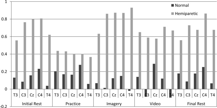
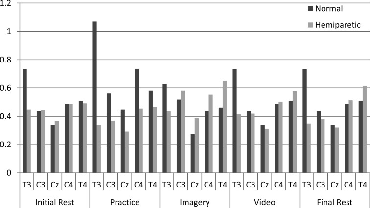
The data in relation to age, and cognitive functional capacity, as well as the scale of imagery are presented in Table 1. All values of the magnitude of absolute power in the alpha band of HV were lower than those of RHV (Fig. 1). Regarding the imagery data scale, despite the individuals finding it easy to imagine, the electroencephalographic results were not the same in HV. This was reflected in the lower value of the absolute power at C3. The analysis verified higher values at CZ, C4 and T4, corresponding to the right hemisphere. In the beta band, in the practice task, an increase in the magnitude of power at T3 of HV (Fig. 2) was observed. In contrast, in RHV, there was no increase in absolute power values in practice compared to rest, but at T4 greater activation was observed.
Table 1. General data of the subjects.
| Subject | Age | MEEM | Barthel index | Scale imagery |
|
|---|---|---|---|---|---|
| Kinesthetic | Visual | ||||
| Hemiparetic | 61 | 23 | 74 | 6 | 7 |
| Normal | 60 | 21 | 100 | 6 | 6 |
Fig. 1.
Comparison of the absolute power of the alpha band across the five tasks
Fig. 2.
Comparison of the absolute power of the beta band across the five tasks
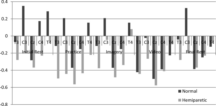
The analysis of the gamma band showed there was increased activity in RHV during the imagery task, specifically at T4 (Fig. 3). During all other tasks, we observed a reduction in the magnitude of the gamma band power. During Initial Rest HV showed high power at C3 that corresponded to representation of the hand. In general, HV showed lower cortical activation during all tasks at all measurement sites; however, at C3, there was an increase in the magnitude of the absolute power during the following tasks: Initial Rest, Practice, Imagery and Final Rest. The HV also showed increased power at T4 during tasks of Initial Rest, Practice, and Imagery, and reduced activity during Rest and Final Video. HV showed higher magnitude at C4 during Initial Rest, but in the other tasks only reductions were observed. HV showed reduced cortical activation at all the other measurement sites during all of the tasks.
Fig. 3.
Comparison of the absolute power of the gamma band across the five tasks
DISCUSSION
The present study aimed to compare between RHV and HV, the values of absolute electroencephalographic in the alpha, beta and gamma frequency bands of the central cortical areas, resulting from the execution of the motor gesture of feeding, the activation of the mirror neuron system, and imagery. For this study, the central electrode sites of C3, CZ, C4, T3 and T4 were chosen in order to represent the motor areas related to the planning and execution of movement, and the sensory areas corresponding to the areas of the body activated whenever any sense or sensory receptor is specifically stimulated8).
The frequency bands were chosen because of their particular characteristics. The alpha band is inversely related to cortical activation7, 9), cognitive processes and wakefulness required during task execution10, 11). This band contains frequencies between 8 and 13 Hz12). Changes in alpha band power seem to be associated with cognitive tasks of different levels of complexity13). Alpha band power is also associated with highly specific perception, attention and memory processes10). The increase in the absolute power of the alpha band observed in this study appears to be related to the mental consolidation of the task9). Alpha rhythm activation is inversely related to the proportion of the population of cortical neurons activated during perception, motor and cognitive processes14). Thus, we suggest that HV responded adequately to the demands of the tasks, while RHV showed lower mental activity in all tasks, possibly because of brain injury. The lower values of the absolute power verified at C3, demonstrate that HV showed greater mental activity during Imagery and Video than RHV. The results of HV showed higher activities at CZ, C4 and T4, corresponding to the right hemisphere. This shows that due to lesion in the left hemisphere, RHV had cortical activation suggestive of prior motion inexperience. It has been reported that cortical activation occurs in the contralateral hemisphere during the execution and learning of movement13).
The beta frequency band spans 12 to 30 Hz. From a functional perspective, beta frequencies are commonly associated with motor activities11, 12). When this band is presented in cortical activity, suggests mental activity and sharp attention9). The beta rhythm seems to be linked to movement precisely in the frontal and central areas of the cortex that are related to the planning and execution of voluntary movements, and it can be influenced by exercise13, 15). The beta band is related to motor activities, especially in the frontal and central cortical areas which are, responsible for activating conditions of planning and execution of voluntary movements12, 13). The magnitude of the absolute power at T3 increased during the Practice by HV (Fig. 2), possibly because T3 is representative of secondary motor areas which work as planners and organizers of the sequence of movements performed by the hands of the subject. In contrast, in RHV, there was no increase in power values during rest, but at T4 greater activation was observed, presumably to assist the work of T3 during Practice12). This would indicate synchronization between the same associated regions of the left and right sides of the brain contralateral to the motor task requested. Beta waves appear in association with maximum attention of the dominant hemisphere, which possibly lends itself to motor planning strategy, as shown predominantly at T38).
The gamma band is related to memory, integrating factors and integrating spatial/temporal proprioception9). According to Teixeira, its frequency range is 30–100 Hz16). However, Uhlhaas demonstrated frequencies from 60 to 200 Hz, which were correlated to specific cognitive functions8). These frequencies were associated with pace, waking as a preparatory state to movement, and processing of diffuse cortical information before the motor event happen10). The increase in the absolute power values of RHV at T4, which is located in the right hemisphere and corresponds to the area responsible for motor control of the hand, during Imagery, indicate an evoked memory of the movement. This shows preservation of the ability to imagine the tasks in RHV, and use of the hemisphere opposite to the brain injury to assist in achieving the movement6, 10).
HV during Initial Rest showed high power at C3 corresponding to representation of the hand. Probably this was due to prior knowledge of the task; however, during Practice, the increase was not significant compared to the other conditions. The magnitude of power of the gamma band is related to the state of preparation for movement and memory; however, the results of this study indicate that there was increased activation in this frequency in RHV10, 17), possibly because neural activity was lowered by the presence of brain injury.
The analysis of absolute power of the alpha, beta and gamma bands allows observation of changes in patterns of consolidation in mental tasks, motor planning, and evocation of memory, which may be associated with brain injury in hemiparetic subjects. Studies indicate that the applicability of motor imagery associated with other physiotherapy resources enhances treatment targeting motor functionalization of patients with stroke18). Future studies should investigate experimental models to complement the present findings, associating different tasks and retention periods, in order to obtain more information about the patterns of cortical activation.
REFERENCES
- 1.Xiaomeng X, Min L, Yongjun J: The paradox role of regulatory T cells in ischemic stroke. The Scientific World Journal, 2013, Article ID 174373. [DOI] [PMC free article] [PubMed] [Google Scholar]
- 2.Jakaitis F, Santos DG, Abrantes CV, et al. : Role of physical therapy of aquatic fitness in stroke patients. J Neurosci, 2012, 20: 204–209 [Google Scholar]
- 3.Braun A, Herber V, Michaelsen SM, et al. : Relationship among physical activity level, balance and quality of life in individuals with hemiparesis. Br J Sports Med, 2012, 18: 30–34 [Google Scholar]
- 4.Barcala L, Colella F, Araujo MC, et al. : Balance analysis in hemiparetics patients after training with Wii Fit program. Phys Ther Mov, 2011, 24: 337–343 [Google Scholar]
- 5.Stecklow MV, Infantosi AF, Cagy M: EEG changes during sequences of visual and kinesthetic motor imagery. Arq Neuropsiquiatr, 2010, 68: 556–561 [DOI] [PubMed] [Google Scholar]
- 6.Ferreira CP: Does the brain have anything to do with morality? Mirror neurons, empathy and neuromorality. Physis, 2011, 21: 471–490 [Google Scholar]
- 7.Stecklow MV, Infantosi AF, Cagy M: [Changes in the electroencephalogram alpha band during visual and kinesthetic motor imagery]. Arq Neuropsiquiatr, 2007, 65: 1084–1088 [DOI] [PubMed] [Google Scholar]
- 8.Uhlhaas PJ, Pipa G, Neuenschwander S, et al. : A new look at gamma? High- (>60 Hz) γ-band activity in cortical networks: function, mechanisms and impairment. Prog Biophys Mol Biol, 2011, 105: 14–28 [DOI] [PubMed] [Google Scholar]
- 9.Machado S, Portella CE, Silva JG, et al. : Changes in quantitative EEG absolute power during the task of catching an object in free fall. Arq Neuropsiquiatr, 2007, 65: 633–636 [DOI] [PubMed] [Google Scholar]
- 10.Portella CE, Silva JG, Bastos VH, et al. : [Procedural learning and anxiolytic effects: electroencephalographic, motor and attentional measures]. Arq Neuropsiquiatr, 2006, 64: 478–484 [DOI] [PubMed] [Google Scholar]
- 11.Bonini-Rocha AN, Chiaramonte M, Zaro MA, et al. : Observation cognitive evidences of the motor learning in the performance of young guitarists monitored by electroencephalogram: a pilot study. Sci Cogn, 2009, 14: 103–120 [Google Scholar]
- 12.Sauseng P, Klimesch W: What does phase information of oscillatory brain activity tell us about cognitive processes? Neurosci Biobehav Rev, 2008, 32: 1001–1013 [DOI] [PubMed] [Google Scholar]
- 13.Bastos VH, Cunha M, Veiga H, et al. : Analysis of the distribution of power as a function of cortical learning typewriting skill. Br J Sports Med, 2004, 10: 494–499 [Google Scholar]
- 14.Bonini-Rocha AN, Timm MI, Chiaramonte M, et al. : Metodologia para observação e quantificação de sinais de EEG relativos a evidências cognitivas de aprendizagem motora. Scis Cogn, 2008, 13: 27–50 (in Portuguese). [Google Scholar]
- 15.Moraes H, Ferreira C, Deslandes A, et al. : Beta and alpha electroencephalographic activity changes after acute exercise. Arq Neuropsiquiatr, 2007, 65: 637–641 [DOI] [PubMed] [Google Scholar]
- 16.Teixeira S, Velasques B, Machado S, et al. : γ band oscillations in parietooccipital areas during performance of a sensorimotor integration task: a qEEG coherence study. Arq Neuropsiquiatr, 2011, 69: 304–309 [DOI] [PubMed] [Google Scholar]
- 17.Smith ME, McEvoy LK, Gevins A: Neurophysiological indices of strategy development and skill acquisition. Brain Res Cogn Brain Res, 1999, 7: 389–404 [DOI] [PubMed] [Google Scholar]
- 18.Holz EM, Glennon M, Prendergast K, et al. : Theta-gamma phase synchronization during memory matching in visual working memory. Neuroimage, 2010, 52: 326–335 [DOI] [PubMed] [Google Scholar]


