
Fig. 6. PTH 1-34 regulates Wnt/β-catenin signaling during tibia fracture healing in young and aged mice.
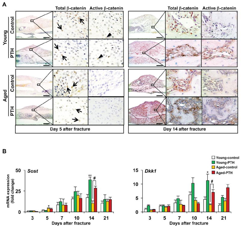
(A) Immunohistochemistry was performed to detect total and active non-phospho-β-catenin in day 5 and 14 tibia fracture calluses in both young and aged mice treated with vehicle or PTH 1-34. Total (arrows) and active (arrowheads) β-catenin staining was present in immature chondrocytes in the callus as well as in osteoblastic cells lining newly formed bone surfaces. PTH 1-34 treatment increased active β-catenin staining in the fracture calluses of both young and aged mice at day 14. (B) Real-time RT-PCR was performed on total RNA to measure the gene expression Sost and Dkk1 during fracture healing in both young and aged mice treated with vehicle or PTH 1-34. Expression was normalized to β-actin (n=4). Data are presented as mean ± standard error (SEM). Statistical analysis was performed using bootstrap-adjusted t-tests and statistical significance denoted as follows: *p<0.05 and **p<0.01 compared to Young control group, or #p<0.05 and ##p<0.01 for Aged control vs. Aged PTH 1-34 groups.