



Abstract
Esophageal cancer treatment continues to be a topic of wide debate. Based on improvements in chemotherapy drugs, surgical techniques, and radiotherapy advances, esophageal cancer treatment approaches are becoming more specific to the stage of the tumor and the overall performance status of the patient. While surgery continues to be the standard treatment option for localized disease, the current direction favors multimodality treatment including both radiation and chemotherapy with surgery. In the next few years, we will continue to see improvements in radiation techniques and proton treatment, with more minimally invasive surgical approaches minimizing postoperative side effects, and the discovery of molecular biomarkers to help deliver more specifically targeted medication to treat esophageal cancers.
Keywords: Esophageal Cancer, Chemoradiation, IMRT, Protons
Introduction
Esophageal cancer is the 6th most common cancer in the United States and worldwide1. While the rates of new diagnoses and rates of death from all cancers combined have declined significantly in most recent years in the United States, the incidence of esophageal cancer is on the rise in this country2. This disease is highly aggressive with a 5 year overall survival of 10%. It is estimated that in 2010, 16,640 Americans will be diagnosed with esophageal cancer, and 14,500 will die from it3.
The majority of esophageal cancers are adenocarcinoma (AC) or squamous cell carcinoma (SCC). These histologies differ in incidence, geographic distribution, ethnic pattern and etiology. SCC remains the predominant disease of the developing world1. It is associated with tobacco, alcohol and certain dietary carcinogens4. In contrast, AC has become the predominant form of esophageal carcinoma in the developed world 1. It is associated with chronic gastroesophageal reflux disease, Barrett’s esophagus, and increasing body mass index. Past several decades has seen a dramatic shift from SCC to AC in the United States5, 6. Between 1975 and 2004, Brown et al. reported a 463% increase in incidence among white US males and 335% increase among while US females using SEER data7.
Current staging of esophageal cancer is based on tumor/node/metastasis classification using the 7th edition of the American Joint Committee on Cancer8. This represents a major change in staging compared to the previous 2007 edition which had separate stage groupings for SCC and AC. Although it continues to serve as an important prognostic tool, the current staging is still based on outcomes from patients treated with esophagectomy alone9. Several groups have shown in retrospective setting that degree of treatment response to neoadjuvant therapy is a strong survival prognosticator9, 10. Thus, the limited utility of the current staging system when it comes to resected patients treated with neoadjuvant therapy should not be overlooked. In addition, inclusion of proximal 5 cm of stomach in the esophageal staging is problematic because staging (example, laparoscopy) and treatment approaches are different. The advances in imaging that have enabled us to more accurately stage patients by non-surgical methods have the potential to improve outcomes and change treatment recommend will be further discussed in this article.
Staging/Work up
Imaging
The clinical staging of esophageal cancer is usually performed using endoscopy/endoscopic ultrasound (EUS) and computed tomography (CT)11, 12. Integrated positron emission tomography (PET) and computer tomography with [18F]-fluoro-2-deoxy-D-glucose (FDG) is being increasingly used in the initial staging of patients with esophageal cancer and improves the accuracy of staging13, 14. The following sections review and clarify aspects of the imaging performed in the clinical staging of patients with esophageal cancer.
Primary Tumor
EUS with fine needle aspiration biopsy is the optimal modality for the diagnosis and evaluation of local invasion (T1 through T4) of the primary tumor (sensitivity of EUS, CT, and FDG-PET 80%, 50% and 57%, respectively, specificity 70%, 83%, and 85%, respectively)11, 12. CT and PET-CT imaging have a limited role in the diagnosis of the primary tumor. The inability to differentiate between T1, T2, and T3 and poor ability to identify invasion of adjacent structures that precludes resection (T4), are major limitations in the use of CT in the evaluation of the primary tumor. Additionally, although the intensity of FDG uptake and depth of tumor invasion are positively related, this association is poor15, 16. The limitations of esophageal tumor detection and evaluation by PET imaging are greatest in the assessment of superficial esophageal carcinomas17. Little et al., reported that only 31 of 58 (53%) patients with superficial tumors had increased FDG uptake in the primary esophageal tumor (median SUV 3.5, range 2.1 to 16.6)17. Additionally, because of the poor sensitivity in detecting nodal metastases and low prevalence of distant metastases, PET imaging is not indicated in the overall T, N and M staging of superficial esophageal tumors.
In the evaluation of the primary esophageal tumor, FDG-PET may have a potential role in the determination of the length of the tumor18. The accurate delineation of the superior and inferior extent of viable esophageal tumor is important in radiotherapy planning and tumor length is also a strong independent prognosticator19, 20. Currently, tumor length is measured by EUS but accurate delineation can be difficult. Mamede et al., have recently reported that FDG-PET-derived tumor metabolic length correlates well with tumor length assessed by EUS and surgical pathology results18.
Regional Lymph Nodes
EUS with or without transesophageal endoscopic biopsy of nodes is the optimal modality for the detection of regional lymph node metastases11-14. CT has poor sensitivity and specificity in the detection of regional lymph node metastases. The addition of FDG-PET to the imaging algorithm of patients with esophageal cancer has not significantly improved the accuracy of regional nodal staging compared with the currently standard combined use of CT and EUS14, 21. In a meta-analysis of 12 studies concerning the value of FDG-PET in the preoperative staging of patients with esophageal cancer, the pooled sensitivity and specificity for the detection of loco-regional nodal disease were 51% and 84%, respectively22.
Metastatic Disease
Distant metastases are common in patients with esophageal cancer and CT and FDG-PET are useful in the detection of these metastases (sensitivity and specificity 71% and 93% and 52% and 91%, respectively)11, 12. Although CT imaging of the chest and abdomen is typically performed routinely in the staging of esophageal cancer, PET-CT imaging may be more accurate in the detection of distant metastatic disease at presentation. In this regard, Lowe et al, reported the sensitivity and specificity for detecting distant metastases to be 81% and 82% for CT, 73% and 86% for EUS, and 81% and 91% for PET13. Furthermore, recent studies suggest that the addition of PET imaging to the conventional pre-operative staging algorithm prevents inappropriate esophageal resection due to the detection of unsuspected metastases14, 23, 24. However, while FDG-PET improves the selection of patients with esophageal cancer for curative resection, the diagnostic benefit after comprehensive conventional staging may be limited. In this regard, in a prospective study, van Westreenen et al. reported that FDG-PET imaging performed after a preoperative staging protocol that included multidetector CT, EUS, and sonography of the neck, revealed distant metastases in only 8 of 199 (4%) of the patients with esophageal cancer and prevented unnecessary resection in only 3%25. Furthermore, there was also a high rate of false-positive PET findings (7.5%) that resulted in unnecessary additional investigations.
Minimally invasive techniques
Endoscopic treatment includes endoscopic resection (ER) and endoscopic ablation. The most important advantage of ER over ablative therapies is that EMR/ESD will result in a resected specimen for histopathological assessment. ER provides the most accurate assessment of invasion depth, grade of differentiation and lymphovascular invasion. ER can therefore serve both as a diagnostic as well as a therapeutic tool. ER is very effective with 5-year disease-specific survival reported to be 98% 26.
To eliminate the malignant potential after ER, eradication of the residual Barrett’s mucosa is strongly advocated. In a study of 11 patients who underwent RFA for Barrett’s esophagus (BE) (6 of them previously treated with ER for visible lesions), complete endoscopic and histologic eradication was seen in all patients at a median follow-up of 14 months 27. In another study from the same group, 12 patients with BE (11 with HGD and 1 with LGD) underwent stepwise RFA ablation. Seven of those patients had previous ER of visible lesions. Complete endoscopic and histologic removal of BE was achieved in all patients at a median follow-up of 14 months 28.
A number of studies have evaluated the effectiveness of RFA in the treatment of dysplastic and non-dysplastic Barrett’s in the absence of neoplasia. In a single-center study of RFA for LGD (n=39) and HGD (n=24), overall complete response of intestinal metaplasia (CR-IM) was 79% with CR-dysplasia (CR-D) of 89%. The median follow-up was 23 months. For the LGD cohort, CR-D was 95%, and for the HGD cohort, CR-D was 79% 29. In the only published controlled trial of BE with either LGD (n=64) or HGD (n=63), patients were randomized 2:1 to RFA or a sham procedure. Eleven patients had ER prior to RFA. In an ITT analysis, CR-IM was seen in 77% of patients treated with RFA as compared to 2.3% in the control group (P<0.001). Among all patients, esophageal cancer developed in significantly more patients in the control group than in the ablation group (9.3% vs. 1.2%, p=0.045).
The second, new ablative modality for the treatment of BE and early esophageal adenocarcinoma is cryoablation. Cryotherapy involves the application of extreme cold in order to achieve tissue destruction. In a study of 30 high-risk patients with HGD or intramucosal adenocarcinoma (IMCA), elimination of cancer or downgrading of HGD was seen in 68% of patients in the HGD group and in 80% of those in the IMCA group, at a median follow-up of 12 months 30. In a retrospective study of cryotherapy in 98 patients with HGD, CR-HGD was seen in 97% of patients, and CR-IM in 57%. The mean number of treatments was 4 and median follow-up was 10 months 31.
Surgical Resection
Surgical intervention continues to be the standard treatment for early stage esophageal cancers. Three to five-year disease free survival is generally poor, at 6-35%, and depends on several factors, including the level of resection performed at the time of surgery 32,33,34,35. The RTOG trial 8911 (Intergroup 133) found that 32% of patients with R0 resection were alive and disease free at five years compared to 5% who underwent R1 resection. Due to poor survival rates from surgery, there have been many attempts to refine surgical approaches for esophageal cancers. There are several approaches that can be used which include a transhiatal esophagectomy, right-thoracotomy (Ivor-Lewis procedure), left thoractomy, and radical (en bloc) resection. The two most commonly used in North American are the transhiatal and Ivor-Lewis techniques. Briefly, the transhiatal approach uses an upper midline laparatomy and a left neck incision to provide exposure to the esophagus. Once the disease is removed, a cervical anastomosis is made, often with gastric pull up 36. Advantages of the procedure include lower morbidity with fewer respiratory side effects. One major disadvantage is the lack of exposure of the middle esophagus.
The abdominal approach, also known as the Ivor-Lewis procedure, combines a laparatomy with a right thoracotomy with its anastomosis located in the thoracic esophagus. While this procedure requires an additional thoracotomy compared to the transhiatal approach, its primary advantage is improved exposure of the middle esophagus which its counterpart lacks. The two procedures have been compared in the past. One major study published in the New England Journal of Medicine found that between patients who underwent transhiatal esophagectomy compared to transthoraric esophagectomy with extended en bloc lymphadenectomy, the transhiatal approach had lower morbidity and improved five-year long term survival37. More recently, a study using SEER also discovered that transhiatal esophagectomy confers an early survival advantage. While initially their observed five-year survival rates were also found to be better in the transhiatal approach, the difference in this study lacked statistical significance when adjustments were made for patient and tumor differences38.
However, based on the high failure rates of surgical intervention alone, many studies have evaluated a trimodality approach with chemotherapy and radiation, followed by surgery. Currently, surgery alone is typically recommended for noncervical T1bN0 and occasional T2N0 esophageal cancer. Overall in the last two decades, several studies have favored multimodality approaches to esophageal cancer; these are discussed in detail below.
Radiation Therapy
Primary Radiation
There have been several studies in the last two decades showing poor survival outcomes in patients with esophageal cancer treated with radiation therapy alone. Prior to this however, radiation therapy for many years was the standard alternative to surgery in patients with localized, inoperable esophageal cancers. Several studies looking at external-beam radiotherapy treatment alone found significantly low 5-year survival rates ranging from 0% to 10%39, 40. These studies look at conventional doses (~50 Gy at 1.8 to 2.0 Gy per fraction). The RTOG 85-01 study, a long-term prospective randomized trial, compared RT alone to combined modality therapy, looking at overall survival41. Radiation alone was inferior with a three-year survival of 0%.
Since then, there have been a few additional studies looking at RT alone. One recent randomized study performed in China compared conventional radiotherapy with additional late course accelerated hyperfractionation compared to radical surgery alone in patients with resectable esophageal cancer 42. There were 135 patients enrolled in the surgery arm and 134 in the radiotherapy only group. Total dose of radiation received was 68.4-71.0 Gy total with initial conventional therapy given at 50-50.4 Gy at 1.8-2.0 Gy/d followed by a boost of 18-21 Gy delivered at 1.5 Gy twice daily. They found comparable rates of 1-, 3-, and 5-year progression free survival rates at 73.3%, 39.7%, and 20.6% in the radiation group respectively compared to 75.9%, 43.7%, and 23.1% in the surgical arm. They also found 1-,3-, and 5-year overall survival rates of 88.6%, 56.2%, and 34.7% in the radiation only group respectively versus 93.3%, 61.5%, and 36.9% in the surgery group. The standard of care continues to be multimodality care, but with current improvements in radiation delivery and increased numbers of patients with inoperable cancers who may also not tolerate chemotherapy, radiation alone may be a viable option.
Adjuvant Radiation Therapy
Given the poor local control seen in patients treated with surgery alone has promoted the investigation of adjuvant radiation to enhanced local control. A recent review by Schreider et al compared the outcome of 1046 patients diagnosed with T3-4NoMo or T1-4N1Mo esophageal cancer and treated with surgery43. Of these patients 683 (65.3%) percent received surgery alone while the other 363 (34.7) received postoperative radiation (PORT). Patients receiving PORT with T3N1 or T4No disease demonstrated both significant improvement in median and 3-year OS (p < 0.001) and DSS (p < 0.001), respectively. Furthermore PORT was the most significant predictor of improved OS (hazard ratio 0.70, 95% confidence interval 0.59-0.83, p < 0.001), an impressive finding given the selection bias present in retrospective reviews and the high risk of systemic spread seen in patients with advanced disease. However, given the large fields needed to encompass the surgically manipulated field, radiation doses are limited as such concurrent chemotherapy, discussed below, may offer further therapeutic advantages.
Radiation versus Chemoradiation
The antitumor effect of concurrent chemoradiation compared to radiation therapy alone has been well documented for many cancers, including esophageal. An early phase III trial published in 1992 44 randomized patients with esophageal cancer into two treatment arms, radiation alone (64 Gy) and radiation (50 Gy) plus combined fluorouracil and cisplatin. When comparing the radiation group to the bimodality therapy group, outcome survivals at 1- and 2-years were 33% and 10% versus 50% and 38% respectively. As expected, side effects were also greater in the chemoradiation group. The majority of these were hematologic and those involving the oral cavity, pharynx, and esophagus. RTOG 85-01 41, also found a significant difference in overall survival with concurrent chemoradiation compared to radiation alone. 5-year overall survival for the bimodality therapy group compared to radiation alone was 26% and 0% respectively. They also found that most patients failed secondary to having persistent localized disease which appeared to be controlled better with concurrent therapy. Other studies also have confirmed similar findings in outcomes survival, favoring chemoradiation compared to radiation alone 45, 46.
Chemotherapy
Rational for Systemic Therapy
Since esophageal cancer is a systemic illness, systemic therapy can be justified as an investigative approach. However, currently we lack effective systemic agents and we have no tools to individualize therapy (biology agents or cytotoxic agents) based on the molecular make up of tumors. However, the need to develop effective, rational therapy is real. In the absence of our ability to individualize therapy, the alternative approach of the use of “most active” combination chemotherapy has been investigated. The most commonly used agents include: fluoropyrimidines and platinum compound.
Preoperative Chemotherapy
There are two important preoperative phase III randomized trials that warrant comments. The first trial was conducted in the United States by the Intergroup 47. In this trial, 440 patients with potentially resectable carcinoma of the esophagus were randomized to surgery alone (n=227) followed by observation or preoperative chemotherapy with FU and cisplatin (n=213) followed by surgery followed by observation. There was no survival advantage observed in the group that had preoperative chemotherapy. In a larger study conducted by the Medical Research Council 48, 802 patients with resectable esophageal cancer were randomized to surgery alone (n=402) followed by observation or preoperative chemotherapy (n=400) with FU and cisplatin followed by surgery followed by observation. Overall survival was longer for the preoperative chemotherapy group (p=0.004). The subgroup analysis showed that patients with squamous cell carcinoma did not benefit. The MRC study was updated with a longer follow-up 49 and although, it shows that the overall p value remained significant (p=0.03), the overall benefit seemed to diminish. In addition, this time the subgroup analysis demonstrated that neither squamous cell carcinoma nor adenocarcinoma patients benefited. Thus, it would be reasonable to conclude that in patients with potentially resectable esophageal cancer, preoperative chemotherapy has limited or no impact on the survival of these patients.
Postoperative Chemotherapy
Currently, there is a lack of definitive data proving survival benefits with postoperative chemotherapy in node-positive patients. The only randomized trial comparing surgery alone to surgery with postoperative chemotherapy was a study performed in Japan looking at patients with esophageal squamous cell carcinoma50. 5-year survival rates for postoperative chemotherapy compared to surgery alone were found to be significant at 55% vs 45% respectively. However, overall survival was not found to be significant. One major weakness of the study was that only two courses of chemotherapy were given in the postoperative chemotherapy arm and up to 25% of these patients did not complete treatment.
Chemoradiation
There are no randomized studies of esophagectomy versus chemoradiation as primary management of esophageal cancer. Review of the literature indicates esophagectomy alone has limited applicability, significant morbidity, higher mortality, and a lesser chance for cure than chemoradiation for patients with squamous cell cancer of the esophagus51. For patients treated by esophagectomy alone the 5-year survival rate of 10% or less, which is comparable to patients treated with chemoradiation alone. However quality of life, especially swallwoing function appear to be improved with the omission of surgery52. Histology is also important here as patient with squamous cell cancer of the esophagus do particularly well with chemoradiation as such many advocate that surgery should only be reserved for recurrent disease in patients with this histological subtype51.
Definitive
For many patient surgical resection is not an option due to comorbidities, as such these patient are often treated with chemoradiation alone. Coia et al, published a series of 90 patient treated prospectively with chemoradiation for unresectable disease53. Patient with stage I or II disease received a dose of 60Gy while patient with stage III or IV received a palliative dose of 50Gy. The overall mean survival for patient with early stage disease away 18 months with 3 & 5 year actuarial survival of 29% and 18% respectively. The 3-year actuarial local failure rate was stage dependent; 24% for stage I and 45% for stage II. They also demonstrated that aggressive chemoradiation of 60Gy changed the failure pattern from local failure to one dominated by distant metastasis, of the 29 patient who failed 14 (48%) had any component of local failure, whereas 21 (72%) had a distant failure as a component of failure. The question of avoiding surgery is commonly discussed in the treatment of the elderly with esophageal cancer, due to concern of further morbidity. This topic was addressed in a review by Abrams et al that specifically looked at the outcomes of pates with stage I or II cancer over the age of 65 treated with surgery versus chemoradiation. In multivariate analyses, chemoradiation was associated with worse disease-specific survival (hazard ratio [HR], 2.08; 95% confidence interval [CI], 1.64-2.64) and overall survival (HR, 1.92; 95%CI, 1.58-2.34)54. However, there was no significant difference for patients with squamous cell carcinoma (HR, 1.33; 95%CI, 0.98-1.80). Perhaps the major benefit to definitive chemoradiation will not be from local control but from enhanced quality of life. In a study by Gill et al, the local failure rate following preoperative chemoradiation and esophagectomy was 12% compared with 17% with chemoradiation alone, yet over 60% of patients had normal swallowing following chemoradiation alone compared with 48% following chemoradiation and esophagectomy52.
Despite this encouraging data and that fact that radiation targeting, planning and delivery has advanced greatly over the 30 year period that data was acquired, our definite radiation dose has Ironically now been reduced due to historical president from trials such RTOG 94-05, which demonstrated increased toxicity in most situation w us the same radiation dose of unresectable disease with is used I for neoadjuvant setting55, 56. A recent retrospective review from MD Anderson of 66 patients with unresectable esophageal cancer treated with chemoradiation therapy alone demonstrated that 24 had local-regional failure (37%); notably, 18 of those failures (75%) were located within the gross tumor volume (GTV). This finding suggests that although current therapies can be quite effective in some cases, local disease control, specifically within the GTV, remains a problem. Logically, the demonstrated benefits of radiation dose escalation for tumors at other anatomic sites in terms of improved local control and survival57-59 could potentially by expected to apply to esophageal cancer as well.
Preoperative Chemoradiation
Preoperative Chemoradiation versus surgery Alone
Several randomized trials have been performed looking at preoperative chemoradiation versus surgery alone (Table 1)33, 34, 60-62. One of the first major studies to address this question was the EORTC trial conducted in France comparing stage I and II squamous-cell cancers of the esophagus treated with chemoradiation followed by surgery versus surgery alone. They found patients treated with chemoradiation and surgery had greater disease free survival rates, longer periods of local free disease, a decreased rate of cancer-related deaths, and higher rates of curative resection 33. Another study looking at trimodality therapy and surgery alone for adenocarcinoma and found an increased median survival rate in the multimodality arm (16 months) compared to the surgery alone group (11 months) 34. They also discovered a significant survival advantage at 3 years under the multimodality approach. These early studies and several others have favored the trimodality approach which physicians have followed in the treatment of esophageal cancers.
Table 1.
Randomized trials of preoperative chemoradiation followed by surgery, versus surgery only
| Study | Patients,n | Tumor histology | Treatment group | pCR rate,% | Median survival time | Survival rates,% | Local failure rates |
|
|---|---|---|---|---|---|---|---|---|
| 3-year | 5-year | |||||||
| Bosset et al. [33] | 282 | 100% SCC | Preop CRT | 26 | 19 mo | 36 | – | – |
| Surgery | 0 | 19 mo | 36 | – | – | |||
| Walsh et al. [34] | 113 | 100% Adeno | Preop CRT | 25 | 16 mo | 32 | – | – |
| Surgery | 0 | 11 mo | 6 | – | – | |||
| Burmeister et al. [60] | 256 | 61% Adeno | Preop CRT | 15 | 22 mo | – | – | – |
| Surgery | 0 | 19 mo | – | – | – | |||
| Tepper et al. [61] | 56 | 75% Adeno | Preop CRT | 40 | 4.48 y | 63 | 39 | 4 |
| Surgery | 0 | 1.79 y | 22 | 16 | 4 | |||
| Gaast et al. [62] | 320 | 75% Adeno | Preop CRT | 33 | 49 mo | 59 | – | – |
| Surgery | 0 | 26 mo | 48 | – | – | |||
Several studies however have found no improvement in overall survival when comparing the trimodality approach to surgery alone. The RTOG 8911 study was a long-term phase III randomized trial which analyzed resectable patients with T1-2NxM0 esophageal cancer who received either surgery alone or pre-operative chemotherapy with cisplatin and 5-FU prior to surgery 32. RTOG 8911 found no significant difference in MS or OS at 4 years when comparing the two treatment arms. Another recent phase III trial in Australia compared patients either treated with surgery alone or chemoradiation followed by surgery 60. They found no significant difference in progression-free survival nor overall survival between the two groups. They performed a subgroup analysis and suspected some improvement in overall survival for squamous cell carcinoma but no conclusions were made due to the studies low power. They did conclude however that the chemoradiotherapy with surgery group had more complete resections with clear margins, fewer positive lymph node compared to surgery alone.
Contrary to the last studies discussed, two more recent trials favored trimodality therapy. The first, a phase III trial comparing trimodality therapy with surgery alone, found significant differences in median survival (4.48 vs 1.79 years), five-year survival (39% vs 16%), favoring trimodality therapy63. In addition, a recently published meta-analysis looking at 10 trials with a total of 1209 patients either undergoing preoperative chemoradiotherapy plus surgery or surgery alone found significantly improved two-year survival rates in the chemoradiation plus surgery arm64. This applied to both squamous cell carcinoma and adenocarcinoma. The CROSS study presented in 2010 was a phase II study looking at surgery versus trimodality therapy in T2-3/N0-1 esophageal cancers62. 363 patients were enrolled in the study. Median overall survival for CRT + surgery compared to surgery alone was 49 and 26 months respectively. One, two and three-year survival rates were 82%, 67%, 59% and 70%, 52%, 48% for the trimodality and surgery alone arms respectively. Subgroup analysis of CROSS suggests that impact of preoperative therapy on survival is high on squamous cell carcinoma but none on the adenocarcinoma patients. While there have been many conflicting studies as to the benefit of surgery alone compared to trimodality therapy, based on several more recent phase III studies showing benefit for chemoradiation followed by surgery, the continued standard of practice favors a trimodality approach.
Preoperative Chemoradiation versus Chemoradiation Alone
There are currently no phase 3 trials comparing the two strategies and this will likely not happen in the near future. Optimizing therapy based on molecular biomarkers or imaging techniques is what is needed.
Postoperative Chemoradiation versus Surgery Alone
Given the high failure rates seen after surgery alone has lead to investigation of adjuvant therapy. Given the high rates of both local failure and systemic disease combined chemoradiation strategies make particular sense. While there is no modern phase III trial addressing this question directly the INT 0116 trial is often quoted as 20% of the patients, from this from this predominately gastric study, had G/E junction tumors65. Of the 556 patients randomized between surgery alone and surgery plus postoperative chemoradiation the median survival; was 27 months for surgery compared with 36 months in the chemoradiation group. Grade three toxicity was present in 41 percent of patients treated with chemoradiation and three patients 1% dies from toxicity.
A more recent Phase II trial investigated the utility of postoperative chemotherapy in a population of pure esophageal or G/E junction population with poor prognosis66. This study was limited to advanced patients with either T3N1 or M1a disease in order to investigate if local postoperative chemoradiation would be of benefit in these patients a very high risk of distant disease. 50 patient were enrolled only four patient 8% require hospitalization for neutropenia and no treatment related deaths were reported. The 4 years projected overall survival was 51% freedom from recurrence 50%, distant metastatic control 56%, and locoregional control 86%. This study suggests that even advance stage and patients with M1a disease adjuvant chemoradiation is well tolerated and positively impacts outcomes. As we move forward patient selection based on prognostic factors or biologic markers may help make personalized medicine a reality allowing us to predict which patient are destine for local failure and warrant adjuvant chemoradiation.
Molecular biology
The field of molecular biology and esophageal cancer is continuously evolving. There are a number of potential targets that have not yet been exploited and current trials are underway. Due to the heterogeneity of esophageal cancers, we need to uncover the molecular biology of it in order to better understand how to treat and manage the disease.
Future Directions
Radiation techniques
The future direction of radiation for the treatment will come from improved tumor targeting, improved treatment planning, and enhance sparing of normal tissues. For the treatment of resectable disease, radiations utility is for sterilizing of microscopic/lymphatic disease, and given that many of these patients are younger and have early stage disease effort at reducing both cardiac dose and pulmonary dose, may help reduce future morbidity. As tumor staging has improved with both EUS and PET studies will need to be done to addressee the appropriate treatment volumes, for example in controversial areas such as celiac coverage in a patient with T2No disease. However in patients with unresectable disease the challenges are quite different. Given the high infield failure rate, particularly in the GTV, some have proposed that pre-operative radiation doses are too low for definitive treatment of unresectable disease. With the rapid advances that have taken place in radiation planning it is quite possible that the outcomes of RTOG 94-05 would be different if more modern technique had been used. Several groups have demonstrated that the implementation of intensity-modulated radiation therapy (IMRT) can provide additional flexibility to modify dose distributions and improve normal tissue sparing67. Although IMRT is clearly useful for reducing the dose to critical structures, it is also beneficial for increasing the dose to volumes at high risk. Moreover, the simultaneous integrated boost (SIB) technique offers the advantage of simultaneously delivering a higher dose to the primary tumor (at 2.2 Gy or 2.3 Gy per fraction) while conventional lower doses are used to treat subclinical disease or electively treated regions (at 1.8 Gy or 2.0 Gy per fraction). A recent dosimetric study comparing the dose distributions between 2D-CRT (50.4Gy) and SIB-IMRT escalating dose to the GTV to 64.8Gy, achieving a 28% increase in dose to the tumor (p = 0.001) while at the same time reducing the mean heart dose by 30% (p = 0.001), the mean total lung dose by 23% (p = 0.007), and the lung V20 by 37% (p = 0.004)56. Clearly prospective studied will need to be done before we can evaluated if dose escalation provide a benefit to our patients, yet as systemic therapies improve the benefit of local control will become further important.
Protons
The location of the esophagus in the posterior aspect of the mediastinum places the structure in close proximity to several critical structures, including the heart, lungs, and spinal cord. As a result, there is significant potential to reduce toxicity with the incorporation of more conformal techniques. Dosimetric planning studies comparing IMRT to Intensity modulated protons therapy (IMPT) have demonstrated significant reductions in the amount of normal lung, heart and liver68. However, it remains to be seen if these benefits translated into reduce toxicity. The initial experience with protons in Esophageal cancer seems to support this. Liao et al. demonstrated that when compared to 52 patients treated with intensity modulated radiation therapy (IMRT), the complete pathologic response rate was higher with proton therapy. In addition, the treatment related toxicities of esophagitis, dysphagia, and stricture were less with proton therapy, and these results reached statistical significance69. Results from the University of Tsukuba in Japan examining proton therapy with or without x-rays also showed promising results, with local control rates at 5 years of 57% and also reporting no treatment interruptions, but with 15% of patients found to have post-radiation ulcers within 3 months of completing treatment70. Updated data from this same institution found that utilizing total doses of 70-90 cobalt-60 Gray equivalents (CGE), the complete clinical response rate was 78%, with no cases of treatment interruption for hematologic toxicity or esophagitis71. As further dosimetric and clinical studies are published, outcomes are likely to continue to improve as clinicians discover how to maximize the potential benefit of this very promising technique.
Fig 1.

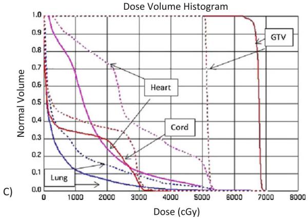
Dose volume comparison of classic 2D-CRT plan versus SIB-IMRT for dose escalation. (A) Axial, sagittal, and coronal view of a two-dimensional conformal radiotherapy (2D-CRT) plan to deliver 50.4 Gy to a patient with esophageal cancer (similar to the plans used in Intergroup 0123). (B) Simultaneous integrated boost intensity-modulated radiotherapy plan with the gross tumor volume being treated to 64.8 Gy and the planning target volume to 50.4 Gy. (C) Dose–volume histogram of an individual patient comparing a two-dimensional conformal radiotherapy (2D-CRT) plan (dashed line) to 50.4 Gy (similar to that used in Intergroup 0123) to a simultaneous integrated boost (SIB)-intensity-modulated radiotherapy (IMRT) plan (solid line) in which the gross tumor volume is treated to 64.8 Gy and the planning target volume to 50.4 Gy.
Contributor Information
James Welsh, Division of Radiation Oncology, UT MD Anderson Cancer Center, 1515 Holcombe Blvd., Unit 97, Houston, TX 77030.
Arya Amini, University of California, Irvine, University Dr, Irvine, CA 92697, aryaa@uci.edu.
Anna Likhacheva, Radiation Oncology, The University of Texas MD Anderson Cancer Center, 1515 Holcombe Blvd. Unit 0097, Houston, TX 77030, alikhacheva@mdanderson.org.
Jeremy Erasmus, Jr, Diagnostic Radiology, The University of Texas MD Anderson Cancer Center, 1515 Holcombe Blvd. Unit 1478, Houston, TX 77030, Phone (713) 792-5878 office, jerasmus@mdanderson.org.
Daniel Gomez, Radiation Oncology Department, The University of Texas MD Anderson Cancer Center, 1515 Holcombe Blvd., Unit 1150, Houston, TX 77030, Phone: (713) 563-8961, dgomez@mdanderson.org.
Marta Davila, Gastroenterology, Hepat,& Nutr, The University of Texas MD Anderson Cancer Center, 1515 Holcombe Blvd., Unit 1466, Houston, TX 77030, Phone: (713) 563-8906, mdavila@mdanderson.org.
Reza J Mehran, Thoracic & Cardiovasc Surgery, The University of Texas MD Anderson Cancer Center, 1515 Holcombe Blvd. Unit 1489, Houston, TX 77030, Phone: (713) 563-3908 office, rjmehran@mdanderson.org.
Ritsuko Komaki, Radiation Oncology Department, The University of Texas MD Anderson Cancer Center, 1515 Holcombe Blvd. Unit 0097, Houston, TX 77030, Phone (713) 563-2300 office, rkomaki@mdanderson.org.
Zhongxing Liao, Radiation Oncology Department, The University of Texas MD Anderson Cancer Center, 1515 Holcombe Blvd. Unit 0097, Houston, TX 77030, Phone (713) 563-2300, zliao@mdanderson.org.
Wayne L Hofstetter, Thoracic & Cardiovasc Surgery, The University of Texas MD Anderson Cancer Center, 1515 Holcombe Blvd. Unit 1489, Houston, TX 77030, Phone: (713) 563-9130 office, whofstetter@mdanderson.org.
Manoop Bhutani, Gastroenterology, Hepat,& Nutr, The University of Texas MD Anderson Cancer Center, 1515 Holcombe Blvd., Unit 1466, Houston, TX 77030, Phone: (713) 794-5073, manoop.bhutani@mdanderson.org.
Jaffer A Ajani, GI Medical Oncology, The University of Texas MD Anderson Cancer Center, 1515 Holcombe Blvd., Unit 0426, Houston, TX 77030, Phone: (713) 792-2828, jajani@mdanderson.org.
References
- 1.Kamangar F, Dores GM, Anderson WF. Patterns of cancer incidence, mortality, and prevalence across five continents: defining priorities to reduce cancer disparities in different geographic regions of the world. J Clin Oncol. 2006;24:2137–50. doi: 10.1200/JCO.2005.05.2308. [DOI] [PubMed] [Google Scholar]
- 2. http://www.cancer.gov.
- 3.Cancer Facts and Figures. American Cancer Society; 2010. [Google Scholar]
- 4.Brown LM, Swanson CA, Gridley G, Swanson GM, Silverman DT, Greenberg RS, Hayes RB, Schoenberg JB, Pottern LM, Schwartz AG, Liff JM, Hoover R, Fraumeni JF., Jr. Dietary factors and the risk of squamous cell esophageal cancer among black and white men in the United States. Cancer Causes Control. 1998;9:467–74. doi: 10.1023/a:1008861806923. [DOI] [PubMed] [Google Scholar]
- 5.Islami F, Kamangar F, Aghcheli K, Fahimi S, Semnani S, Taghavi N, Marjani HA, Merat S, Nasseri-Moghaddam S, Pourshams A, Nouraie M, Khatibian M, Abedi B, Brazandeh MH, Ghaziani R, Sotoudeh M, Dawsey SM, Abnet CC, Taylor PR, Malekzadeh R. Epidemiologic features of upper gastrointestinal tract cancers in Northeastern Iran. Br J Cancer. 2004;90:1402–6. doi: 10.1038/sj.bjc.6601737. [DOI] [PMC free article] [PubMed] [Google Scholar]
- 6.PJ DEJ, Wolters LM, Steyerberg EW, H VAND, Kusters JG, Kuipers EJ, Siersema PD. Environmental risk factors in the development of adenocarcinoma of the oesophagus or gastric cardia: a cross-sectional study in a Dutch cohort. Aliment Pharmacol Ther. 2007;26:31–9. doi: 10.1111/j.1365-2036.2007.03344.x. [DOI] [PubMed] [Google Scholar]
- 7.Brown LM, Devesa SS, Chow WH. Incidence of adenocarcinoma of the esophagus among white Americans by sex, stage, and age. J Natl Cancer Inst. 2008;100:1184–7. doi: 10.1093/jnci/djn211. [DOI] [PMC free article] [PubMed] [Google Scholar]
- 8.Rice TW, Rusch VW, Ishwaran H, Blackstone EH. Cancer of the esophagus and esophagogastric junction: data-driven staging for the seventh edition of the American Joint Committee on Cancer/International Union Against Cancer Cancer Staging Manuals. Cancer. 116:3763–73. doi: 10.1002/cncr.25146. [DOI] [PubMed] [Google Scholar]
- 9.Barbour AP, Jones M, Gonen M, Gotley DC, Thomas J, Thomson DB, Burmeister B, Smithers BM. Refining esophageal cancer staging after neoadjuvant therapy: importance of treatment response. Ann Surg Oncol. 2008;15:2894–902. doi: 10.1245/s10434-008-0084-y. [DOI] [PubMed] [Google Scholar]
- 10.Rizk NP, Venkatraman E, Bains MS, Park B, Flores R, Tang L, Ilson DH, Minsky BD, Rusch VW. American Joint Committee on Cancer staging system does not accurately predict survival in patients receiving multimodality therapy for esophageal adenocarcinoma. J Clin Oncol. 2007;25:507–12. doi: 10.1200/JCO.2006.08.0101. [DOI] [PubMed] [Google Scholar]
- 11.Puli SR, Reddy JB, Bechtold ML, Antillon D, Ibdah JA, Antillon MR. Staging accuracy of esophageal cancer by endoscopic ultrasound: a meta-analysis and systematic review. World J Gastroenterol. 2008;14:1479–90. doi: 10.3748/wjg.14.1479. [DOI] [PMC free article] [PubMed] [Google Scholar]
- 12.van Vliet EP, Heijenbrok-Kal MH, Hunink MG, Kuipers EJ, Siersema PD. Staging investigations for oesophageal cancer: a meta-analysis. Br J Cancer. 2008;98:547–57. doi: 10.1038/sj.bjc.6604200. [DOI] [PMC free article] [PubMed] [Google Scholar]
- 13.Lowe VJ, Booya F, Fletcher JG, Nathan M, Jensen E, Mullan B, Rohren E, Wiersema MJ, Vazquez-Sequeiros E, Murray JA, Allen MS, Levy MJ, Clain JE. Comparison of positron emission tomography, computed tomography, and endoscopic ultrasound in the initial staging of patients with esophageal cancer. Mol Imaging Biol. 2005;7:422–30. doi: 10.1007/s11307-005-0017-0. [DOI] [PubMed] [Google Scholar]
- 14.Pfau PR, Perlman SB, Stanko P, Frick TJ, Gopal DV, Said A, Zhang Z, Weigel T. The role and clinical value of EUS in a multimodality esophageal carcinoma staging program with CT and positron emission tomography. Gastrointest Endosc. 2007;65:377–84. doi: 10.1016/j.gie.2006.12.015. [DOI] [PubMed] [Google Scholar]
- 15.Cerfolio RJ, Bryant AS. Maximum standardized uptake values on positron emission tomography of esophageal cancer predicts stage, tumor biology, and survival. Ann Thorac Surg. 2006;82:391–4. doi: 10.1016/j.athoracsur.2006.03.045. discussion 394-5. [DOI] [PubMed] [Google Scholar]
- 16.Rizk N, Downey RJ, Akhurst T, Gonen M, Bains MS, Larson S, Rusch V. Preoperative 18[F]-fluorodeoxyglucose positron emission tomography standardized uptake values predict survival after esophageal adenocarcinoma resection. Ann Thorac Surg. 2006;81:1076–81. doi: 10.1016/j.athoracsur.2005.09.063. [DOI] [PubMed] [Google Scholar]
- 17.Little SG, Rice TW, Bybel B, Mason DP, Murthy SC, Falk GW, Rybicki LA, Blackstone EH. Is FDG-PET indicated for superficial esophageal cancer? Eur J Cardiothorac Surg. 2007;31:791–6. doi: 10.1016/j.ejcts.2007.01.037. [DOI] [PubMed] [Google Scholar]
- 18.Mamede M, Abreu ELP, Oliva MR, Nose V, Mamon H, Gerbaudo VH. FDG-PET/CT tumor segmentation-derived indices of metabolic activity to assess response to neoadjuvant therapy and progression-free survival in esophageal cancer: correlation with histopathology results. Am J Clin Oncol. 2007;30:377–88. doi: 10.1097/COC.0b013e31803993f8. [DOI] [PubMed] [Google Scholar]
- 19.Bolton WD, Hofstetter WL, Francis AM, Correa AM, Ajani JA, Bhutani MS, Erasmus J, Komaki R, Maru DM, Mehran RJ, Rice DC, Roth JA, Vaporciyan AA, Walsh GL, Swisher SG. Impact of tumor length on long-term survival of pT1 esophageal adenocarcinoma. J Thorac Cardiovasc Surg. 2009;138:831–6. doi: 10.1016/j.jtcvs.2009.02.003. [DOI] [PubMed] [Google Scholar]
- 20.Yendamuri S, Swisher SG, Correa AM, Hofstetter W, Ajani JA, Francis A, Maru D, Mehran RJ, Rice DC, Roth JA, Walsh GL, Vaporciyan AA. Esophageal tumor length is independently associated with long-term survival. Cancer. 2009;115:508–16. doi: 10.1002/cncr.24062. [DOI] [PubMed] [Google Scholar]
- 21.Okada M, Murakami T, Kumano S, Kuwabara M, Shimono T, Hosono M, Shiozaki H. Integrated FDG-PET/CT compared with intravenous contrast-enhanced CT for evaluation of metastatic regional lymph nodes in patients with resectable early stage esophageal cancer. Ann Nucl Med. 2009;23:73–80. doi: 10.1007/s12149-008-0209-1. [DOI] [PubMed] [Google Scholar]
- 22.van Westreenen HL, Westerterp M, Bossuyt PM, Pruim J, Sloof GW, van Lanschot JJ, Groen H, Plukker JT. Systematic review of the staging performance of 18F-fluorodeoxyglucose positron emission tomography in esophageal cancer. J Clin Oncol. 2004;22:3805–12. doi: 10.1200/JCO.2004.01.083. [DOI] [PubMed] [Google Scholar]
- 23.Katsoulis IE, Wong WL, Mattheou AK, Damani N, Chambers J, Livingstone JI. Fluorine-18 fluorodeoxyglucose positron emission tomography in the preoperative staging of thoracic oesophageal and gastro-oesophageal junction cancer: a prospective study. Int J Surg. 2007;5:399–403. doi: 10.1016/j.ijsu.2007.05.009. [DOI] [PubMed] [Google Scholar]
- 24.Meyers BF, Downey RJ, Decker PA, Keenan RJ, Siegel BA, Cerfolio RJ, Landreneau RJ, Reed CE, Balfe DM, Dehdashti F, Ballman KV, Rusch VW, Putnam JB., Jr. The utility of positron emission tomography in staging of potentially operable carcinoma of the thoracic esophagus: results of the American College of Surgeons Oncology Group Z0060 trial. J Thorac Cardiovasc Surg. 2007;133:738–45. doi: 10.1016/j.jtcvs.2006.09.079. [DOI] [PubMed] [Google Scholar]
- 25.van Westreenen HL, Westerterp M, Sloof GW, Groen H, Bossuyt PM, Jager PL, Comans EF, van Dullemen HM, Fockens P, Stoker J, van der Jagt EJ, van Lanschot JJ, Plukker JT. Limited additional value of positron emission tomography in staging oesophageal cancer. Br J Surg. 2007;94:1515–20. doi: 10.1002/bjs.5708. [DOI] [PubMed] [Google Scholar]
- 26.Ell C, May A, Pech O, Gossner L, Guenter E, Behrens A, Nachbar L, Huijsmans J, Vieth M, Stolte M. Curative endoscopic resection of early esophageal adenocarcinomas (Barrett’s cancer) Gastrointest Endosc. 2007;65:3–10. doi: 10.1016/j.gie.2006.04.033. [DOI] [PubMed] [Google Scholar]
- 27.Gondrie JJ, Pouw RE, Sondermeijer CM, Peters FP, Curvers WL, Rosmolen WD, Krishnadath KK, Ten Kate F, Fockens P, Bergman JJ. Stepwise circumferential and focal ablation of Barrett’s esophagus with high-grade dysplasia: results of the first prospective series of 11 patients. Endoscopy. 2008;40:359–69. doi: 10.1055/s-2007-995567. [DOI] [PubMed] [Google Scholar]
- 28.Gondrie JJ, Pouw RE, Sondermeijer CM, Peters FP, Curvers WL, Rosmolen WD, Ten Kate F, Fockens P, Bergman JJ. Effective treatment of early Barrett’s neoplasia with stepwise circumferential and focal ablation using the HALO system. Endoscopy. 2008;40:370–9. doi: 10.1055/s-2007-995589. [DOI] [PubMed] [Google Scholar]
- 29.Sharma VK, Jae Kim H, Das A, Wells CD, Nguyen CC, Fleischer DE. Circumferential and focal ablation of Barrett’s esophagus containing dysplasia. Am J Gastroenterol. 2009;104:310–7. doi: 10.1038/ajg.2008.142. [DOI] [PubMed] [Google Scholar]
- 30.Dumot JA, Vargo JJ, 2nd, Falk GW, Frey L, Lopez R, Rice TW. An open-label, prospective trial of cryospray ablation for Barrett’s esophagus high-grade dysplasia and early esophageal cancer in high-risk patients. Gastrointest Endosc. 2009;70:635–44. doi: 10.1016/j.gie.2009.02.006. [DOI] [PubMed] [Google Scholar]
- 31.Shaheen NJ, Greenwald BD, Peery AF, Dumot JA, Nishioka NS, Wolfsen HC, Burdick JS, Abrams JA, Wang KK, Mallat D, Johnston MH, Zfass AM, Smith JO, Barthel JS, Lightdale CJ. Safety and efficacy of endoscopic spray cryotherapy for Barrett’s esophagus with high-grade dysplasia. Gastrointest Endosc. 71:680–5. doi: 10.1016/j.gie.2010.01.018. [DOI] [PMC free article] [PubMed] [Google Scholar]
- 32.Kelsen DP, Winter KA, Gunderson LL, Mortimer J, Estes NC, Haller DG, Ajani JA, Kocha W, Minsky BD, Roth JA, Willett CG. Long-term results of RTOG trial 8911 (USA Intergroup 113): a random assignment trial comparison of chemotherapy followed by surgery compared with surgery alone for esophageal cancer. J Clin Oncol. 2007;25:3719–25. doi: 10.1200/JCO.2006.10.4760. [DOI] [PubMed] [Google Scholar]
- 33.Bosset JF, Gignoux M, Triboulet JP, Tiret E, Mantion G, Elias D, Lozach P, Ollier JC, Pavy JJ, Mercier M, Sahmoud T. Chemoradiotherapy followed by surgery compared with surgery alone in squamous-cell cancer of the esophagus. N Engl J Med. 1997;337:161–7. doi: 10.1056/NEJM199707173370304. [DOI] [PubMed] [Google Scholar]
- 34.Walsh TN, Noonan N, Hollywood D, Kelly A, Keeling N, Hennessy TP. A comparison of multimodal therapy and surgery for esophageal adenocarcinoma. N Engl J Med. 1996;335:462–7. doi: 10.1056/NEJM199608153350702. [DOI] [PubMed] [Google Scholar]
- 35.Urba SG, Orringer MB, Turrisi A, Iannettoni M, Forastiere A, Strawderman M. Randomized trial of preoperative chemoradiation versus surgery alone in patients with locoregional esophageal carcinoma. J Clin Oncol. 2001;19:305–13. doi: 10.1200/JCO.2001.19.2.305. [DOI] [PubMed] [Google Scholar]
- 36.Ellis FH, Jr., Gibb SP, Watkins E., Jr. Esophagogastrectomy. A safe, widely applicable, and expeditious form of palliation for patients with carcinoma of the esophagus and cardia. Ann Surg. 1983;198:531–40. doi: 10.1097/00000658-198310000-00013. [DOI] [PMC free article] [PubMed] [Google Scholar]
- 37.Hulscher JB, van Sandick JW, de Boer AG, Wijnhoven BP, Tijssen JG, Fockens P, Stalmeier PF, ten Kate FJ, van Dekken H, Obertop H, Tilanus HW, van Lanschot JJ. Extended transthoracic resection compared with limited transhiatal resection for adenocarcinoma of the esophagus. N Engl J Med. 2002;347:1662–9. doi: 10.1056/NEJMoa022343. [DOI] [PubMed] [Google Scholar]
- 38.Chang AC, Ji H, Birkmeyer NJ, Orringer MB, Birkmeyer JD. Outcomes after transhiatal and transthoracic esophagectomy for cancer. Ann Thorac Surg. 2008;85:424–9. doi: 10.1016/j.athoracsur.2007.10.007. [DOI] [PubMed] [Google Scholar]
- 39.Sun DR. Ten-year follow-up of esophageal cancer treated by radical radiation therapy: analysis of 869 patients. Int J Radiat Oncol Biol Phys. 1989;16:329–34. [PubMed] [Google Scholar]
- 40.Okawa T, Kita M, Tanaka M, Ikeda M. Results of radiotherapy for inoperable locally advanced esophageal cancer. Int J Radiat Oncol Biol Phys. 1989;17:49–54. doi: 10.1016/0360-3016(89)90369-6. [DOI] [PubMed] [Google Scholar]
- 41.Cooper JS, Guo MD, Herskovic A, Macdonald JS, Martenson JA, Jr., Al-Sarraf M, Byhardt R, Russell AH, Beitler JJ, Spencer S, Asbell SO, Graham MV, Leichman LL, Radiation Therapy Oncology Group Chemoradiotherapy of locally advanced esophageal cancer: long-term follow-up of a prospective randomized trial (RTOG 85-01) JAMA. 1999;281:1623–7. doi: 10.1001/jama.281.17.1623. [DOI] [PubMed] [Google Scholar]
- 42.Sun XD, Yu JM, Fan XL, Ren RM, Li MH, Zhang GL. Randomized clinical study of surgery versus radiotherapy alone in the treatment of resectable esophageal cancer in the chest. Zhonghua Zhong Liu Za Zhi. 2006;28:784–7. [PubMed] [Google Scholar]
- 43.Schreiber D, Rineer J, Vongtama D, Wortham A, Han P, Schwartz D, Choi K, Rotman M. Impact of postoperative radiation after esophagectomy for esophageal cancer. J Thorac Oncol. 5:244–50. doi: 10.1097/JTO.0b013e3181c5e34f. [DOI] [PubMed] [Google Scholar]
- 44.Herskovic A, Martz K, al-Sarraf M, Leichman L, Brindle J, Vaitkevicius V, Cooper J, Byhardt R, Davis L, Emami B. Combined chemotherapy and radiotherapy compared with radiotherapy alone in patients with cancer of the esophagus. N Engl J Med. 1992;326:1593–8. doi: 10.1056/NEJM199206113262403. [DOI] [PubMed] [Google Scholar]
- 45.al-Sarraf M, Martz K, Herskovic A, Leichman L, Brindle JS, Vaitkevicius VK, Cooper J, Byhardt R, Davis L, Emami B. Progress report of combined chemoradiotherapy versus radiotherapy alone in patients with esophageal cancer: an intergroup study. J Clin Oncol. 1997;15:277–84. doi: 10.1200/JCO.1997.15.1.277. [DOI] [PubMed] [Google Scholar]
- 46.Smith TJ, Ryan LM, Douglass HO, Jr., Haller DG, Dayal Y, Kirkwood J, Tormey DC, Schutt AJ, Hinson J, Sischy B. Combined chemoradiotherapy vs. radiotherapy alone for early stage squamous cell carcinoma of the esophagus: a study of the Eastern Cooperative Oncology Group. Int J Radiat Oncol Biol Phys. 1998;42:269–76. doi: 10.1016/s0360-3016(98)00232-6. [DOI] [PubMed] [Google Scholar]
- 47.Kelsen DP, Ginsberg R, Pajak TF, Sheahan DG, Gunderson L, Mortimer J, Estes N, Haller DG, Ajani J, Kocha W, Minsky BD, Roth JA. Chemotherapy followed by surgery compared with surgery alone for localized esophageal cancer. N Engl J Med. 1998;339:1979–84. doi: 10.1056/NEJM199812313392704. [DOI] [PubMed] [Google Scholar]
- 48.Surgical resection with or without preoperative chemotherapy in oesophageal cancer: a randomised controlled trial. Lancet. 2002;359:1727–33. doi: 10.1016/S0140-6736(02)08651-8. [DOI] [PubMed] [Google Scholar]
- 49.Allum WH, Stenning SP, Bancewicz J, Clark PI, Langley RE. Long-term results of a randomized trial of surgery with or without preoperative chemotherapy in esophageal cancer. J Clin Oncol. 2009;27:5062–7. doi: 10.1200/JCO.2009.22.2083. [DOI] [PubMed] [Google Scholar]
- 50.Ando N, Iizuka T, Ide H, Ishida K, Shinoda M, Nishimaki T, Takiyama W, Watanabe H, Isono K, Aoyama N, Makuuchi H, Tanaka O, Yamana H, Ikeuchi S, Kabuto T, Nagai K, Shimada Y, Kinjo Y, Fukuda H. Surgery plus chemotherapy compared with surgery alone for localized squamous cell carcinoma of the thoracic esophagus: a Japan Clinical Oncology Group Study--JCOG9204. J Clin Oncol. 2003;21:4592–6. doi: 10.1200/JCO.2003.12.095. [DOI] [PubMed] [Google Scholar]
- 51.Coia LR. Chemoradiation: A Superior Alternative for the Primary Management of Esophageal Carcinoma. Semin Radiat Oncol. 1994;4:157–164. doi: 10.1053/SRAO00400157. [DOI] [PubMed] [Google Scholar]
- 52.Gill PG, Denham JW, Jamieson GG, Devitt PG, Yeoh E, Olweny C. Patterns of treatment failure and prognostic factors associated with the treatment of esophageal carcinoma with chemotherapy and radiotherapy either as sole treatment or followed by surgery. J Clin Oncol. 1992;10:1037–43. doi: 10.1200/JCO.1992.10.7.1037. [DOI] [PubMed] [Google Scholar]
- 53.Coia LR, Engstrom PF, Paul AR, Stafford PM, Hanks GE. Long-term results of infusional 5-FU, mitomycin-C and radiation as primary management of esophageal carcinoma. Int J Radiat Oncol Biol Phys. 1991;20:29–36. doi: 10.1016/0360-3016(91)90134-p. [DOI] [PubMed] [Google Scholar]
- 54.Abrams JA, Buono DL, Strauss J, McBride RB, Hershman DL, Neugut AI. Esophagectomy compared with chemoradiation for early stage esophageal cancer in the elderly. Cancer. 2009;115:4924–33. doi: 10.1002/cncr.24536. [DOI] [PMC free article] [PubMed] [Google Scholar]
- 55.Minsky BD, Pajak TF, Ginsberg RJ. INT 0123 (Radiation Therapy Oncology Group 94-05) phase III trial of combined-modality therapy for esophageal cancer: high-dose versus standard-dose radiation therapy. J Clin Oncol. 2002;20:1167–74. doi: 10.1200/JCO.2002.20.5.1167. [DOI] [PubMed] [Google Scholar]
- 56.Welsh J, Palmer MB, Ajani JA, Liao Z, Swisher SG, Hofstetter WL, Allen PK, Settle SH, Gomez D, Likhacheva A, Cox JD, Komaki R. Esophageal Cancer Dose Escalation using a Simultaneous Integrated Boost Technique. Int J Radiat Oncol Biol Phys. doi: 10.1016/j.ijrobp.2010.10.023. [DOI] [PMC free article] [PubMed] [Google Scholar]
- 57.Pollack A, Zagars GK, Starkschall G, Antolak JA, Lee JJ, Huang E, von Eschenbach AC, Kuban DA, Rosen I. Prostate cancer radiation dose response: results of the M. D. Anderson phase III randomized trial. International Journal of Radiation Oncology*Biology*Physics. 2002;53:1097–1105. doi: 10.1016/s0360-3016(02)02829-8. [DOI] [PubMed] [Google Scholar]
- 58.Martel MK, Ten Haken RK, MB H. Estimation of tumor control probability parameters from 3-D dose distributions of non-small cell lung cancer patients. Lung Cancer. 1999;31:37. doi: 10.1016/s0169-5002(99)00019-7. [DOI] [PubMed] [Google Scholar]
- 59.Kong F-M, Ten Haken RK, Schipper MJ, Sullivan MA, Chen M, Lopez C, Kalemkerian GP, Hayman JA. High-dose radiation improved local tumor control and overall survival in patients with inoperable/unresectable non-small-cell lung cancer: Long-term results of a radiation dose escalation study. International Journal of Radiation Oncology*Biology*Physics. 2005;63:324–333. doi: 10.1016/j.ijrobp.2005.02.010. [DOI] [PubMed] [Google Scholar]
- 60.Burmeister BH, Smithers BM, Gebski V, Fitzgerald L, Simes RJ, Devitt P, Ackland S, Gotley DC, Joseph D, Millar J, North J, Walpole ET, Denham JW. Surgery alone versus chemoradiotherapy followed by surgery for resectable cancer of the oesophagus: a randomised controlled phase III trial. Lancet Oncol. 2005;6:659–68. doi: 10.1016/S1470-2045(05)70288-6. [DOI] [PubMed] [Google Scholar]
- 61.Tepper J, Krasna MJ, Niedzwiecki D, Hollis D, Reed CE, Goldberg R, Kiel K, Willett C, Sugarbaker D, Mayer R. Phase III Trial of Trimodality Therapy With Cisplatin, Fluorouracil, Radiotherapy, and Surgery Compared With Surgery Alone for Esophageal Cancer: CALGB 9781. J Clin Oncol. 2008;26:1086–1092. doi: 10.1200/JCO.2007.12.9593. [DOI] [PMC free article] [PubMed] [Google Scholar]
- 62.Gaast A, et al. Effect of preoperative concurrent chemoradiotherapy on survival of patients with resectable esophageal or esophagogastric junction cancer: Results from a multicenter randomized phase III study. J Clin Oncol. 2010:28. [Google Scholar]
- 63.Tepper J, Krasna MJ, Niedzwiecki D, Hollis D, Reed CE, Goldberg R, Kiel K, Willett C, Sugarbaker D, Mayer R. Phase III trial of trimodality therapy with cisplatin, fluorouracil, radiotherapy, and surgery compared with surgery alone for esophageal cancer: CALGB 9781. J Clin Oncol. 2008;26:1086–92. doi: 10.1200/JCO.2007.12.9593. [DOI] [PMC free article] [PubMed] [Google Scholar]
- 64.Gebski V, Burmeister B, Smithers BM, Foo K, Zalcberg J, Simes J. Survival benefits from neoadjuvant chemoradiotherapy or chemotherapy in oesophageal carcinoma: a meta-analysis. Lancet Oncol. 2007;8:226–34. doi: 10.1016/S1470-2045(07)70039-6. [DOI] [PubMed] [Google Scholar]
- 65.Macdonald JS, Smalley SR, Benedetti J, Hundahl SA, Estes NC, Stemmermann GN, Haller DG, Ajani JA, Gunderson LL, Jessup JM, Martenson JA. Chemoradiotherapy after surgery compared with surgery alone for adenocarcinoma of the stomach or gastroesophageal junction. N Engl J Med. 2001;345:725–30. doi: 10.1056/NEJMoa010187. [DOI] [PubMed] [Google Scholar]
- 66.Adelstein DJ, Rice TW, Rybicki LA, Saxton JP, Videtic GM, Murthy SC, Mason DP, Rodriguez CP, Ives DI. Mature results from a phase II trial of postoperative concurrent chemoradiotherapy for poor prognosis cancer of the esophagus and gastroesophageal junction. J Thorac Oncol. 2009;4:1264–9. doi: 10.1097/JTO.0b013e3181b26f8e. [DOI] [PubMed] [Google Scholar]
- 67.Marks LB, Ma J. Challenges in the clinical application of advanced technologies to reduce radiation-associated normal tissue injury. Int J Radiat Oncol Biol Phys. 2007;69:4–12. doi: 10.1016/j.ijrobp.2007.05.010. [DOI] [PubMed] [Google Scholar]
- 68.Welsh J, Riley B, Palmera M, Amina M, Komakia R, Gomez D, Liao Z, Hofstetter W, Ajani J, Cox J. Intensity Modulated Proton Therapy Allows Dose Escalation and Normal-Tissue Sparing in Locally Advanced Distal Esophageal Tumors. International Journal of Radiation Oncology*Biology*Physics. 2010:78. doi: 10.1016/j.ijrobp.2010.07.2001. [DOI] [PMC free article] [PubMed] [Google Scholar]
- 69.Xiaomao GL, Komaki R, Wen H, Chang JY, O’Reilly M, Jeter M, Bucci K, Cox JD. Concurrent chemotherapy and proton beam therapy for esophageal cancer. Int J Radiat Biol. 2009;75:S289–S290. Z.X. [Google Scholar]
- 70.Sugahara S, Tokuuye K, Okumura T, Nakahara A, Saida Y, Kagei K, Ohara K, Hata M, Igaki H, Akine Y. Clinical results of proton beam therapy for cancer of the esophagus. Int J Radiat Oncol Biol Phys. 2005;61:76–84. doi: 10.1016/j.ijrobp.2004.04.003. [DOI] [PubMed] [Google Scholar]
- 71.Mizumoto M, Sugahara S, Nakayama H, Hashii H, Nakahara A, Terashima H, Okumura T, Tsuboi K, Tokuuye K, Sakurai H. Clinical results of proton-beam therapy for locoregionally advanced esophageal cancer. Strahlenther Onkol. 186:482–8. doi: 10.1007/s00066-010-2079-4. [DOI] [PubMed] [Google Scholar]