Abstract
Azide-enabled adenovirus for gene delivery: We demonstrate here a novel two-step “click” labeling process in which adenoviral particles are first metabolically labeled during production with unnatural azido sugars. Subsequent chemoselective modification allows access to viruses decorated with a broad array of effector functionality. Adenoviruses modified with folate, a known cancer-targeting motif, demonstrated a marked increase in gene delivery to a murine cancer cell line.
Virus mediated gene delivery has been limited by the inability to easily access a wide array of surface displayed functionality. Here we demonstrate the chemoselective attachment of a cancer selective small molecule, folate, to the surface of human adenovirus type 5 (hAd5). Modification of the viral surface is mediated by the metabolic incorporation of an azido sugar, O-linked N-acetylglucosamine (O-GlcNAz), into the fiber protein during virus production. Folate decorated hAd5 demonstrate a significant increase in transgene delivery to murine breast cancer cells.
Viral surface engineering has significant potential to tune the virus-host-interface enabling gene replacement therapy, oncolytic therapy and vaccine development.1–3 Despite being a long-standing objective, targeting therapeutically relevant tissues remains a significant hurdle.2 A contributing factor is the lack of general, effective methods to introduce targeting ligands onto viral surfaces that do not compromise virus production or infectivity. Currently the majority of targeting efforts rely upon genetic manipulation of virus surface proteins. However, due to the delicate nature of viral protein assembly and infection, such changes often offset particle production and cellular transduction.4,5 In order to sidestep virion assembly issues and the limitations in available functionality inherent to genetic manipulation, chemical modification of full viral particles has been explored.6 The bulk of chemical modification approaches utilize mono- or bi- functional PEG molecules, which has the potential to both de- and retarget vectors to specific tissues. As these approaches are dependent upon naturally occuring residues, they lack the precision and control of genetic manipulation.7 Alternatively, cysteines, inserted into adenoviral capsid proteins at genetically amenable sites, allow the selective modification of viral particles.8 Here, we demonstrate the selective incorporation of an azido sugar, N-azidoacetylglucosamine (GlcNAz), at Ser-109 of the adenoviral fiber protein. Proximal to the primary natural targeting motif, the introduced azide allows chemoselective modification of full, infective adenovirus particles with peptides, fluorophores and small molecule targeting elements. Folate modification enabled efficient gene delivery to breast cancer cells normally refractive to adenoviral infection.
Unnatural amino acids have been introduced into simple capsid protein assemblies derived from bacteriophage, plant viruses or hepatitis B.9–11 Advantageously, such non-covalent homopolymeric structures are easy to produce from e. coli and, as a result, straightforward to engineer. In addition, a number of papers have described the modification of natural residues on the surfaces of viruses or virus-like particles with azides or alkynes.12,13 While this approach does not provide any of the selectivity advantages of unnatural amino acid incorporation, it does allow access to “click” reactions and, as a result, higher levels of modification. These chemically addressable nanoparticles are excellent candidates for imaging and drug delivery applications; however their simple architecture prohibits infection and gene delivery into mammalian cells.
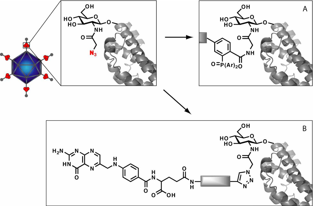
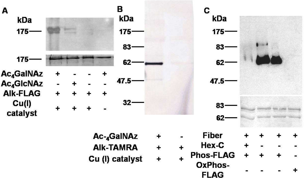
Metabolic incorporation of azide bearing sugars into mammalian cell surface proteins has been used for identification, functional analysis and imaging purposes.14,15 As viruses used for therapeutic applications are generated in eukaryotic cell lines, use of unnatural sugars during the propagation cycle is expected to potentially substitute for monosaccharides within viral glycoproteins. As a result of low pathogenic potential, ease of production and established safety protocols, human adenovirus type 5 has been developed extensively as vectors for gene replacement and oncolytic therapy.16 Human Ad5 is reported to have a single O-GlcNAc residue on Ser-109 of the fiber protein (Figure 1), which exists as a homotrimer on twelve vertices of the virus capsid.17,18 Replacing this sugar residue with an azido analog would allow for specific placement of an azide on the viral coat. Advantageously, Ser-109 is located proximal to the natural primary targeting motif on the fiber knob. Thus chemoselective placement of targeting moieties at this site is expected to be effective for re-directing viral particles. Towards this goal, E1 deleted Ad5 particles were propagated on HEK 293 cells. During the infection period, the culture media was supplemented with 50 µM Ac4GalNAz (Supplementary Scheme 1) or Ac4GlcNAz (Supplementary Scheme 2). Both precursor sugars are metabolic precursors of UDP-GlcNAz, a known substrate for O-GlcNAc Transferase.19 Viruses produced from the infected 293 cells were purified on double CsCl gradients and probed for incorporation of azido sugar with an engineered peptide epitope FLAG (DYKDDDDK), bearing either an N-terminal alkyne or a modified triaryl phosphine motif (Figure 1). Purified viruses were modified with FLAG epitope and tetramethylrhodamine (TAMRA) probes using the copper accelerated azide-alkyne cycloaddition (CuAAC),20,21 commonly known as the “click” chemistry, or the Staudinger ligation reaction.22,23 Initial characterization of the O-GlcNAc site of hAd5 indicated that it was enzymatically inaccessible in complete virion. In order to determine if an installed O-GlcNAz was chemically accessible, whole viruses were chemically treated, and not denatured, prior to analysis. While western analysis of viruses grown with either Ac4GalNAz or Ac4GlcNaz sugar demonstrated effective labeling of the fiber protein (MW 61.5 kD) with O-GlcNAz (Figure 2A), Ac4GalNAz mediated labeling was markedly higher. In order to confirm both the identity and placement of the installed azide, modification analysis of both denatured virus and partially purified fiber was carried out. Modification of the denatured virus with a fluorescent probe under click conditions demonstrated exclusive labeling of a protein at 62 kD (Figure 2B). This corresponds to either fiber or protein IIIa, which co-migrate. Fiber was partially purified from complete virus and treated with a hexosaminidase known to remove both O-GlcNAc and O-GlcNAz.19 Subsequent chemical modification demonstrated almost a complete loss of azide-dependent signal (Figure 2C). Cumulatively, these results strongly indicate the fiber bears an O-GlcNAz modification, which is the exclusive source of virus labeling.
Figure 1.
A cartoon illustrating incorporated O-GlcNAz residue on the adenoviral fiber protein and its subsequent chemical modification with ligands. Potential sites of O-GlcNAz incorporation are indicated in red circles. Either “click” (A) or Staudinger ligation (B) chemistries were used to decorate metabolically labeled adenovirus.
Figure 2.
Qualitative analysis of azido sugar incorporation into the fiber protein of hAd5. A) Anti-FLAG Western of non-denatured viral particles treated with alkyne-FLAG under CuAAC conditions (100 mM Tris pH = 8; 1 mM CuBr; 3 mM Bathophenanthroline disulphonic acid disodium salt; 400 µM alk-FLAG; 12 hr; RT). Top: anti-FLAG Western. Bottom: Coomassie. B) Fluorescent analysis of denatured O-GlcNAz-enabled viral particles treated with alkyne-TAMRA under CuAAC conditions (ex: 532; em: 580 BP 30) C) Hexosaminidase treatment (5 µg/µL Hex-C; 37°C overnight) and subsequent Staudinger modification of partially purified O-GlcNAz labeled fiber protein (100 mM Tris pH = 8; 400 µM phos-FLAG; 2 hr; RT). Top: anti-FLAG Western. Bottom: Coomassie.
In contrast to genetic engineering, we expected the introduction of O-GlcNAz to have little impact on adenoviral physiology. Virus production in the presence of either azido sugar demonstrated no difference on producer cell viability. In addition, no reduction in particle production was apparent as a result of azide incorporation (Supplementary Figure S1A). Similarly, infectivity of O-GlcNAz labeled virus particles as assessed via standard plaque forming assay, remained unaffected (Supplementary Figure S1B).
Fluorescently labeled particles were analyzed to determine the number of chemically addressable azides on the viral surface. Quantification of the number of on each virus by fluorescent gel scanning demonstrated exclusive fiber labeling in agreement with Western analysis (Supplementary Figure S2) and an average attachment of 21.9 ± 1.5 dyes per particle (Supplementary Table 1), consistent with previous O-GlcNAc characterization indicating ~50% occupancy at Ser109.18
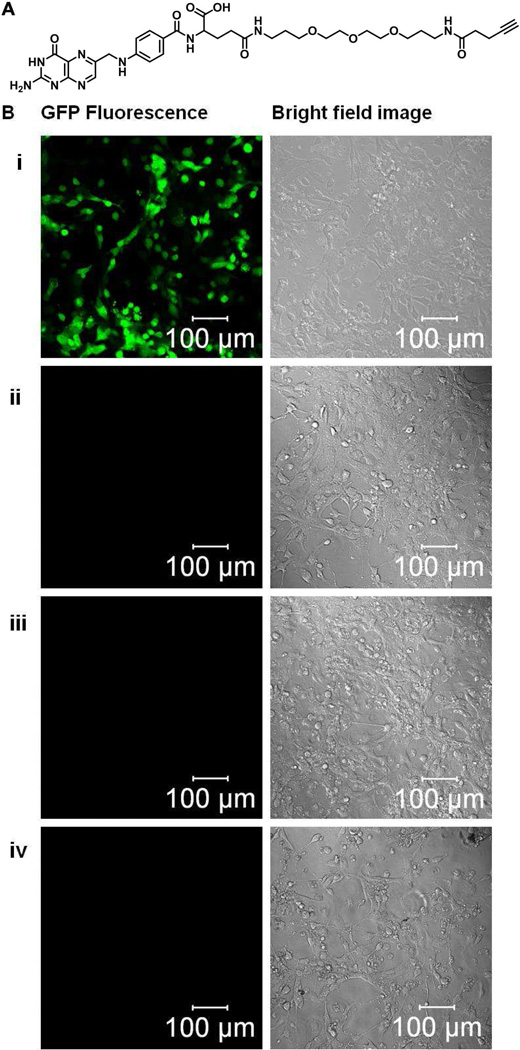
Low CAR expression is exhibited in a number of cancers, including ovarian, pancreatic and gastrointestinal cancers, generally limiting the effectiveness of hAd5 for oncolysis.24–27 As a result of tumor associated overexpression and limited abundance in normal tissues, the folate receptor has become an appealing target for cancer therapy.28,29 In addition, folate conjugates are easily produced and generally have minor impact on receptor affinity. In order to explore O-GlcNAz mediated folate modification we synthesized an alkyne probe containing a folic acid residue tethered by a small linker (Figure 3A and Supplementary scheme 3). O-GlcNAz enabled adenovirus, encoding a GFP transgene in the E1 deleted region of the viral genome regulated by a CMV promoter, was conjugated to alkynefolate via CuAAC. Mouse breast carcinoma cells (4T1), known to express moderate to high levels of folate receptors30 and naturally refractive to human adenovirus, were cultured in folate deficient media for 2 weeks. 24 hours after replating, the cells were infected with folate decorated virus at an MOI of 50. One day after infection the GFP expression was visualized. The results demonstrate a high level of GFP expression within the 4T1 cells infected with the folate labeled virus (Figure 3B). Human Ad5 produced without azido sugar, but treated with the folate reagent under CuAAC conditions, failed to produce significant transgene expression. In addition, free folate completely abrogated infection of folate modified hAd5, indicating folate receptor meditated gene delivery. Quantification of infection was assessed using a Synergy 2 fluoresence plate reader (excitation 485 ± 10 nm; emission 528 ± 10 nm) which showed a 3–4 fold increase of GFP expression in cells infected with metabolically and chemically modified virions versus unmodified virus. Infection assayed in the presence of increasing free folate concentration demonstrated dose dependent transfection inhibition of 4T1 cells (Supplemental Figure S3).
Figure 3.
Effective gene transduction of 4T1 cells with retargeted Ad5. A) Structure of alkyne-folate ligand. See Synthesis scheme 3 in supplemental information. B) GFP fluorescence microscopy images of alkyne-folate modified metabolically labeled Ad5 virus and metabolically unlabeled Ad5 infected 4T1 cells. Microscopy was carried out on Glass Bottom dishes using a Zeiss LSM 510 fluorescence microscope. Cells were infected at an MOI of 50pfu/cell and images taken 24hours post infection. (lanes i: 4T1 cells infected with folate modified Ac4GalNAz labeled Ad5; lane ii: 4T1 cells infected with metabolically unlabeled Ad5 (no Ac4GalNAz); lane iii: 4T1 cells infected with folate unmodified Ac4GalNAz labeled Ad5 (no alkyne-folate); lane iv: 4T1 cells infected with folate modified Ac4GalNAz labeled Ad5 pre-treated with 1 mg/L folic acid).
In summary we have demonstrated that adenoviruses can be chemoselectively labeled through a two-step process. Metabolic labeling with azido sugars yields adenoviral particles with site-specific placement of a chemically accessible azide without loss in either viral production or infectivity. Subsequent chemical modification of these particles allows the facile appendage of a variety of functionality from peptides to fluorophores to small molecules targeting moieties. The remarkable ease and specificity of this approach in combination with its non-perturbing nature make it accessible to a wide range of researchers. Further, while the broad application of adenoviral vectors make them particularly attractive targets, this approach is not limited to adenoviruses, but is expected to be generally applicable to the wide variety of viruses with peripheral glycoproteins, including many oncolytic vectors currently under development (retroviruses, lentiviruses, poxviruses and herpes viruses).
Supplementary Material
Acknowledgements
Support fort his work was provided by the NSF (CBET-1080909, ISC) and the NIH (5R01AI041636, PH). We would like to thank Dr. Guo-Wei Tian for support with fluorescence imaging. We are grateful to Prof. Nicole Sampson and Prof. Orlando Schärer for help with instrumentation.
Footnotes
Supporting Information Available
Details of azido sugar substrates and targeting folate molecule synthesis, peptide synthesis, cell culture condition, adenovirus production and purification as well as experimental details of labelling, detection, characterization and targeting of modified viruses. The material is available free of charge via the internet at http://pubs.acs.org.
Reference
- 1.Waehler R, Russell SJ, Curiel DT. Nat. Rev. Genet. 2007;8:573. doi: 10.1038/nrg2141. [DOI] [PMC free article] [PubMed] [Google Scholar]
- 2.Hedley S, Chen J, Mountz J, Li J, Curiel D, Korokhov N, Kovesdi I. Cancer Immunol. Immunother. 2006;55:1412. doi: 10.1007/s00262-006-0158-2. [DOI] [PMC free article] [PubMed] [Google Scholar]
- 3.Matthews Q, Yang P, Wu Q, Belousova N, Rivera A, Stoff-Khalili M, Waehler R, Hsu H-C, Li Z, Li J, Mountz J, Wu H, Curiel D. Virol. J. 2008;5:98. doi: 10.1186/1743-422X-5-98. [DOI] [PMC free article] [PubMed] [Google Scholar]
- 4.Magnusson MKHSS, Henning P, Boulanger P, Lindholm L. J. Gene Med. 2002;4:356. doi: 10.1002/jgm.285. [DOI] [PubMed] [Google Scholar]
- 5.Henning P, Lundgren E, Carlsson M, Frykholm K, Johannisson J, Magnusson MK, Tang E, Franqueville L, Hong SS, Lindholm L, Boulanger P. J. Gen. Virol. 2006;87:3151. doi: 10.1099/vir.0.81992-0. [DOI] [PubMed] [Google Scholar]
- 6.Croyle MA, Yu Q-C, Wilson JM. Hum. Gene Ther. 2000;11:1713. doi: 10.1089/10430340050111368. [DOI] [PubMed] [Google Scholar]
- 7.Kreppel F, Kochanek S. Mol. Ther. 2007;16:16. doi: 10.1038/sj.mt.6300321. [DOI] [PubMed] [Google Scholar]
- 8.Kreppel F, Gackowski J, Schmidt E, Stefan K. Mol. Ther. 2005;12:107. doi: 10.1016/j.ymthe.2005.03.006. [DOI] [PubMed] [Google Scholar]
- 9.Carrico ZM, Romanini DW, Mehl RA, Francis MB. Chem. Commun. 2008:1205. doi: 10.1039/b717826c. [DOI] [PubMed] [Google Scholar]
- 10.Strable E, Prasuhn DE, Udit AK, Brown S, Link AJ, Ngo JT, Lander G, Quispe J, Potter CS, Carragher B, Tirrell DA, Finn MG. Bioconj. Chem. 2008;19:866. doi: 10.1021/bc700390r. [DOI] [PMC free article] [PubMed] [Google Scholar]
- 11.Gupta SS, Raja KS, Kaltgrad E, Strable E, Finn MG. Chem. Comm. 2005:4315. doi: 10.1039/b502444g. [DOI] [PubMed] [Google Scholar]
- 12.Bruckman MA, Kaur G, Lee LA, Xie F, Sepulveda J, Breitenkamp R, Zhang X, Joralemon M, Russell TP, Emrick T, Wang Q. Chem Bio Chem. 2008;9:519. doi: 10.1002/cbic.200700559. [DOI] [PubMed] [Google Scholar]
- 13.Hong V, Presolski S, Ma C, Finn M. Angewandte Chemie International Edition. 2009;48:9879. doi: 10.1002/anie.200905087. [DOI] [PMC free article] [PubMed] [Google Scholar]
- 14.Chang PV, Prescher JA, Hangauer MJ, Bertozzi CR. J. Am. Chem. Soc. 2007;129:8400. doi: 10.1021/ja070238o. [DOI] [PMC free article] [PubMed] [Google Scholar]
- 15.Agard NJ, Bertozzi CR. Acct. Chem. Res. 2009;42:788. doi: 10.1021/ar800267j. [DOI] [PMC free article] [PubMed] [Google Scholar]
- 16.Christoph V, Stefan K. J. Gene Med. 2004;6:S164. [Google Scholar]
- 17.Mullis KG, Haltiwanger RS, Hart GW, Marchase RB, Engler JA. J. Virol. 1990;64:5317. doi: 10.1128/jvi.64.11.5317-5323.1990. [DOI] [PMC free article] [PubMed] [Google Scholar]
- 18.Cauet G, Strub J-M, Leize E, Wagner E, Dorsselaer AV, Lusky M. Biochem. 2005;44:5453. doi: 10.1021/bi047702b. [DOI] [PubMed] [Google Scholar]
- 19.Vocadlo DJ, Hang HC, Kim E-J, Hanover JA, Bertozzi CR. Proc. Natl. Acad. Sci. USA. 2003;100:9116. doi: 10.1073/pnas.1632821100. [DOI] [PMC free article] [PubMed] [Google Scholar]
- 20.Agard NJ, Baskin JM, Prescher JA, Lo A, Bertozzi CR. ACS Chem. Biol. 2006;1:644. doi: 10.1021/cb6003228. [DOI] [PubMed] [Google Scholar]
- 21.Gupta SS, Kuzelka J, Singh P, Lewis WG, Manchester M, Finn MG. Bioconj. Chem. 2005;16:1572. doi: 10.1021/bc050147l. [DOI] [PubMed] [Google Scholar]
- 22.Kiick KL, Saxon E, Tirrell DA, Bertozzi CR. Proc. Natl. Acad. Sci. USA. 2002;99:19. doi: 10.1073/pnas.012583299. [DOI] [PMC free article] [PubMed] [Google Scholar]
- 23.Saxon E, Bertozzi CR. Science. 2000;287:2007. doi: 10.1126/science.287.5460.2007. [DOI] [PubMed] [Google Scholar]
- 24.Glasgow JN, Everts M, Curiel DT. Cancer Gene Ther. 2006;13:830. doi: 10.1038/sj.cgt.7700928. [DOI] [PMC free article] [PubMed] [Google Scholar]
- 25.Yamamoto M, Curiel DT. Mol. Ther. 2009;18:243. doi: 10.1038/mt.2009.266. [DOI] [PMC free article] [PubMed] [Google Scholar]
- 26.Jogler C, Hoffmann D, Theegarten D, Grunwald T, Uberla K, Wildner O. J. Virol. 2006;80:3549. doi: 10.1128/JVI.80.7.3549-3558.2006. [DOI] [PMC free article] [PubMed] [Google Scholar]
- 27.Shashkova EV, May SM, Barry MA. Virology. 2009;394:311. doi: 10.1016/j.virol.2009.08.038. [DOI] [PMC free article] [PubMed] [Google Scholar]
- 28.Low PS, Kularatne SA. Curr. Opin. Chem. Biol. 2009;13:256. doi: 10.1016/j.cbpa.2009.03.022. [DOI] [PubMed] [Google Scholar]
- 29.Oh IK, Mok H, Park TG. Bioconj. Chem. 2006;17:721. doi: 10.1021/bc060030c. [DOI] [PubMed] [Google Scholar]
- 30.Russell-Jones G, McTavish K, McEwan J, Rice J, Nowotnik D. J. Inorg. Biochem. 2004;98:1625. doi: 10.1016/j.jinorgbio.2004.07.009. [DOI] [PubMed] [Google Scholar]
Associated Data
This section collects any data citations, data availability statements, or supplementary materials included in this article.


