Abstract
Hypothesis
Screening for esophageal adenocarcinoma has focused on identifying Barrett esophagus (BE) in patients with severe, long-standing symptoms of gastroesophageal reflux disease(GERD). Unfortunately, 95%of patients who develop esophageal adenocarcinoma are unaware of the presence of BE before their cancer diagnosis, which means they never had been selected for screening. One possible explanation is that no correlation exists between the severity of GERD symptoms and cancer risk. We hypothesize that severe GERD symptoms are not associated with an increase in the prevalence of BE, dysplasia, or cancer in patients undergoing primary endoscopic screening.
Design
Cross-sectional study.
Setting
University hospital.
Patients
A total of 769 patients with GERD.
Interventions
Primary screening endoscopy performed from November 1, 2004, through June 7, 2007.
Main Outcomes Measures
Symptom severity, proton pump inhibitor therapy, and esophageal adenocarcinogenesis (ie, BE, dysplasia, or cancer).
Results
Endoscopy revealed adenocarcinogenesis in 122 patients. An increasing number of severe GERD symptoms correlated positively with endoscopic findings of esophagitis (odds ratio, 1.05; 95% confidence interval, 1.01-1.09). Conversely, an increasing number of severe GERD symptoms were associated with decreased odds of adenocarcinogenesis (odds ratio, 0.94; 95% confidence interval, 0.89-0.98). Patients taking proton pump inhibitors were 61.3% and 81.5% more likely to have adenocarcinogenesis if they reported no severe typical or atypical GERD symptoms, respectively, compared with patients taking proton pump inhibitors, who reported that all symptoms were severe.
Conclusions
Medically treated patients with mild or absent GERD symptoms have significantly higher odds of adenocarcinogenesis compared with medically treated patients with severe GERD symptoms. This finding may explain the failure of the current screening paradigm in which the threshold for primary endoscopic examination is based on symptom severity.
On the basis of several epidemiologic studies in the late 1990s,1-5 recommendations have been made to screen white men older than 50 years with longstanding and severe symptoms of gastroesophageal reflux disease (GERD) for Barrett esophagus (BE), the known precursor lesion to esophageal adenocarcinoma.6 Despite these recommendations, 95% of patients who develop esophageal adenocarcinoma have not received endoscopic screening or been diagnosed as having BE, and most patients present with advanced disease and little chance for cure.7 Indications for screening1,8-11 historically included long-standing GERD and persistent severe symptoms despite adequate medical therapy, but these criteria have failed physicians in their attempt to stratify patients for prevention and early detection. In fact, the most recent guidelines state that screening has not been effective in even the highest-risk group and should, therefore, be “individualized to the patient.”11
One possible explanation for the failure of the current screening paradigm is that no correlation has been observed between the severity of GERD symptoms and cancer risk; because their symptoms are absent, insignificant, or atypical, these patients do not seek medical care or are not selected for endoscopic screening, and occult disease progression ensues. In addition, because proton pump inhibitors (PPIs) are extremely effective in reducing or eliminating symptoms, patients may be overlooked for screening under the false assumption that their disease is adequately treated. We hypothesize that severe GERD symptoms, particularly in patients who are taking PPIs, are not associated with an increase in the prevalence of BE, dysplasia, or cancer in patients undergoing primary endoscopic screening.
Methods
Overview
We performed an institutional review board–approved cross-sectional analysis of typical and atypical GERD symptoms and endoscopic findings in patients at Oregon Health & Science University (OHSU) and the Portland Veterans Administration Medical Center (PVAMC). All patients underwent primary screening endoscopy (ie, esophagogastroduodenoscopy), defined as the first ever esophagogastroduodenoscopy, and completed the validated GERD Health-Related Quality of Life questionnaire (GERD-HRQL)12 and the Reflux Symptom Index (RSI)13 from November 1, 2004, through June 7, 2007. Three distinct cohorts of patients were included to capture the full spectrum of possible GERD symptoms (Table 1).
Table 1. Number of Study Patients by Indication for Screening Endoscopya.
| Indicationb | Eligible | Excluded | Total Enrolled |
|---|---|---|---|
| Any indication | 460 | 45c | 415 |
| Typical GERD symptoms | 274 | 173d | 101 |
| Atypical GERD symptoms | 523 | 270e | 253 |
| Total No. of Patients | 1257 | 488 | 769 |
Abbreviation: GERD, gastroesophageal reflux disease.
Exclusion criteria: prior antireflux surgery, pregnancy, esophageal diverticulum, anticoagulation therapy, history of recurrent epistaxis, esophageal varices, otolaryngologic malignant neoplasms, prior laryngeal surgery, or trauma to the larynx.
Because of personnel constraints, only approximately 1 in 5 patients seen in the clinic was approached for study enrollment.
Reasons for exclusion: inadequate endoscopic examination (n = 15) and incomplete questionnaires (n = 30).
Reasons for exclusion: declined to participate (n = 132), unable to schedule or complete both endoscopies (n = 20), excluded after randomization at primary physician request (n = 3), medical contraindications at time of endoscopy (n = 3), and incomplete questionnaires (n = 15).
Reasons for exclusion: declined to participate (n = 149), unable to provide consent (n = 70), inability to schedule or complete endoscopy (n = 25), and incomplete questionnaires (n = 26).
The primary cohort was composed of adult patients referred to gastroenterology clinics who were subsequently scheduled for a clinically indicated upper endoscopy. Patients were enrolled regardless of the indication for the procedure. The second cohort of patients, all with typical GERD symptoms (ie, heartburn, regurgitation, or dysphagia) undergoing their first endoscopy or BE surveillance, had been targeted specifically for a randomized controlled trial studying small-caliber endoscopic screening without sedation.14 All patients in this cohort also had been scheduled for a clinically indicated upper endoscopic test (based on GERD symptoms) before being enrolled. The third cohort consisted of patients with atypical reflux symptoms (ie, hoarseness, throat-clearing, excess mucus, globus sensation, or cough) in the general otolaryngology clinics at OHSU and PVAMC who were enrolled in a BE prevalence study; all patients with nonmalignant ear, nose, and throat conditions completed the RSI at the time of their initial appointment. The patients who scored 2 or higher of 5 points for any 2 symptoms or 3 or higher of 5 points for any 1 symptom were approached for enrollment. Patients with a history of prior upper endoscopy or BE were excluded from all 3 cohorts. A total of 769 patients had GERD-HRQL and RSI questionnaires and a detailed medication history obtained; the questionnaires were administered prior to endoscopy.
Symptom Assessment and Severity Definitions
The severity of each patient's typical and atypical GERD symptoms was quantified using the GERD-HRQL questionnaire12 and the RSI,13 respectively (Table 2). The former is a disease-specific instrument whose validity and reliability have been assessed compared with generic quality-of-life scales (ie, the 36-Item Short Form Health Survey)15 and to physiologic factors of GERD.16 The latter is used to assess the severity of atypical symptoms of GERD, specifically laryngopharyngeal reflux symptoms such as hoarseness and cough. The validity and reliability of this measure have been established for patients with laryngopharyngeal reflux who were treated with PPIs and antireflux surgery.13 The GERD-HRQL questionnaire and the RSI provide complementary information regarding GERD symptoms.
Table 2. Gastroesophageal Reflux Disease Symptom Questionnaires.
| GERD Health-Related Quality of Life Questionnaire12 | ||||||
| 1. How bad is your heartburn? | □No symptoms (0) | |||||
| 2. Do you have heartburn when lying down? | □Symptoms noticeable but not bothersome (1) | |||||
| 3. Do you have heartburn when standing up? | □Symptoms bothersome every day (2) | |||||
| 4. Do you have heartburn after meals? | □Symptoms affect daily activities (3) | |||||
| 5. Does heartburn change your diet? | □Symptoms are incapacitating—unable to do daily activities (4) | |||||
| 6. Does heartburn wake you from sleep? | ||||||
| 7. Do you have difficulty swallowing? | ||||||
| 8. Do you have pain with swallowing? | ||||||
| 9. Do you have bloating or gassy feelings? | ||||||
|
| ||||||
| Reflux Symptom Index13 | ||||||
| Within the last month, how did the following problems affect you? | 0 = No problem→5 = severe problem | |||||
| Please circle the number that describes how you felt. | ||||||
| 1. Hoarseness or a problem with your voice | 0 | 1 | 2 | 3 | 4 | 5 |
| 2. Clearing your throat | 0 | 1 | 2 | 3 | 4 | 5 |
| 3. Excess throat mucus or postnasal drip | 0 | 1 | 2 | 3 | 4 | 5 |
| 4. Difficulty swallowing food, liquids, or pillsa | 0 | 1 | 2 | 3 | 4 | 5 |
| 5. Coughing after you eat or after lying down | 0 | 1 | 2 | 3 | 4 | 5 |
| 6. Breathing difficulties or choking episodes | 0 | 1 | 2 | 3 | 4 | 5 |
| 7. Troublesome or annoying cough | 0 | 1 | 2 | 3 | 4 | 5 |
| 8. Sensations of something sticking in your throat or a lump in your throat | 0 | 1 | 2 | 3 | 4 | 5 |
Abbreviation: GERD, gastroesophageal reflux disease.
Not used when responses from the GERD Health-Related Quality of Life questionnaire and Reflux Symptom Index were combined to avoid duplication of dysphagia data.
Symptom severity was designated as “no symptoms,” “mild symptoms,” or “severe symptoms” based on the patient's response to each item in the GERD-HRQL questionnaire and the RSI (outlined in the severity schema in Table 3). On the basis of this classification system, the total number of severe GERD-HRQL (with 0-9 possible) and RSI (with 0-8 possible) symptoms were calculated independently as continuous variables. Finally, by combining the total number of severe symptoms from each (with dysphagia included only once; range, 0-16), a composite for each category of symptoms (ie, typical and atypical) was calculated.
Table 3. Symptom Severity Schema.
| Symptom Severity | GERD-HRQL Symptom Severity Definition | RSI |
|---|---|---|
| No symptoms | No symptoms | 0 |
| Mild symptoms | Noticeable but not bothersome | 1 |
| 2 | ||
| Severe symptoms | Bothersome every day | 3 |
| Affect daily activities | 4 | |
| Incapacitating | 5 |
Abbreviations: GERD-HRQL, Gastroesophageal Reflux Disease Health-Related Quality of Life questionnaire; RSI, Reflux Symptom Index.
Endoscopic Evaluation
Endoscopy was performed by gastroenterologists at OHSU and PVAMC as part of standard clinical practice or by trained clinician investigators (R.W.O. and B.A.J.) within the studies outlined previously. All endoscopic findings were entered into the Clinical Outcomes Research Initiative database with details of patient history, including demographic and lifestyle variables and medical history. Barrett esophagus was suspected during endoscopy when the squamocolumnar junction was located proximal to the anatomical esophagogastric junction (ie, the most proximal extent of the gastric folds).17 Intestinal metaplasia of the gastric cardia mucosa was not considered BE. Biopsy specimens were obtained in 4 quadrants every 2 cm throughout the entire Barrett segment using large biopsy forceps.18,19 Esophageal biopsy specimens were evaluated by staff pathologists at OHSU and PVAMC. The diagnosis of BE required the unequivocal presence of goblet cells within the columnar epithelium. Standard diagnostic criteria were used to identify columnar epithelial dysplasia and invasive cancer.20 Esophagitis was documented using the Los Angeles classification system,21,22 and the hiatal hernia size was measured as the distance in centimeters between the crural impressions and the anatomical esophagogastric junction.
Statistical Analyses
Statistical analyses were performed with SPSS for Windows (SPSS Inc, Chicago, Illinois) and SAS 9.1 TS Level 1M2 (SAS Institute Inc, Cary, North Carolina). Means, standard deviations, and ranges were presented for continuous variables. Proportions were presented for categorical variables. The value of 80 missing questionnaire responses for 62 study participants was estimated using the mean (rounded to the nearest integer) of each study participant's actual responses for that specific measure (ie, GERD-HRQL or RSI). This imputation allowed for the creation of summary GERD symptom severity measures, although it underestimated the variability of each participant's symptoms.
Associations between the outcome (ie, Barrett metaplasia, dysplasia, or adenocarcinoma) and covariates were assessed using the Pearson χ2 test and the Mantel-Haenszel test of trend, where appropriate. A multiple logistic regression model was created to model the relationship between symptom severity and the study outcome. Variables that were significant in univariate analysis with P < .25, known confounders (specifically, age, sex, and race/ethnicity), and probable biologic importance (specifically, presence of hiatal hernia and use of PPIs) were included in the model.
Pearson 2-tailed correlation statistics were calculated for all potential confounders. Of pairs that had at least moderate correlation (r>0.70), only 1 variable was included in the preliminary model. Backward stepwise selection was used to derive the preliminary effects model. The criteria for variable removal was determined as a Wald statistic with P> .10. Interactions between each GERD symptom severity variable and age, sex, and the current use of PPIs were assessed and retained in the final model at P< .05. The final model's goodness of fit was assessed with the Hosmer-Lemeshow test.23 Duration of symptoms was available only for the patients within the second (ie, patients with typical GERD) and third (ie, patients with atypical GERD) cohorts. To determine the association among symptom severity, duration of symptoms, and the study outcome, the analysis was repeated with the primary cohort (ie, any reason for endoscopy) excluded.
Results
Demographic Factors Associated with Esophageal Adenocarcinogenesis
Most patients were male (65.4%), white (94.3%), and non-Hispanic (97.8%). Typical GERD symptoms were present at the time of assessment in 67.1% of patients, and 57.2% were using a PPI at the time of primary endoscopy. One hundred seventy-nine of the 769 patients (23.3%) were found during endoscopy to have esophagitis, and 365 (47.5%) had a hiatal hernia with a mean (SD) size of 2.72 (1.72) cm.
Several factors, including the use of PPIs, were associated with increased odds for the presence of esophageal adenocarcinogenesis (Table 4). One hundred twenty-two (15.9%) of the patients had esophageal adenocarcinogenesis: BE in 99 (12.9%) (99 of 769), esophageal dysplasia in 17 (2.2%), and esophageal adenocarcinoma in 6 (0.8%). Esophageal adenocarcinogenesis was present in 31 of 253 otolaryngology clinic patients (12.3%) and 91 of 516 gastroenterology clinic patients (17.6%) (P = .06). Typical and/or atypical GERD symptoms were present for longer than 10 years in 130 of 367 (35.4%).
Table 4. Sociodemographic Characteristics of Case and Control Individuals: Crude Odds for Esophageal Adenocarcinogenesis by Known and Potential Risk Factors.
| Esophageal Adenocarcinogenesis | ||||
|---|---|---|---|---|
|
|
||||
| Characteristic | Total (N = 769) | Yes (n = 122 [15.9%]) | No (n = 647 [84.1%]) | Crude OR (95% CI) |
| Age, mean (SD), y | 57.1 (13.7) | 61.6 (11.6) | 56.3 (13.9) | 1.03 (1.02-1.05) |
| Sex, No. (%) | 6.56 (3.46-12.44) | |||
| Male | 503 (65.4) | 111 (91.0) | 392 (77.9) | |
| Female | 266 (34.6) | 11 (9.0) | 255 (95.9) | |
| Race/ethnicity, No. (%)a | 3.36 (1.03-10.95) | |||
| White | 708 (92.1) | 115 (94.3) | 593 (83.8) | |
| Other than white | 55 (7.2) | 3 (2.5) | 52 (94.5) | |
| Use of proton pump inhibitors, No. (%)b | 2.41 (1.55-3.72) | |||
| Yes | 440 (57.2) | 89 (73.0) | 351 (79.8) | |
| No | 325 (42.3) | 31 (25.4) | 294 (90.5) | |
| Hiatal hernia, No. (%) | 3.63 (2.36-5.57) | |||
| Yes | 365 (47.5) | 89 (24.4) | 276 (75.6) | |
| No | 404 (52.5) | 33 (8.2) | 371 (91.8) | |
| Duration of symptoms, mean (SD), yc | 2.66 (1.57-4.50) | |||
| No. of patients | 367 | 76 | 291 | |
| ≤10 | 237 (65) | 35 (14.8) | 202 (85.2) | |
| >10 | 130 (35) | 41 (31.5) | 89 (68.5) | |
Abbreviations: CI, confidence interval; OR, odds ratio.
Six participants (0.8%) were missing data regarding this characteristic.
Four participants (0.5%) were missing data regarding this characteristic.
Duration of symptoms was obtained only for the typical gastroesophageal reflux disease and atypical gastroesophageal reflux disease patient cohorts. Symptoms included typical and/or atypical gastroesophageal reflux disease.
Association Between Gerd Symptom Severity and Esophagitis
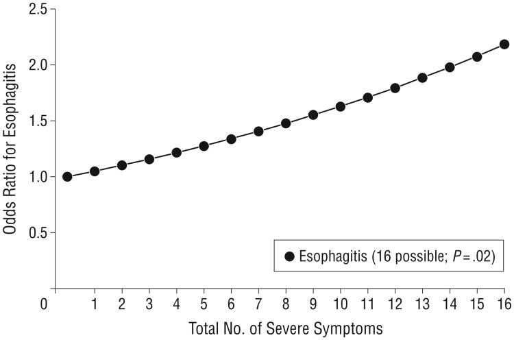
Regardless of PPI use status, an increased number of severe GERD symptoms correlated positively with the identification of esophagitis (odds ratio [OR], 1.05; 95% confidence interval [CI], 1.01-1.09) when compared with those with mild or no symptoms (Figure 1). However, when stratified by typical and atypical symptoms, only an increased number of severe typical GERD symptoms correlated positively with the presence of esophagitis (OR, 1.08; 95% CI, 1.02-1.1).
Figure 1.
Correlation between the number of severe symptoms and the presence of erosive esophagitis. The odds of esophagitis increase as the number of severe symptoms increases. This finding is expected because esophageal acidification is responsible for the sensation of heartburn, and effective acid suppression has been proven to reduce or eliminate symptoms and heal erosions.24
Association Between Gerd Symptom Severity and Esophageal Adenocarcinogenesis
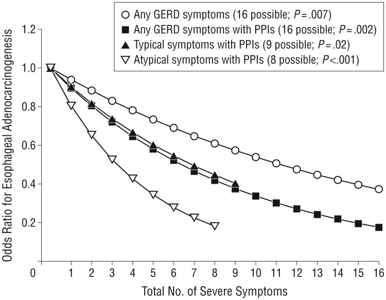
Patients with no severe symptoms were 63% (OR, 0.94; 95% CI, 0.89-0.98) more likely to have esophageal adenocarcinogenesis than patients reporting that all GERD symptoms were severe. This relationship persisted when typical and atypical symptoms were analyzed separately (OR, 0.92 [95% CI, 0.86-0.99]; and 0.91 [0.84-0.99], respectively).
Gerd Symptom Severity, Odds of Esophageal Adenocarcinogenesis, and Ppi Use
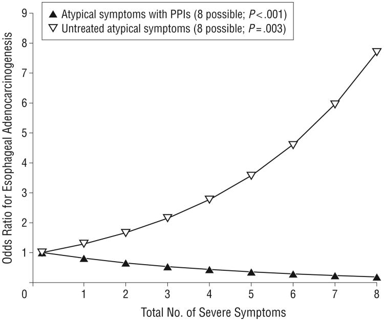
After adjusting for age, sex, race/ethnicity, presence of hiatal hernia, clinic, and institution of recruitment, the total number of severe GERD symptoms was inversely and significantly associated with the odds of esophageal adenocarcinogenesis among patients using PPIs (OR, 0.90; 95% CI, 0.84-0.96). Within this model, no significant association was observed between symptom severity and adenocarcinogenesis in patients not using PPIs (OR, 1.06; 95% CI, 0.96-1.18) (Table 5 and Figure 2).
Table 5. Adjusted Risk of Esophageal Adenocarcinogenesis by GERD Symptom Severity and PPI Usea.
| PPI User | PPI Nonuser | |||||
|---|---|---|---|---|---|---|
|
|
|
|||||
| Symptom | Mean (SD)[Range] | Adjusted OR (95% CI) | P Value (Wald Statistic) | Mean (SD) | Adjusted OR (95% CI) | P Value (Wald Statistic) |
| Total No. of severe symptoms | 6.33 (4.33) [0-16] | 0.90 (0.84-0.96) | .002 | 4.99 (4.15) | 1.06 (0.96-1.18) | .24 |
| No. of severe typical symptoms | 2.79 (2.97) [0-9] | 0.90 (0.83-0.99) | .02 | NA | NA | NA |
| No. of severe atypical symptoms | 3.60 (2.56) [0-8] | 0.81 (0.72-0.91) | <.001 | 2.97 (2.46) | 1.29 (1.09-1.54) | .003 |
Abbreviations: CI, confidence interval; GERD, gastroesophageal reflux disease; NA, not applicable; OR, odds ratio; PPI, proton pump inhibitor.
Multivariate logistic regression model adjusted for age (categorized with cutoffs at ages 58 and 68 years), sex, race/ethnicity (white vs other than white), use of PPIs, presence of hiatal hernia, clinic where recruited (gastroenterology vs otolaryngology), and institution where recruited (Oregon Health & Science University vs Portland Veterans Administration Medical Center). Three multivariate models included an interaction term between the GERD symptom severity variable and the use of PPIs and so were reported stratified by PPI use. Odds ratios were calculated by logistic regression.
Figure 2.
Correlation between the number of gastroesophageal reflux disease (GERD), typical, and atypical severe symptoms and the presence of esophageal adenocarcinogenesis in patients. As the number of severe typical or atypical reflux symptoms increased, a significantly decreased risk was observed for the presence of Barrett esophagitis, dysplasia, and cancer. This finding was most pronounced in patients taking proton pump inhibitors (PPIs).
When analyzed by typical (OR, 0.90; 95% CI, 0.83-0.99) and atypical symptoms (0.81; 0.72-0.91), this inverse association persisted among PPI users. Patients taking PPIs were 61.3% and 81.5% more likely to have esophageal adenocarcinogenesis if they reported no severe typical or atypical GERD symptoms, respectively. In patients with atypical GERD symptoms who were not using PPIs, the converse was true. For these patients, the odds of esophageal adenocarcinogenesis at screening endoscopy increased by 29% (OR, 1.29; 95% CI, 1.09-1.54) as the number of severe GERD symptoms increased (Figure 3).
Figure 3.
Correlation between the number of atypical severe symptoms and the presence of esophageal adenocarcinogenesis. Patients with an increasing number of severe atypical gastroesophageal reflux disease (GERD) symptoms who were not taking proton pump inhibitors (PPIs) composed the group most likely to have Barrett esophagitis, dysplasia, or cancer. This finding is surprising because most of these patients were recruited from otolaryngology clinics and rarely would have been targeted for screening endoscopy.
Gerd Symptom Severity, Odds of Esophageal Adenocarcinogenesis, and Symptom Duration
The association between the number of severe typical and/or atypical GERD symptoms and esophageal adenocarcinogenesis was then determined while controlling for duration of symptoms and PPI use. Patients with a 10-year or longer history of typical or atypical GERD symptoms were 3-fold more likely to have esophageal adenocarcinogenesis (OR, 3.02; 95% CI, 1.70-5.40) compared with a duration of symptoms of less than 10 years. Despite this, the inverse association with the total number of severe GERD symptoms persisted (OR, 0.82; 95% CI, 0.74-0.92). The relationship was even more pronounced when the analysis was stratified for duration of symptoms of more than 10 years in patients who were using PPIs (OR, 0.70; 95% CI, 0.58-0.84).
Comment
This study explored the relationship between typical and atypical GERD symptom severity and the odds of esophageal adenocarcinogenesis at the time of primary endoscopic screening. The results demonstrate that patients with minimal or no GERD symptoms who are using PPIs have increased odds of esophageal adenocarcinogenesis compared with those with severe symptoms, especially when GERD symptoms have been present for more than 10 years. These findings highlight one of many potential causes for the ineffectiveness of current screening efforts and implicate PPIs as a factor by which patients may be rendered asymptomatic in the face of continued mutagenic exposures.
The importance of further defining potential risk factors for esophageal adenocarcinoma is clear. Since 1975, the incidence of this cancer has increased significantly.25-27 Using the presence of long-standing severe GERD symptoms to guide screening strategies has failed to reduce the number of patients presenting with incurable disease. Some have suggested that the nearly epidemic use of antisecretory medications, coupled with the insensate nature of the Barrett epithelium,28-31 may be reducing or eliminating GERD symptoms in the patients who are at highest risk, thus rendering them susceptible to occult disease progression. In addition, for a given degree of exposure to refluxate, the perception of GERD symptom severity may be highly variable among patients, which may contribute to occult disease progression in minimally symptomatic patients.
Although we are unable to determine causality from this study, the results have biologic plausibility. Acid suppression therapy is effective at eliminating symptoms despite unremitting reflux events.32 Vela and colleagues32 demonstrated that acid suppression in GERD patients did not result in a decrease in the total number of reflux episodes but rather in a shift from acidic to nonacidic reflux coupled with symptom resolution. In fact, as many as 50% of patients with BE who are taking PPIs will have persistent weak acid esophageal exposure despite complete symptom resolution.33-35 Because bile acids become lipid soluble in the presence of a weak acid environment (ie, pH 3-5), they can move through the cell membrane, where they likely activate the caudal-related homeobox gene (Cdx-2 [GenBank NM_007673]) to promote BE.36,37
Lagergren and colleagues1 established symptomatic GERD as a risk factor for esophageal adenocarcinoma in their population-based, case-control study of esophageal adenocarcinoma cases in the 1990s. They established that long-standing and severe GERD symptoms were associated with an adjusted OR for esophageal adenocarcinoma of 43.5-fold compared with no symptoms. However, in their study, symptom severity was defined by symptom frequency as opposed to patient perception of severity. Although frequency most likely serves as a proxy for severity, it is not necessarily indicative of clinically severe GERD. Indeed, physicians and patients rarely consider mild symptoms, even if they occur frequently, as indicating severe GERD because the symptoms are tolerable and easily managed with medications.
Although cross-sectional and matched case-control studies3,8,38 of patients referred for upper endoscopy have shown that the age of onset, male sex, hiatal hernia, and duration of GERD symptoms are associated with BE, the relationship between the severity of GERD symptoms and BE has been less understood, and reports give conflicting results. For example, Locke and colleagues39 found no association between the presence of BE and GERD symptom severity in a large community-based population referred for esophagogastroduodenoscopy, although Eloubeidi and Provenzale40 discovered in their study of US Armed Services veterans that patients with BE were more likely to report less severe symptoms (adjusted OR, 0.13; 95% CI, 0.04-0.42) than patients with clinical GERD and no BE.
Of interest, in our study, patients with severe atypical symptoms who were not medically treated had significantly increased odds of esophageal adenocarcinogenesis compared with those with no severe symptoms. This finding is important because patients who are being evaluated for symptoms such as hoarseness, throat-clearing, excess mucus, globus sensation, or cough may not have reflux of gastrointestinal contents considered to be a possible cause of their symptoms. As such, they do not undergo esophageal screening and are not treated with PPIs. This group of patients may represent those who have little or no heartburn or regurgitation and are thus referred for evaluation to an otolaryngologist rather than a gastroenterologist. This supposition is supported by the fact that the prevalences of adenocarcinogenesis in otolaryngology and gastroenterology patients were statistically similar in this study.
As a cross-sectional investigation, this study reports only correlations at a single time point among the variables of interest, and it cannot establish causation. Given this limitation, this study reflects the patient population encountered clinically, with a wide range of GERD symptom severity at various stages of esophageal injury. In addition, the analysis did not control for all known risk factors for BE and esophageal adenocarcinoma, such as obesity41 and the duration of GERD symptoms.3,8 Inclusion of these potential confounders may have revealed a different relationship between GERD symptom severity and esophageal adenocarcinogenesis. Although the individual questionnaires are validated, the symptom severity scale developed for this study was not. However, given the lack of a commonly accepted measurement of GERD symptom severity, it was necessary to create internal scales to achieve the aims of this investigation.
Despite these concerns, this study exhibited a high degree of content validity. For example, the rate of esophageal adenocarcinogenesis discovered in this screening population was consistent with that reported for the general GERD population by other authors.42-47 Also, previously established risk factors for esophageal adenocarcinogenesis, such as age, sex, ethnicity, and presence of hiatal hernia, were confirmed and reproduced in this study. Although this study is the first, to our knowledge, to examine the relationship among the severity of GERD symptoms, PPI use, and esophageal adenocarcinogenesis, others have demonstrated an increased risk of esophageal adenocarcinogenesis for patients taking PPIs. Those studies attributed this finding to confounding by intent, whereby PPI use was simply a marker for the severity of the patient's GERD rather than a direct causative factor.39,48,49 Finally, consistent with the published literature, our results demonstrated a positive correlation between symptom severity and the presence of esophagitis.24,50,51
In summary, patients with medically controlled GERD symptoms who underwent primary screening endoscopy had significantly increased odds of BE, dysplasia, and cancer compared with medically treated patients with severe GERD symptoms. Also, patients with untreated severe atypical symptoms are at increased risk for esophageal adenocarcinogenesis. These findings highlight potential causes for the ineffectiveness of the current esophageal cancer screening paradigm and suggest that, rather than recommending BE screening only in patients with long-standing, poorly controlled GERD, patients with long-standing but well-controlled symptoms of typical or atypical GERD may be a better population to target. In addition, patients who present initially to the otolaryngology clinic with severe atypical-predominate symptoms should be strongly considered for primary screening endoscopy.
Larger-scale prospective studies, ideally having a validated measure of symptom severity, will enable us to determine the prevalence of BE stratified by symptom duration, antisecretory medication use, and current symptoms severity and lead to stronger guidance in recommendations for screening endoscopy.
Acknowledgments
Funding/Support: This study was supported by the Robert Anthony McHugh Research Fund for the Prevention and Early Detection of Esophageal Cancer, American Surgical Association Foundation Fellowship Award (Dr O'Rourke); and by National Institutes of Health (NIH) grants UL1 RR024140, K23 DK066165-01, and R21 DK081161 (Dr Jobe); by NIH grant K08 DK074397 (Dr O'Rourke); and by NIH grant U01 DK57132.
Footnotes
Author Contributions: Study concept and design: Wichienkuer, Awais, Morris, and Jobe. Acquisition of data: O'Rourke, Hunter, Morris, and Jobe. Analysis and interpretation of data: Nason, Wichienkuer, Schuchert, Luketich, O'Rourke, Morris, and Jobe. Drafting of the manuscript: Nason and Jobe. Critical revision of the manuscript for important intellectual content: Nason, Awais, Schuchert, Luketich, O'Rourke, Hunter, Morris, and Jobe. Statistical analysis: Nason, Wichienkuer, and Morris. Obtained funding: Hunter and Jobe. Administrative, technical, and material support: Nason, O'Rourke, Hunter, and Jobe. Study supervision: Hunter and Jobe.
Financial Disclosure: None reported.
Previous Presentation: These data were presented at the 81st Annual Meeting of the Pacific Coast Surgical Association Meeting; February 14, 2010; Kapalua, Hawaii.
Contributor Information
Katie S. Nason, Division of Thoracic and Foregut Surgery, University of Pittsburgh, Pittsburgh, Pennsylvania.
Promporn Paula Wichienkuer, Departments of Public Health and Preventive Medicine, Oregon Health & Science University, Portland.
Omar Awais, Division of Thoracic and Foregut Surgery, University of Pittsburgh, Pittsburgh, Pennsylvania.
Matthew J. Schuchert, Division of Thoracic and Foregut Surgery, University of Pittsburgh, Pittsburgh, Pennsylvania.
James D. Luketich, Division of Thoracic and Foregut Surgery, University of Pittsburgh, Pittsburgh, Pennsylvania.
Robert W. O'Rourke, Departments of Surgery, Oregon Health & Science University, Portland.
John G. Hunter, Departments of Surgery, Oregon Health & Science University, Portland.
Cynthia D. Morris, Departments of Informatics and Clinical Epidemiology, Oregon Health & Science University, Portland.
Blair A. Jobe, Division of Thoracic and Foregut Surgery, University of Pittsburgh, Pittsburgh, PennsylvaniaDepartments of Surgery, Oregon Health & Science University, Portland.
References
- 1.Lagergren J, Bergstrōm R, Lindgren A, Nyrén O. Symptomatic gastroesophageal reflux as a risk factor for esophageal adenocarcinoma. N Engl J Med. 1999;340(11):825–831. doi: 10.1056/NEJM199903183401101. [DOI] [PubMed] [Google Scholar]
- 2.Chow WH, Blot WJ, Vaughan TL, et al. Body mass index and risk of adenocarcinomas of the esophagus and gastric cardia. J Natl Cancer Inst. 1998;90(2):150–155. doi: 10.1093/jnci/90.2.150. [DOI] [PubMed] [Google Scholar]
- 3.Lieberman DA, Oehlke M, Helfand M. Gastroenterology Outcomes Research Group in Endoscopy. Risk factors for Barrett's esophagus in community-based practice: GORGE consortium. Am J Gastroenterol. 1997;92(8):1293–1297. [PubMed] [Google Scholar]
- 4.Drewitz DJ, Sampliner RE, Garewal HS. The incidence of adenocarcinoma in Barrett's esophagus: a prospective study of 170 patients followed 4.8 years. Am J Gastroenterol. 1997;92(2):212–215. [PubMed] [Google Scholar]
- 5.Vaughan TL, Davis S, Kristal A, Thomas DB. Obesity, alcohol, and tobacco as risk factors for cancers of the esophagus and gastric cardia: adenocarcinoma versus squamous cell carcinoma. Cancer Epidemiol Biomarkers Prev. 1995;4(2):85–92. [PubMed] [Google Scholar]
- 6.Baquet CR, Commiskey P, Mack K, Meltzer S, Mishra SI. Esophageal cancer epidemiology in blacks and whites: racial and gender disparities in incidence, mortality, survival rates and histology. J Natl Med Assoc. 2005;97(11):1471–1478. [PMC free article] [PubMed] [Google Scholar]
- 7.Dulai GS, Guha S, Kahn KL, Gornbein J, Weinstein WM. Preoperative prevalence of Barrett's esophagus in esophageal adenocarcinoma: a systematic review. Gastroenterology. 2002;122(1):26–33. doi: 10.1053/gast.2002.30297. [DOI] [PubMed] [Google Scholar]
- 8.Eisen GM, Sandler RS, Murray S, Gottfried M. The relationship between gastroesophageal reflux disease and its complications with Barrett's esophagus. Am J Gastroenterol. 1997;92(1):27–31. [PubMed] [Google Scholar]
- 9.Sampliner RE. Practice Parameters Committee of the American College of Gastroenterology. Updated guidelines for the diagnosis, surveillance, and therapy of Barrett's esophagus. Am J Gastroenterol. 2002;97(8):1888–1895. doi: 10.1111/j.1572-0241.2002.05910.x. [DOI] [PubMed] [Google Scholar]
- 10.Sampliner RE. Practice Parameters Committee of the American College of Gastroenterology. Practice guidelines on the diagnosis, surveillance, and therapy of Barrett's esophagus. Am J Gastroenterol. 1998;93(7):1028–1032. doi: 10.1111/j.1572-0241.1998.00362.x. [DOI] [PubMed] [Google Scholar]
- 11.Wang KK, Sampliner RE. Practice Parameters Committee of the American College of Gastroenterology. Updated guidelines 2008 for the diagnosis, surveillance and therapy of Barrett's esophagus. Am J Gastroenterol. 2008;103(3):788–797. doi: 10.1111/j.1572-0241.2008.01835.x. [DOI] [PubMed] [Google Scholar]
- 12.Velanovich V. The development of the GERD-HRQL symptom severity instrument. Dis Esophagus. 2007;20(2):130–134. doi: 10.1111/j.1442-2050.2007.00658.x. [DOI] [PubMed] [Google Scholar]
- 13.Belafsky PC, Postma GN, Koufman JA. Validity and reliability of the reflux symptom index (RSI) J Voice. 2002;16(2):274–277. doi: 10.1016/s0892-1997(02)00097-8. [DOI] [PubMed] [Google Scholar]
- 14.Jobe BA, Hunter JG, Chang EY, et al. Office-based unsedated small-caliber endoscopy is equivalent to conventional sedated endoscopy in screening and surveillance for Barrett's esophagus: a randomized and blinded comparison. Am J Gastroenterol. 2006;101(12):2693–2703. doi: 10.1111/j.1572-0241.2006.00890.x. [DOI] [PubMed] [Google Scholar]
- 15.Velanovich V. Comparison of generic (SF-36) vs. disease-specific (GERD-HRQL) quality-of-life scales for gastroesophageal reflux disease. J Gastrointest Surg. 1998;2(2):141–145. doi: 10.1016/s1091-255x(98)80004-8. [DOI] [PubMed] [Google Scholar]
- 16.Velanovich V, Karmy-Jones R. Measuring gastroesophageal reflux disease: relationship between the Health-Related Quality of Life score and physiologic parameters. Am Surg. 1998;64(7):649–653. [PubMed] [Google Scholar]
- 17.Wallner B, Sylvan A, Janunger KG. Endoscopic assessment of the “Z-line” (squamocolumnar junction) appearance: reproducibility of the ZAP classification among endoscopists. Gastrointest Endosc. 2002;55(1):65–69. doi: 10.1067/mge.2002.119876. [DOI] [PubMed] [Google Scholar]
- 18.Reid BJ, Blount PL, Feng Z, Levine DS. Optimizing endoscopic biopsy detection of early cancers in Barrett's high-grade dysplasia. Am J Gastroenterol. 2000;95(11):3089–3096. doi: 10.1111/j.1572-0241.2000.03182.x. [DOI] [PubMed] [Google Scholar]
- 19.Levine DS, Blount PL, Rudolph RE, Reid BJ. Safety of a systematic endoscopic biopsy protocol in patients with Barrett's esophagus. Am J Gastroenterol. 2000;95(5):1152–1157. doi: 10.1111/j.1572-0241.2000.02002.x. [DOI] [PubMed] [Google Scholar]
- 20.Lewin KJ, Appleman HD. Atlas of Tumor Pathology. Vol. 18. Washington, DC: Armed Forces Institute of Pathology, American Registry of Pathology; 1996. Tumors of the esophagus and stomach. [Google Scholar]
- 21.Nasseri-Moghaddam S, Razjouyan H, Nouraei M, et al. Inter- and intra-observer variability of the Los Angeles classification: a reassessment. Arch Iran Med. 2007;10(1):48–53. published correction appears in Arch Iran Med. 2007;10(3):429. [PubMed] [Google Scholar]
- 22.Lundell LR, Dent J, Bennett JR, et al. Endoscopic assessment of oesophagitis: clinical and functional correlates and further validation of the Los Angeles classification. Gut. 1999;45(2):172–180. doi: 10.1136/gut.45.2.172. [DOI] [PMC free article] [PubMed] [Google Scholar]
- 23.Hosmer DW, Lemeshow S. Applied Logistic Regression. Hoboken, NJ: John Wiley & Sons, Inc; 2000. [Google Scholar]
- 24.Pilotto A, Franceschi M, Leandro G, et al. Clinical features of reflux esophagitis in older people: a study of 840 consecutive patients. J Am Geriatr Soc. 2006;54(10):1537–1542. doi: 10.1111/j.1532-5415.2006.00899.x. [DOI] [PubMed] [Google Scholar]
- 25.Devesa SS, Blot WJ, Fraumeni JF., Jr Changing patterns in the incidence of esophageal and gastric carcinoma in the United States. Cancer. 1998;83(10):2049–2053. [PubMed] [Google Scholar]
- 26.Brown LM, Devesa SS, Chow WH. Incidence of adenocarcinoma of the esophagus among white Americans by sex, stage, and age. J Natl Cancer Inst. 2008;100(16):1184–1187. doi: 10.1093/jnci/djn211. [DOI] [PMC free article] [PubMed] [Google Scholar]
- 27.Jemal A, Siegel R, Ward E, Murray T, Xu J, Thun MJ. Cancer statistics, 2007. CA Cancer J Clin. 2007;57(1):43–66. doi: 10.3322/canjclin.57.1.43. [DOI] [PubMed] [Google Scholar]
- 28.Korkmaz M, Tarhan E, Unal H, Selcuk H, Yilmaz U, Ozluoglu L. Esophageal mucosal sensitivity: possible links with clinical presentations in patients with erosive esophagitis and laryngopharyngeal reflux. Dig Dis Sci. 2007;52(2):451–456. doi: 10.1007/s10620-006-9514-5. [DOI] [PubMed] [Google Scholar]
- 29.Fass R, Yalam JM, Camargo L, Johnson C, Garewal HS, Sampliner RE. Increased esophageal chemoreceptor sensitivity to acid in patients after successful reversal of Barrett's esophagus. Dig Dis Sci. 1997;42(9):1853–1858. doi: 10.1023/a:1018850824287. [DOI] [PubMed] [Google Scholar]
- 30.Grade A, Pulliam G, Johnson C, Garewal H, Sampliner RE, Fass R. Reduced chemoreceptor sensitivity in patients with Barrett's esophagus may be related to age and not to the presence of Barrett's epithelium. Am J Gastroenterol. 1997;92(11):2040–2043. [PubMed] [Google Scholar]
- 31.Niemantsverdriet EC, Timmer R, Breumelhof R, Smout AJ. The roles of excessive gastrooesophageal reflux, disordered oesophageal motility and decreased mucosal sensitivity in the pathogenesis of Barrett's oesophagus. Eur J Gastroenterol Hepatol. 1997;9(5):515–519. doi: 10.1097/00042737-199705000-00019. [DOI] [PubMed] [Google Scholar]
- 32.Vela MF, Camacho-Lobato L, Srinivasan R, Tutuian R, Katz PO, Castell DO. Simultaneous intraesophageal impedance and +pH measurement of acid and nonacid gastroesophageal reflux: effect of omeprazole. Gastroenterology. 2001;120(7):1599–1606. doi: 10.1053/gast.2001.24840. [DOI] [PubMed] [Google Scholar]
- 33.Gerson LB, Boparai V, Ullah N, Triadafilopoulos G. Oesophageal and gastric pH profiles in patients with gastrooesophageal reflux disease and Barrett's oesophagus treated with proton pump inhibitors. Aliment Pharmacol Ther. 2004;20(6):637–643. doi: 10.1111/j.1365-2036.2004.02127.x. [DOI] [PubMed] [Google Scholar]
- 34.Vallböhmer D, DeMeester SR, Peters JH, et al. Cdx-2 expression in squamous and metaplastic columnar epithelia of the esophagus. Dis Esophagus. 2006;19(4):260–266. doi: 10.1111/j.1442-2050.2006.00586.x. [DOI] [PubMed] [Google Scholar]
- 35.Ouatu-Lascar R, Fitzgerald RC, Triadafilopoulos G. Differentiation and proliferation in Barrett's esophagus and the effects of acid suppression. Gastroenterology. 1999;117(2):327–335. doi: 10.1053/gast.1999.0029900327. [DOI] [PubMed] [Google Scholar]
- 36.Burnat G, Rau T, Elshimi E, Hahn EG, Konturek PC. Bile acids induce overexpression of homeobox gene CDX-2 and vascular endothelial growth factor (VEGF) in human Barrett's esophageal mucosa and adenocarcinoma cell line. Scand J Gastroenterol. 2007;42(12):1460–1465. doi: 10.1080/00365520701452209. [DOI] [PubMed] [Google Scholar]
- 37.Kazumori H, Ishihara S, Rumi MAK, Kadowaki Y, Kinoshita Y. Bile acids directly augment caudal related homeobox gene Cdx2 expression in oesophageal keratinocytes in Barrett's epithelium. Gut. 2006;55(1):16–25. doi: 10.1136/gut.2005.066209. [DOI] [PMC free article] [PubMed] [Google Scholar]
- 38.Gerson LB, Edson R, Lavori PW, Triadafilopoulos G. Use of a simple symptom questionnaire to predict Barrett's esophagus in patients with symptoms of gastroesophageal reflux. Am J Gastroenterol. 2001;96(7):2005–2012. doi: 10.1111/j.1572-0241.2001.03933.x. [DOI] [PubMed] [Google Scholar]
- 39.Locke GR, Zinsmeister AR, Talley NJ. Can symptoms predict endoscopic findings in GERD? Gastrointest Endosc. 2003;58(5):661–670. doi: 10.1016/s0016-5107(03)02011-x. [DOI] [PubMed] [Google Scholar]
- 40.Eloubeidi MA, Provenzale D. Clinical and demographic predictors of Barrett's esophagus among patients with gastroesophageal reflux disease: a multivariable analysis in veterans. J Clin Gastroenterol. 2001;33(4):306–309. doi: 10.1097/00004836-200110000-00010. [DOI] [PubMed] [Google Scholar]
- 41.Lagergren J, Bergström R, Nyrén O. Association between body mass and adenocarcinoma of the esophagus and gastric cardia. Ann Intern Med. 1999;130(11):883–890. doi: 10.7326/0003-4819-130-11-199906010-00003. [DOI] [PubMed] [Google Scholar]
- 42.Salem SB, Kushner Y, Marcus V, Mayrand S, Fallone CA, Barkun AN. The potential impact of contemporary developments in the management of patients with gastroesophageal reflux disease undergoing an initial gastroscopy. Can J Gastroenterol. 2009;23(2):99–104. doi: 10.1155/2009/859271. [DOI] [PMC free article] [PubMed] [Google Scholar]
- 43.Nandurkar S, Locke GR, III, Murray JA, et al. Rates of endoscopy and endoscopic findings among people with frequent symptoms of gastroesophageal reflux in the community. Am J Gastroenterol. 2005;100(7):1459–1465. doi: 10.1111/j.1572-0241.2005.41115.x. [DOI] [PubMed] [Google Scholar]
- 44.Dickman R, Mattek N, Holub J, Peters D, Fass R. Prevalence of upper gastrointestinal tract findings in patients with noncardiac chest pain versus those with gastroesophageal reflux disease (GERD)-related symptoms: results from a national endoscopic database. Am J Gastroenterol. 2007;102(6):1173–1179. doi: 10.1111/j.1572-0241.2007.01117.x. [DOI] [PubMed] [Google Scholar]
- 45.Fan X, Snyder N. Prevalence of Barrett's esophagus in patients with or without GERD symptoms: role of race, age, and gender. Dig Dis Sci. 2009;54(3):572–577. doi: 10.1007/s10620-008-0395-7. [DOI] [PubMed] [Google Scholar]
- 46.Poelmans J, Feenstra L, Demedts I, Rutgeerts P, Tack J. The yield of upper gastrointestinal endoscopy in patients with suspected reflux-related chronic ear, nose, and throat symptoms. Am J Gastroenterol. 2004;99(8):1419–1426. doi: 10.1111/j.1572-0241.2004.30066.x. [DOI] [PubMed] [Google Scholar]
- 47.Toruner M, Soykan I, Ensari A, Kuzu I, Yurdaydin C, Özden A. Barrett's esophagus: prevalence and its relationship with dyspeptic symptoms. J Gastroenterol Hepatol. 2004;19(5):535–540. doi: 10.1111/j.1440-1746.2003.03342.x. [DOI] [PubMed] [Google Scholar]
- 48.Duan L, Wu AH, Sullivan-Halley J, Bernstein L. Antacid drug use and risk of esophageal and gastric adenocarcinomas in Los Angeles County. Cancer Epidemiol Biomarkers Prev. 2009;18(2):526–533. doi: 10.1158/1055-9965.EPI-08-0764. [DOI] [PubMed] [Google Scholar]
- 49.García Rodríguez LA, Lagergren J, Lindblad M. Gastric acid suppression and risk of oesophageal and gastric adenocarcinoma: a nested case control study in the UK. Gut. 2006;55(11):1538–1544. doi: 10.1136/gut.2005.086579. [DOI] [PMC free article] [PubMed] [Google Scholar]
- 50.Okamoto K, Iwakiri R, Mori M, et al. Clinical symptoms in endoscopic reflux esophagitis: evaluation in 8031 adult subjects. Dig Dis Sci. 2003;48(12):2237–2241. doi: 10.1023/b:ddas.0000007857.15694.15. [DOI] [PubMed] [Google Scholar]
- 51.Vakil NB, Traxler B, Levine D. Dysphagia in patients with erosive esophagitis: prevalence, severity, and response to proton pump inhibitor treatment. Clin Gastroenterol Hepatol. 2004;2(8):665–668. doi: 10.1016/s1542-3565(04)00289-7. [DOI] [PubMed] [Google Scholar]


