Abstract
To maintain tissue homeostasis, cell fate decisions within stem cell lineages have to respond to the needs of the tissue. This coordination of lineage choices with regenerative demand remains poorly characterized. Here we identify a signal from enteroendocrine cells (EEs) that controls lineage specification in the Drosophila intestine. We find that EEs secrete Slit, a ligand for the Robo2 receptor in stem cells (ISCs) that limits ISC commitment to the endocrine lineage, establishing negative feedback control of EE regeneration. We further show that this lineage decision is made within ISCs and requires induction of the transcription factor Prospero in ISCs. Our work identifies a new function for the conserved Slit/Robo pathway in the regulation of adult stem cells, establishing negative feedback control of ISC lineage specification as a critical strategy to preserve tissue homeostasis. Our results further amend the current understanding of cell fate commitment within the Drosophila ISC lineage.
Introduction
While determinants of lineage specification in several somatic stem cell lineages of vertebrate model systems have been identified (Beck and Blanpain, 2012; Rock and Hogan, 2011; Yeung et al., 2011), little is known about how tissue needs are monitored and information about specific missing cell types is relayed to stem cells. The Drosophila posterior midgut has emerged as a powerful genetically tractable system for the characterization of stem cell function and the control of epithelial homeostasis, serving as an ideal model for the identification of such signaling interactions (Biteau et al., 2011; Casali and Batlle, 2009; Jiang and Edgar, 2012; Wang and Hou, 2010). ISCs can regenerate all cell types of the intestinal epithelium, producing, through asymmetric and symmetric divisions, precursor cells (such as enteroblasts, EBs) that differentiate into either enterocytes (ECs) or EEs (de Navascues et al., 2012; Micchelli and Perrimon, 2006; Ohlstein and Spradling, 2006, 2007).
Homeostasis of the intestinal epithelium is maintained both by cell-autonomous control of proliferation and differentiation in the ISC lineage, as well as by cell-cell interactions. One example is the induction of ISC proliferation by damaged ECs, a mechanism that allows regenerating new ECs as needed (Amcheslavsky et al., 2009; Buchon et al., 2009; Chatterjee and Ip, 2009; Cronin et al., 2009; Jiang et al., 2009). So far, it remained unclear if EEs have a similar ability to control the regeneration of their own lineage. The balance between EC and EE differentiation is influenced by Notch signaling. High expression of the Delta ligand in ISCs activates Notch in EBs, promoting EC differentiation. Low Delta expressing ISCs, on the other hand, promote the differentiation of their daughter cells into EEs (Ohlstein and Spradling, 2007). However, the signals that control ISC cell fate decisions, or that regulate the level of Delta expression in ISCs have not been identified to date.
Here we report the identification of Slit/Robo2 signaling as a critical regulator of the balance between the EE and EC lineages. We show that the Slit ligand is expressed in EEs, establishing a retrograde signal that controls cell fate decisions in ISCs. Our results suggest that Robo2 regulates lineage specification by inhibiting the expression of the transcription factor Prospero in ISCs prior to cell division, acting upstream of the establishment of differential Notch signaling.
Results and Discussion
Slit/Robo signaling between enteroendocrine and progenitor cells in the posterior midgut
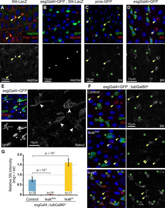
In a screen for new signaling molecules involved in the regulation of tissue homeostasis in the posterior midgut, we identified the secreted ligand Slit as a factor specifically expressed in EEs. Using a LacZ expressing reporter line inserted in the slit locus (SlitPZ05248), we found that the Slit promoter is active in a subset of cells in the intestinal epithelium (Figure S1A), and that these cells represent prospero-positive EEs (Fig. 1A), but not small esg-positive ISCs/EBs or polyploid ECs (Fig. 1B). Immunocytochemistry confirmed that high levels of Slit protein are present in the cytoplasm of prospero-positive and esg-negative diploid EEs (Figure 1C, 1D, S1B). Interestingly, the Slit protein can also be detected on escargot-positive ISCs and EBs (Figure 1D, S1C), suggesting that this secreted molecule diffuses from EEs to these progenitors.
Figure 1. Slit/Robo2 signaling between enteroendocrine and stem/progenitor cells in the posterior midgut.
(A,B) The Slit promoter is active in EEs, as shown by the detection of β-galactosidase in prospero-positive cells, using the SlitPZ05248-LacZ reporter line (A; arrowheads), and inactive in escargot-positive progenitors and polyploid enterocytes (B).
(C,D) The Slit protein is detected in the cytoplasm of prospero-GFP-positive cells (C; arrowheads, and see Supplementary Fig. 2b for the characterization of the pros-GFP reporter) and at the periphery of escargot-positive cells (D; asterisks).
(E) The Robo2 receptor is expressed in esg-positive cells, as shown by immunocytochemistry using a Robo2-specific antibody.
(F) Knock-down of lea/Robo2 in esg-positive cells is sufficient to abolish the accumulation of Slit at the surface of these cells (asterisks) without affecting Slit expression in EEs (arrowheads). Over-expressing Robo2, using the leaEP line, increases the signal at the periphery of ISCs.
(G) Quantification of Slit immunostaining intensity in esg-positive ISCs/EBs compared to esg-negative diploid EEs in similar conditions as Figure 1F.
n represents the number of pairs of diploid cells (one esg-positive and one esg-negative) that were analyzed. p-value from two-tailed Student's t-test. See also Figure S1.
In Drosophila, three Roundabout receptors (Robo1, Robo2/leak and Robo3) have been shown to transduce the Slit signal in different biological contexts (Ypsilanti et al., 2010). To determine whether one of these is a receptor for EE-derived Slit, we first assessed whether they are expressed in the intestine. Using genome-wide transcriptome profiling by RNASeq, we found that only Robo1 and leak/Robo2 transcripts can be detected in dissected intestines (data not shown). Using previously described antibodies (Kidd et al., 1998; Rajagopalan et al., 2000; Simpson et al., 2000), we further found no evidence that Robo1 and Robo3 proteins are expressed in the posterior midgut epithelium (data not shown; Robo1 is expressed in the proventriculus, explaining the detection of the Robo1 mRNA in dissected intestines). Robo2, on the other hand, was detected in esg-positive cells of the posterior midgut (Figure 1E), suggesting that Robo2 might be the ISC- and EB-specific receptor of Slit. To test this hypothesis, we used an inducible system to express a dsRNA construct directed against Robo2 (leaRNAi, which efficiently knocks down Robo2 function (Tayler et al., 2004)) specifically in ISCs and EBs (using the esgGal4 driver combined with a ubiquitously expressed temperature-sensitive Gal80 repressor, tubGal80ts). This manipulation is sufficient to decrease the expression of Robo2 in esg-positive cells and in the intestine (Figure S1C, S1E), and to prevent the accumulation of the Slit protein at the periphery of these cells (Figure 1F, 1G). Conversely, over-expressing Robo2 (using a previously described Gal4-sensitive P-element inserted into the leak locus, leaEP2582) in ISCs and EBs is sufficient to increase the localization of the Slit protein to these cells, without affecting its expression in EEs (Figure 1F, 1G). Altogether, these results indicate that the Slit ligand is secreted by EEs and may transmit a signal from EEs to ISCs and/or EBs through the Robo2 receptor.
The Robo2/Slit pathway regulates the proportion of endocrine cells in the intestinal epithelium
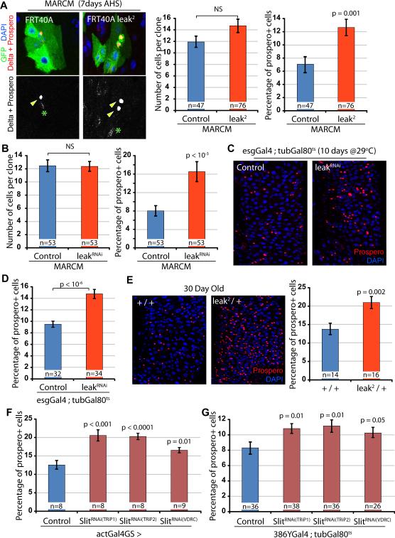
To investigate the function of the Robo2 signaling pathway in the ISC lineage, we generated GFP-labeled ISC clones expressing the Robo2/leaRNAi construct in the posterior midgut, using somatic recombination (MARCM method (Lee and Luo, 1999)). Seven days after induction, leaRNAi expressing ISC clones showed normal growth compared to their wild-type counterparts, indicating that Robo2 is not required for ISC proliferation or self-renewal (Figure 2A). However, we found that the number of prospero-positive cells in leaRNAi clones is significantly higher than in control clones, suggesting that Robo2 may regulate the balance between EE and EC lineages (Figure 2A and Figure S2A). We confirmed this lea loss of function phenotype by generating clones homozygous for the loss-of-function allele lea2. Similar to what we observed using RNAi-mediated knockdown, we found that robo2 homozygosity does not affect ISC proliferation or self-renewal, but significantly increases the proportion of prospero-positive cells in ISC clones (Figure 2B, S2B, S2C). We further used the esgGal4ts system to specifically manipulate the expression of Robo2 in all ISCs and EBs of adult flies. After ten days of expression of the leaRNAi construct in these cells, we observed an increased proportion of prospero-positive cells in the intestinal epithelium (Figure 2C, 2D). Finally, we analyzed the composition of the epithelium of lea2 heterozygous mutants in 30 day old animals (a time sufficient to allow at least one full turn-over of the female intestinal epithelium (Jiang et al., 2009)), and found a significant accumulation of EEs in the midgut of these animals compared to wild-type controls (Figure 2E).
Figure 2. The Slit/Robo2 pathway regulates the proportion of endocrine cells in the intestinal epithelium.
(A,B) MARCM clones expressing a dsRNA directed against leak/Robo2 or homozygous for the mutant allele lea2 contain a greater proportion of prospero-positive cells (arrowheads), 7 days after clone induction by heat shock (AHS), without affecting clone size. Confocal images show representative clones, propero labels EEs, Delta marks ISCs (asterisks).
(C,D) Adult specific knock-down of Robo2 in ISCs and EBs, using the temperature-sensitive esgGal4ts (10 days at 29°C), causes an accumulation of prospero-positive cells in the intestinal epithelium.
(E) Quantification of the proportion of prospero-positive cells in 30 day old lea2 heterozygous and control flies shows an accumulation of EEs in the intestine of mutant animals.
(F,G) Ubiquitous knock-down of Slit (act5cGal4GeneSwitch, 15 days treatment with RU486) is sufficient to induce an increase in the proportion of EEs in the intestinal epithelium. Similar phenotype is observed when SlitRNAi constructs are specifically expressed in EE, using the temperature-sensitive 386YGal4ts driver. Three independent RNAi constructs were tested. n represents the number of clones analyzed in A and B and the number of posterior midguts observed in C-G. p-value from two-tailed Student's t-test. NS, Not Significant. See also Figure S2.
Based on these observations, we hypothesized that EE-derived Slit inhibits the formation of new EEs by promoting Robo2 activity in precursor cells. To test this idea, we first expressed three independent dsRNA constructs directed against Slit in adult flies using an inducible ubiquitous driver (actGal4GeneSwitch). Fifteen days after induction, we observed a significant increase in the proportion of EEs in the posterior midgut for all three RNAi constructs, similar to the phenotype induced by lea loss-of-function (Figure 2F). Next, to directly test the function of the Slit signal in the endocrine lineage, we identified an EE-specific Gal4 line that allows manipulating gene expression of in these cells. We took advantage of a Gal4-containing P-element inserted in the Amontillado gene (386YGal4), which encodes a protease required for the processing of peptide hormones in the fly intestine (Reiher et al., 2011). Similar to its activity in the larval intestine (Reiher et al., 2011), this driver is sufficient to specifically, but weakly express UAS-driven GFP in most prospero-positive and Slit-positive EEs in the adult intestinal epithelium (Figure S2D, S2E). We used this transgenic line to knock-down Slit expression in EEs. Despite the weak activity of the 386YGal4 driver, 10 days after induction, we observed a small but significant increase in the proportion of prospero-positive cells in the posterior midgut epithelium (Figure 2G).
Finally, we tested the effect of over-expressing Robo2/leak in ISC/EBs and over-expressing Slit in EEs, ECs or ISC/EBs on tissue homeostasis. Surprisingly, we found that these manipulations do not affect the composition of the posterior midgut (Figure S2F, S2G). To confirm this result, we co-overexpressed Slit and Robo2/leak in ISC/EBs using the esgGal4 driver and observed no effect on the proportion of EEs in the posterior midgut (data not shown). These finding suggest that while reduced Robo2/Slit signaling promotes EE production, ensuring replenishment of the EE pool when the amount of these cells falls under a critical threshold, endogenous Slit and Robo2 expression levels in the intestinal epithelium are sufficient and not limiting for the inhibition of excessive EE commitment.
Altogether, these results demonstrate that the Slit/Robo2 signaling pathway negatively influences the commitment of ISC daughter cells to the endocrine lineage. The origin of this signal is the EEs themselves, establishing a negative feedback loop, and suggesting that ISCs constantly assess their immediate environment to control the destiny of their progeny and specifically replace missing EEs in the absence of a Slit signal.
The endocrine fate of daughter cells is established in ISCs rather than in EBs
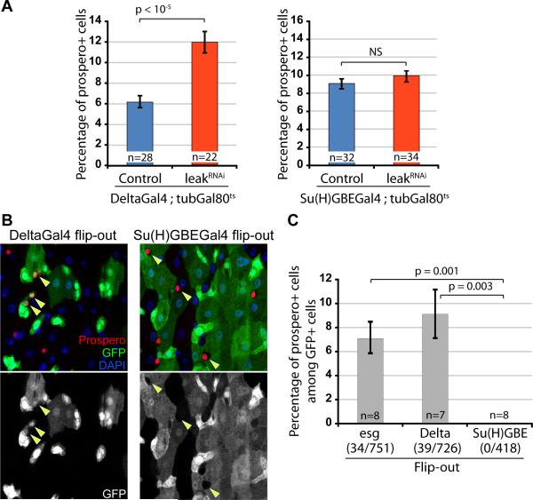
To further refine our understanding of this signaling interaction, we asked whether the Slit/Robo2 signal functions on ISCs or EBs to control commitment to the EE lineage. ISCs can be distinguished from EBs by their differential expression of Dl (in ISCs) and Su(H)GBE reporters (in EBs) (Ohlstein and Spradling, 2007). We knocked-down Robo2 specifically in ISCs and EBs, using the temperature sensitive drivers DeltaGal4ts and Su(H)GBEGal4ts (Zeng et al., 2010). Similar to the results obtained using the esgGal4ts driver, driving the leakRNAi construct with the DeltaGal4ts driver caused a significant increase of the proportion of EEs in the epithelium, while the composition of the intestine was not affected when leakRNAi was expressed using Su(H)GBEGal4ts (Figure 3A). Robo2 signaling thus seems to determine ISC daughter cell identity by acting in ISCs themselves rather than in Su(H)GBEGal4-expressing EBs.
Figure 3. Commitment to the endocrine lineage is established in ISCs rather than enteroblasts.
(A) Adult specific knock-down of Robo2 in ISCs, using the temperature-sensitive DeltaGal4ts drivers (10 days at 29°C), increases the proportion of prospero-positive cells in the posterior midgut. Similar experiment using the EB-specific Su(H)GBEGal4ts driver does not affect the composition of the intestinal epithelium.
(B,C) Flip-out lineage tracing analysis of the progeny of esgGal4 (ISCs and EBs), DeltaGal4 (ISCs only) and Su(H)GBEGal4 (EBs only) expressing cells, 4 days after induction. GFP+Prospero double positive cells (arrowheads) are found in the progeny of esgGal4 and DeltaGal4 positive cells, but absent from the lineage of Su(H)GBEGal4 expressing cells. n indicates the number of gut analyzed, the numbers between parentheses represent the number of pros+ GFP+ cells / total GFP+ cells.
Previous studies have proposed that two types of EBs are generated by ISCs: EC-committed EBs that express high levels of a reporter for Notch activity and EE-committed EBs (Ohlstein and Spradling, 2007). This lineage description was supported by genetic evidence demonstrating that loss of Delta/Notch function in ISCs impairs EC differentiation, while promoting the specification of EEs (Micchelli and Perrimon, 2006; Ohlstein and Spradling, 2006, 2007; Perdigoto et al., 2011). So far, markers for the EE-committed EB population have not been described, and lineage tracing experiments have not yet definitively established the existence of these cells. To test this model, we therefore first analyzed the composition of the progeny of esgGal4 (ISCs and EBs), deltaGal4 (only ISCs (Zeng et al., 2010)) or Su(H)GBEGal4 (only EBs) expressing cells, using an adult-specific lineage tracing strategy in which heritable expression of GFP was induced by recombination initiated from a UAS-linked Flippase. We found that EEs represent 6 to 10% of the progeny of ISCs (Figure 3B, 3C; using both esgGal4 and DeltaGal4), a proportion similar to the one found in the whole intestinal epithelium. Strikingly, however, we found that prospero-expressing EEs are absent from the progeny of Su(H)GBEGal4 expressing cells. This demonstrates that, contrary to the previously accepted model of cell differentiation in the ISC lineage in Drosophila, EBs (defined as ISC daughter cells that show high levels of Notch signaling activity) are not multipotent, as they do not have the capacity to generate EEs, but are rather EC-committed precursors prior to their terminal differentiation.
Prospero expression in ISCs is required for EE-commitment and influenced by Robo2
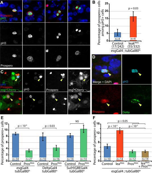
In addition to clarifying the intestinal lineage, these results raise two possibilities regarding the commitment of intestinal progenitors: cell specification to the EC or EE lineage may occur before cell division and ISCs give rise to already distinct daughter cells; or the specification may take place in already formed ISC/precursor pairs, in which the level of expression of Dl in ISCs is regulated to activate or not the Notch signaling pathway in the neighboring cell. Distinguishing between these two models is essential to understand the role of Robo2 in the cell-fate decision. Importantly, we and others have observed that, when Delta/Notch signaling is impaired in ISCs (a genetic manipulation that causes a dramatic accumulation of EEs in the intestinal epithelium (Micchelli and Perrimon, 2006; Ohlstein and Spradling, 2006, 2007)), the EE marker Prospero can be detected in a subset of esg-positive cells (Biteau et al., 2008; Liu et al., 2010; Micchelli and Perrimon, 2006), including in mitotic cells (Figure S3A). Therefore, we tested the notion that prospero-positive ISCs may exist in wild-type animals. To this end, we re-assessed the expression pattern of Prospero in the epithelium of wild-type animals and found that around 6% of the cells positive for the mitotic marker phospho-Histone H3 (pH3) also express Prospero (Figure 4A, 4B), suggesting that these cells may have adopted an endocrine fate. Next, using the esgGal4 and esgLacZ reporters, we found that these pH3+pros+ cells also express the escargot ISC and EB marker (Figure 4C, S3B). Finally, using immunocytochemistry we confirmed that both the Delta and Prospero proteins are detected in this population of mitotic cells (Figure 4D, S3C), demonstrating that these cells are EE-committed dividing progenitors and not dividing terminally differentiated EEs.
Figure 4. Prospero expression in ISCs is regulated by the Robo2 pathway.
(A) Representative images of pros+pH3 double positive cells in the posterior midgut of wild-type flies.
(B) Quantification of the proportion of pros+ cells among dividing pH3+ positive cells in control flies or after knocking-down Robo2 expression in esg-positive cells for 10 days. n represents the number of independent experiments, the numbers indicated below the genotypes represents the total number of pH3+ cells counted.
(C) Representative images of esgGal4>mCherry pH3+ pros+ cells.
(D) Representative image of a mitotic cell expressing both the Delta and Prospero proteins (See Figure S3C for additional examples).
(E) Adult specific knock-down of Prospero in ISCs leads to a decrease in the proportion of EEs in the intestine.
(F) Knock-down of Prospero in esg-positive cells suppresses the increased proportion of EEs induced by leaRNAi expression.
n indicates the number of guts analyzed in B, E and F. p-value from two-tailed Student's t-test. NS, Not Significant. See also Figure S3.
Our previous results suggest that Prospero expression in ISCs, prior to cell division, promotes EE-commitment. To test this model, we used the esgGal4ts, deltaGal4ts and Su(H)GBEGal4ts drivers to knock-down Prospero in ISCs and/or EBs. We found that expression of ProsperoRNAi for 10 days in ISCs (esgGal4+ and DeltaGal4+ cells), but not in Su(H)GBE+ cells, significantly reduced the proportion of EEs in the intestine (Figure 3F), confirming that Prospero expression in ISCs themselves is required for optimal maintenance of the EE lineage.
Since we find that Slit/Robo2 signaling negatively influences the production of EEs, we assessed the influence of Robo2 on the expression of Prospero in mitotic ISCs, and found that the proportion of pH3+pros+ cells is greatly augmented when Robo2 expression is knocked-down (Figure 4E), mirroring the increase in EEs in the progeny of these mutant ISCs. Importantly, Prospero knock-down suppresses this Robo2 loss-of-function phenotype (Figure 4F), confirming that the proposed Robo2-mediated cell fate decision mechanism acts upstream of Prospero expression in ISCs.
Robo2 regulates lineage specification upstream and independently of Notch signaling
Our data support a model in which Slit/Robo2 controls cell fate decisions in the ISC lineage by regulating the specification of ISCs into prospero-expressing EE precursors before or during mitosis. Interestingly, we found that manipulating the activity of Robo2 in ISCs does not affect the phenotype generated by expression of NotchRNAi (in which the formation of EC-committed EBs is specifically inhibited; Figure S4A). In addition, we found no evidence that loss of Robo2 affects Delta expression in ISCs (data not shown). Finally, the activation of the Notch pathway is sufficient to promote differentiation independently of Robo2 signaling (Figure S4B). This supports the idea that Robo2 acts upstream and independently of the activation of the Notch signaling pathway, regulating lineage commitment in ISCs, while Notch specifically controls differentiation of daughter cells into the EC fate. In this model, the absence of Notch signaling results in default commitment of ISC-daughter cells into an EE fate, and lineage commitment thus becomes independent of Robo2/Slit signaling, since EC differentiation is impaired (Figure S4C, S4D).
It is interesting to note that the intensity of the Slit signal is integrated by ISCs to generate an all-or-nothing response: above a defined Slit threshold, Prospero is expressed by around 6% of mitotic ISCs, while below this level, 15-20% of ISCs express Prospero, and no intermediate expression of Prospero can be detected. Further studies will be required to characterize the signaling cascade that controls Prospero expression downstream of the Robo2 receptor in ISCs.
Robo4 has recently been identified as a regulator of hematopoietic stem cell homing in mice (Shibata et al., 2009; Smith-Berdan et al., 2011). In addition, proteins of the Slit and Robo families have been suggested to act as tumor suppressors and be directly involved in the tumorigenesis process (Biankin et al., 2012; Legg et al., 2008; Marlow et al., 2008; Zhou et al., 2011). Our study identifies a new mechanism by which differentiated cells engage this pathway to directly regulate stem cell function and lineage commitment. A role for Slit/Robo signaling in the control of fate decisions in mammalian normal or cancer stem cell lineages has not yet been tested. However, based on the conservation of mechanisms that control Drosophila ISC self-renewal and differentiation (Biteau et al., 2011; Casali and Batlle, 2009; Jiang and Edgar, 2012; Wang and Hou, 2010), it can be anticipated that this feedback control of stem cell fate decisions through Slit/Robo signaling also controls adult tissue homeostasis in higher organisms.
Experimental procedures
Drosophila stocks and culture
The following strains were obtained from the Bloomington Drosophila Stock Center: OregonR, w1118, lea2, leaEP2582, UAS-leaRNAi, UAS-Slit, pros38 (pros-GFP), slitPZ05248, esgk00606, P{GawB}386Y, UAS-Flp [#5254] and act>y>Gal4,UAS-GFP [#4411], UAS-mCherry. UASSlitRNAi(TRiP1) and UAS-SlitRNAi(TRiP2) are from the Transgenic RNAi Project, stocks JF01228 and JF01229. UAS-SlitRNAi(VDRC) and UAS-ProsperoRNAi were obtained from the Vienna Drosophila RNAi Center (transformant ID 20210 and 101477 respectively). The line esgGal4NP5130 was kindly provided by S. Hayashi, DeltaGal4 and Su(H)GBEGal4 by S. Hou, UAS-NotchRNAi by N. Perrimon UAS-Notchintra by M. Rand, actin5cGal4Geneswitch(255) by J. Towers and NP1Gal4 by D.Ferrandon.
The UAS-leaRNAi, UAS-SlitRNAi(VDRC) and UAS-ProsRNAi were validated and successfully used in previous studies (Brierley et al., 2009; Neumuller et al., 2011; Tayler et al., 2004).
Flies were raised on standard yeast and molasses - based food, at 25°C and 65% humidity, on a 12 h light/dark cycle, unless otherwise indicated.
Conditional expression of UAS-linked transgenes
The TARGET system was used to conditionally express UAS-linked transgenes in ISCs and/or EBs. The esgGal4, DeltaGal4 and Su(H)GBEGal4 drivers were combined with a ubiquitously expressed temperature-sensitive Gal80 inhibitor (tub-Gal80ts). Crosses and flies were kept at 18°C (permissive temperature), 3-5 day old adults were then shifted to 29°C to allow expression of the transgenes.
For ubiquitous expression using the actin5cGal4GeneSwitch, adult flies were fed RU486 as described before (Biteau et al., 2010).
Mosaic analysis with a repressible cell marker (MARCM) clones and Flip-out lineage tracing
Positively marked clones were generated by somatic recombination using the following MARCM stock: hsFlp;FRT40A tub-Gal80;tub-Gal4,UAS-GFP (gift from B. Ohlstein). Virgins were crossed to the following lines: FRT40A lea2 or FRT40A;UAS-leaRNAi. 3-5 day old mated female flies were heat-shocked for 45 minutes at 37°C to induce somatic recombination. Flies were transferred to 25°C and clones were observed 7 days after induction.
For Flip-out lineage tracing analysis, the following genotypes were used:
UAS-Flp/+ ; esg-Gal4,UAS-GFP/act5c-FRT-y-FRT-Gal4,UAS-GFP ; tubulin-Gal80ts/+
UAS-Flp/+ ; Su(H)GBE-Gal4,UAS-GFP/act5c-FRT-y-FRT-Gal4,UAS-GFP ; tubulin-Gal80ts/+
UAS-Flp/+ ; act5c-FRT-y-FRT-Gal4,UAS-GFP/+ ; Delta-Gal4,UAS-GFP/tubulin-Gal80ts
Crosses were set up at 18°C. 3-5 day old females were heat-shocked at 37°C for 30 minutes and transferred to 29°C to induce the expression of UAS-driven Flippase and permanently label ISCs and/or EBs and their progeny. The composition of the lineages was analyzed 4 days after labeling.
Immunocytochemistry and Microscopy
Fly intestines were dissected in PBS and fixed at room temperature for 45 minutes in 100 mM glutamic acid, 25 mM KCl, 20 mM MgSO4, 4 mM Sodium Phosphate, 1 mM MgCl2, 4% formaldehyde. All subsequent incubations were done in PBS, 0.5% BSA, 0.1% TritonX-100 at 4°C.
The following primary antibodies were obtained from the Developmental Studies Hybridoma Bank: mouse anti-slit, anti-Delta, anti-Prospero, anti-Armadillo and anti-β-galactosidase and used 1:50, 1:100, 1:250, 1:100 and 1:500 respectively. Rabbit anti-β-galactosidase is from Cappel and used 1:1000; rabbit anti-pH3 from Upstate, 1:1000. The anti-Robo2 antibody was obtained from B. Dickson and used 1:50. The rat anti-Delta antibody was obtained from M. Rand and used 1:200. Fluorescent secondary antibodies were obtained from Jackson Immunoresearch. Hoechst was used to stain DNA.
Confocal images were collected using a Leica SP5 confocal system and processed using the Leica software and Adobe Photoshop CS5.
To quantify the intensity of the slit immunocytochemistry in Figure 1g, the mean pixel intensity for the appropriate color channel of EEs and ISCs was measured using the Adobe Photoshop CS5 software. The intensity of each ISC was normalized to the intensity of the closest EE to take into account differences in staining between intestines and experiments.
Phenotype analysis
For clonal studies, only isolated clones that can be identified with confidence were included in the analysis of clone size and composition.
For all experiments, the data is represented as average +/− SEM. All p-values are calculated using unpaired two-tailed Student's t-test.
Supplementary Material
Highlights.
Slit/Robo2 signaling regulates the production of endocrine cells in the fly midgut
Commitment to the endocrine fate is established in ISCs prior to cell division
Prospero expression in ISCs is required for endocrine cell production
Acknowledgements
We thank M. Nuzzo and the University of Rochester Genomics Research Center for technical assistance; as well as M. Rand, J. Towers, B. Dickson, D. Ferrandon, S. Hayashi, S. Hou, N. Perrimon, the Bloomington Drosophila Stock Center, the Vienna Drosophila RNAi Center and the Developmental Studies Hybridoma Bank for providing essential reagents. This work was supported by an Ellison Medical Foundation/AFAR postdoctoral award and a New Scholar in Aging Research from the Ellison Medical Foundation (AG-NS-0990-13) to B.B., and by grants from the National Institute on General Medical Sciences (NIH RO1 GM100196) and the Ellison Medical Foundation (AG-SS-2224-08) to H.J.
Footnotes
Publisher's Disclaimer: This is a PDF file of an unedited manuscript that has been accepted for publication. As a service to our customers we are providing this early version of the manuscript. The manuscript will undergo copyediting, typesetting, and review of the resulting proof before it is published in its final citable form. Please note that during the production process errors may be discovered which could affect the content, and all legal disclaimers that apply to the journal pertain.
References
- Amcheslavsky A, Jiang J, Ip YT. Tissue damage-induced intestinal stem cell division in Drosophila. Cell stem cell. 2009;4:49–61. doi: 10.1016/j.stem.2008.10.016. [DOI] [PMC free article] [PubMed] [Google Scholar]
- Beck B, Blanpain C. Mechanisms regulating epidermal stem cells. The EMBO journal. 2012;31:2067–2075. doi: 10.1038/emboj.2012.67. [DOI] [PMC free article] [PubMed] [Google Scholar]
- Biankin AV, Waddell N, Kassahn KS, Gingras MC, Muthuswamy LB, Johns AL, Miller DK, Wilson PJ, Patch AM, Wu J, et al. Pancreatic cancer genomes reveal aberrations in axon guidance pathway genes. Nature. 2012;491:399–405. doi: 10.1038/nature11547. [DOI] [PMC free article] [PubMed] [Google Scholar]
- Biteau B, Hochmuth CE, Jasper H. JNK activity in somatic stem cells causes loss of tissue homeostasis in the aging Drosophila gut. Cell stem cell. 2008;3:442–455. doi: 10.1016/j.stem.2008.07.024. [DOI] [PMC free article] [PubMed] [Google Scholar]
- Biteau B, Hochmuth CE, Jasper H. Maintaining tissue homeostasis: dynamic control of somatic stem cell activity. Cell stem cell. 2011;9:402–411. doi: 10.1016/j.stem.2011.10.004. [DOI] [PMC free article] [PubMed] [Google Scholar]
- Biteau B, Karpac J, Supoyo S, Degennaro M, Lehmann R, Jasper H. Lifespan extension by preserving proliferative homeostasis in Drosophila. PLoS genetics. 2010;6:e1001159. doi: 10.1371/journal.pgen.1001159. [DOI] [PMC free article] [PubMed] [Google Scholar]
- Brierley DJ, Blanc E, Reddy OV, Vijayraghavan K, Williams DW. Dendritic targeting in the leg neuropil of Drosophila: the role of midline signalling molecules in generating a myotopic map. PLoS Biol. 2009;7:e1000199. doi: 10.1371/journal.pbio.1000199. [DOI] [PMC free article] [PubMed] [Google Scholar]
- Buchon N, Broderick NA, Chakrabarti S, Lemaitre B. Invasive and indigenous microbiota impact intestinal stem cell activity through multiple pathways in Drosophila. Genes & development. 2009;23:2333–2344. doi: 10.1101/gad.1827009. [DOI] [PMC free article] [PubMed] [Google Scholar]
- Casali A, Batlle E. Intestinal stem cells in mammals and Drosophila. Cell stem cell. 2009;4:124–127. doi: 10.1016/j.stem.2009.01.009. [DOI] [PubMed] [Google Scholar]
- Chatterjee M, Ip YT. Pathogenic stimulation of intestinal stem cell response in Drosophila. Journal of cellular physiology. 2009;220:664–671. doi: 10.1002/jcp.21808. [DOI] [PMC free article] [PubMed] [Google Scholar]
- Cronin SJ, Nehme NT, Limmer S, Liegeois S, Pospisilik JA, Schramek D, Leibbrandt A, Simoes Rde M, Gruber S, Puc U, et al. Genome-wide RNAi screen identifies genes involved in intestinal pathogenic bacterial infection. Science. 2009;325:340–343. doi: 10.1126/science.1173164. [DOI] [PMC free article] [PubMed] [Google Scholar]
- de Navascues J, Perdigoto CN, Bian Y, Schneider MH, Bardin AJ, Martinez-Arias A, Simons BD. Drosophila midgut homeostasis involves neutral competition between symmetrically dividing intestinal stem cells. The EMBO journal. 2012;31:2473–2485. doi: 10.1038/emboj.2012.106. [DOI] [PMC free article] [PubMed] [Google Scholar]
- Jiang H, Edgar BA. Intestinal stem cell function in Drosophila and mice. Curr Opin Genet Dev. 2012;22:354–360. doi: 10.1016/j.gde.2012.04.002. [DOI] [PMC free article] [PubMed] [Google Scholar]
- Jiang H, Patel PH, Kohlmaier A, Grenley MO, McEwen DG, Edgar BA. Cytokine/Jak/Stat signaling mediates regeneration and homeostasis in the Drosophila midgut. Cell. 2009;137:1343–1355. doi: 10.1016/j.cell.2009.05.014. [DOI] [PMC free article] [PubMed] [Google Scholar]
- Kidd T, Brose K, Mitchell KJ, Fetter RD, Tessier-Lavigne M, Goodman CS, Tear G. Roundabout controls axon crossing of the CNS midline and defines a novel subfamily of evolutionarily conserved guidance receptors. Cell. 1998;92:205–215. doi: 10.1016/s0092-8674(00)80915-0. [DOI] [PubMed] [Google Scholar]
- Lee T, Luo L. Mosaic analysis with a repressible cell marker for studies of gene function in neuronal morphogenesis. Neuron. 1999;22:451–461. doi: 10.1016/s0896-6273(00)80701-1. [DOI] [PubMed] [Google Scholar]
- Legg JA, Herbert JM, Clissold P, Bicknell R. Slits and Roundabouts in cancer, tumour angiogenesis and endothelial cell migration. Angiogenesis. 2008;11:13–21. doi: 10.1007/s10456-008-9100-x. [DOI] [PubMed] [Google Scholar]
- Liu W, Singh SR, Hou SX. JAK-STAT is restrained by Notch to control cell proliferation of the Drosophila intestinal stem cells. J Cell Biochem. 2010;109:992–999. doi: 10.1002/jcb.22482. [DOI] [PMC free article] [PubMed] [Google Scholar]
- Marlow R, Strickland P, Lee JS, Wu X, Pebenito M, Binnewies M, Le EK, Moran A, Macias H, Cardiff RD, et al. SLITs suppress tumor growth in vivo by silencing Sdf1/Cxcr4 within breast epithelium. Cancer Res. 2008;68:7819–7827. doi: 10.1158/0008-5472.CAN-08-1357. [DOI] [PMC free article] [PubMed] [Google Scholar]
- Micchelli CA, Perrimon N. Evidence that stem cells reside in the adult Drosophila midgut epithelium. Nature. 2006;439:475–479. doi: 10.1038/nature04371. [DOI] [PubMed] [Google Scholar]
- Neumuller RA, Richter C, Fischer A, Novatchkova M, Neumuller KG, Knoblich JA. Genome-wide analysis of self-renewal in Drosophila neural stem cells by transgenic RNAi. Cell stem cell. 2011;8:580–593. doi: 10.1016/j.stem.2011.02.022. [DOI] [PMC free article] [PubMed] [Google Scholar]
- Ohlstein B, Spradling A. The adult Drosophila posterior midgut is maintained by pluripotent stem cells. Nature. 2006;439:470–474. doi: 10.1038/nature04333. [DOI] [PubMed] [Google Scholar]
- Ohlstein B, Spradling A. Multipotent Drosophila intestinal stem cells specify daughter cell fates by differential notch signaling. Science. 2007;315:988–992. doi: 10.1126/science.1136606. [DOI] [PubMed] [Google Scholar]
- Perdigoto CN, Schweisguth F, Bardin AJ. Distinct levels of Notch activity for commitment and terminal differentiation of stem cells in the adult fly intestine. Development. 2011;138:4585–4595. doi: 10.1242/dev.065292. [DOI] [PubMed] [Google Scholar]
- Rajagopalan S, Nicolas E, Vivancos V, Berger J, Dickson BJ. Crossing the midline: roles and regulation of Robo receptors. Neuron. 2000;28:767–777. doi: 10.1016/s0896-6273(00)00152-5. [DOI] [PubMed] [Google Scholar]
- Reiher W, Shirras C, Kahnt J, Baumeister S, Isaac RE, Wegener C. Peptidomics and peptide hormone processing in the Drosophila midgut. J Proteome Res. 2011;10:1881–1892. doi: 10.1021/pr101116g. [DOI] [PubMed] [Google Scholar]
- Rock JR, Hogan BL. Epithelial Progenitor Cells in Lung Development, Maintenance, Repair, and Disease. Annu Rev Cell Dev Biol. 2011;27:493–512. doi: 10.1146/annurev-cellbio-100109-104040. [DOI] [PubMed] [Google Scholar]
- Shibata F, Goto-Koshino Y, Morikawa Y, Komori T, Ito M, Fukuchi Y, Houchins JP, Tsang M, Li DY, Kitamura T, et al. Roundabout 4 is expressed on hematopoietic stem cells and potentially involved in the niche-mediated regulation of the side population phenotype. Stem Cells. 2009;27:183–190. doi: 10.1634/stemcells.2008-0292. [DOI] [PMC free article] [PubMed] [Google Scholar]
- Simpson JH, Bland KS, Fetter RD, Goodman CS. Short-range and long-range guidance by Slit and its Robo receptors: a combinatorial code of Robo receptors controls lateral position. Cell. 2000;103:1019–1032. doi: 10.1016/s0092-8674(00)00206-3. [DOI] [PubMed] [Google Scholar]
- Smith-Berdan S, Nguyen A, Hassanein D, Zimmer M, Ugarte F, Ciriza J, Li D, Garcia-Ojeda ME, Hinck L, Forsberg EC. Robo4 cooperates with CXCR4 to specify hematopoietic stem cell localization to bone marrow niches. Cell stem cell. 2011;8:72–83. doi: 10.1016/j.stem.2010.11.030. [DOI] [PMC free article] [PubMed] [Google Scholar]
- Tayler TD, Robichaux MB, Garrity PA. Compartmentalization of visual centers in the Drosophila brain requires Slit and Robo proteins. Development. 2004;131:5935–5945. doi: 10.1242/dev.01465. [DOI] [PMC free article] [PubMed] [Google Scholar]
- Wang P, Hou SX. Regulation of intestinal stem cells in mammals and Drosophila. Journal of cellular physiology. 2010;222:33–37. doi: 10.1002/jcp.21928. [DOI] [PubMed] [Google Scholar]
- Yeung TM, Chia LA, Kosinski CM, Kuo CJ. Regulation of self-renewal and differentiation by the intestinal stem cell niche. Cell Mol Life Sci. 2011;68:2513–2523. doi: 10.1007/s00018-011-0687-5. [DOI] [PMC free article] [PubMed] [Google Scholar]
- Ypsilanti AR, Zagar Y, Chedotal A. Moving away from the midline: new developments for Slit and Robo. Development. 2010;137:1939–1952. doi: 10.1242/dev.044511. [DOI] [PubMed] [Google Scholar]
- Zeng X, Chauhan C, Hou SX. Characterization of midgut stem cell- and enteroblast-specific Gal4 lines in drosophila. Genesis. 2010;48:607–611. doi: 10.1002/dvg.20661. [DOI] [PMC free article] [PubMed] [Google Scholar]
- Zhou WJ, Geng ZH, Chi S, Zhang W, Niu XF, Lan SJ, Ma L, Yang X, Wang LJ, Ding YQ, et al. Slit-Robo signaling induces malignant transformation through Hakai-mediated E-cadherin degradation during colorectal epithelial cell carcinogenesis. Cell Res. 2011;21:609–626. doi: 10.1038/cr.2011.17. [DOI] [PMC free article] [PubMed] [Google Scholar]
Associated Data
This section collects any data citations, data availability statements, or supplementary materials included in this article.


