Abstract
AIM: To study the effect of oral steroids upon clinical response and rectal mucosa secretion of eosinophil cationic protein (ECP), myeloperoxidase (MPO), basic fibroblast growth factor (bFGF), vascular endothelial growth factor (VEGF) and albumin in patients with collagenous colitis (CC).
METHODS: A segmental perfusion technique was used to collect perfusates from rectum of CC patients once before and twice (one and four weeks) after the start of steroid treatment. Clinical data was monitored and ECP, MPO, bFGF, VEGF and albumin concentrations were analyzed by immunochemical methods in perfusates and in serum.
RESULTS: Steroids reduced the number of bowel movements by more than five times within one week and all patients reported improved subjective well-being at wk 1 and 4. At the same time, the median concentrations of ECP, bFGF, VEGF and albumin in rectal perfusates decreased significantly. MPO values were above the detection limit in only 3 patients before treatment and in none during treatment. VEGF, bFGF, ECP and albumin concentrations correlated with each other with the exception of ECP and albumin. A decrease of serum ECP and VEGF concentrations was also seen even if the overtime reduction was not significant.
CONCLUSION: Oral steroid treatment in CC patients induced a simultaneous reduction of bowel movements and rectal release of ECP, bFGF, VEGF and albumin, suggesting that these polypeptides and increased mucosal permeability are important components of the pathophysiology in collagenous colitis.
Keywords: Collagenous colitis, Inflammatory mediator, Steroid treatment, Eosinophil cationic protein, Basic fibroblast growth factor, Vascular endothelial growth factor
INTRODUCTION
Collagenous colitis (CC) is characterized by watery diarrhea and an increased subepithelial collagen layer with infiltration of inflammatory cells in the surface epithelium and lamina propria of the colon[1]. The thickened collagen in CC is an amorphous eosinophilic band located beneath the intercryptal epithelial surface and is best visualized using special collagen staining, such as Masson´s trichrome[1]. The collagen composition of the basal membrane of colonic mucosa in CC patients is identical to that of normal individuals[1]. In contrast, the subepithelial collagen layer differs in CC patients and consists of collagen type I and III or VI[2-4].
Intestinal inflammatory cells present in CC are lymphocytes, plasma cells, often eosinophil and sometimes neutrophil granulocytes. In the epithelium, lymphocytes are most frequent and dominated by CD8+ T-cells[5]. In the lamina propria, the inflammatory cell infiltration includes plasma cells, eosinophils, macrophages and mast cells, but rarely neutrophils. Unlike epithelial lymphocytes, the lymphocytes in the lamina propria are mainly composed of CD4+ T-cells[5].
Eosinophils are one of the main components of the inflammatory cell population in colonic mucosa of CC patients[1]. Eosinophils produce not only cytotoxic granule proteins, such as eosinophil cationic protein (ECP), major basic protein (MBP) or eosinophil peroxidase (EPO), but also a wide range of polypeptides, such as transforming growth factor-β1 (TGF-β1), vascular endothelial growth factor (VEGF), etc[6-10]. Eosinophilopoietic cytokines, such as granulocyte-macrophage colony-stimulating factor (GM-CSF) and interleukin-5 (IL-5), activate eosinophils by inhibiting apoptosis and prolonging the life span of eosinophils[7]. Eosinophils interact with fibroblasts and contribute to fibrosis in inflammatory diseases like asthma and inflammatory bowel disease (IBD). Activated fibroblasts/myofibroblasts are regarded as the major contributors of collagen accumulation in the intestinal wall of IBD[11,12] including CC patients[13].
In asthma, basic fibroblast growth factor (bFGF), separately or in combination with other growth factors, perpetuate chronic inflammation and triggers fibrosis leading to airway tissue remodeling[14] including excessive extra cellular matrix deposition in the subepithelial region. The role of bFGF, as one contributing factor in tissue remodeling, is supported by perfusion studies in ulcerative colitis (UC)[15] and CC[16] patients, where the secretion of bFGF was increased.
VEGF is a pleiotropic polypeptide, which has physiological and pathophysiological actions on the extra cellular matrix and may enhance fibrosis through induction of connective tissue growth factor (CTGF)[17]. A recent study on the pathophysiological role of VEGF in CC showed that VEGF, by changing the local matrix metalloproteinase -1 and tissue inhibitor metalloproteinase-1 (MMP-1/TIMP-1) balance, causes the accumulation of immature subepithelial ECM in CC[18].
Glucocorticosteroids have several anti-inflammatory effects, including the ability to block the recruitment of proinflammatory cells like eosinophils, downregulation of the secretion of proinflammatory cytokines, enhancement of apoptosis in eosinophils[19-21] and engulfment of apoptotic eosinophils by phagocytic cells[7]. In asthma, prolonged treatment with inhaled corticosteroids reverses tissue remodeling by downregulating fibroblast-functions[14,20,22]. In our previous perfusion studies, we found increased colorectal concentrations of ECP[23], bFGF[16], VEGF[24] and the permeability marker albumin in CC patients compared with control patients. We put forward the hypothesis that these mediators enhanced chronic inflammation and fibrosis, leading to pathological accumulation of subepithelial collagen. Oral steroid treatment is effective for inducing remission in CC patients[25,26] but the mechanisms leading to relief of symptoms are not fully known. The aim of this study was to elucidate the relationship between steroid treatment and rectal mucosa secretion of ECP, MPO, bFGF, VEGF and albumin in CC patients.
MATERIALS AND METHODS
Patients
Twelve patients (10 women, 2 men; mean age 52 years; range 34-66 years) who fulfilled the diagnostic criteria of CC[1] were recruited between November 2000 and November 2002 at the Department of Medicine, University Hospital, Uppsala, Sweden.
Ethics
Committee of the Medical faculty at Uppsala University approved the study and informed consent was obtained from all participants. No patient had anti- or pro-inflammatory drug treatment during the investigation period, but one patient (number eight) was taking indomethacin (IndomeeR, Merck Sharp and Dohme, Stockholm, Sweden) up until one week before the first perfusion. Patient number seven also had celiac disease and lactose intolerance, while patients five and eight suffered from hypothyroidism. Patients eleven and twelve were smokers.
Design of study
Steroid treatment was initiated after the first perfusion with an oral dose of 40 mg prednisolone daily, which was reduced successively by 5 mg/wk. Three rectal perfusions were fulfilled per patient, one before and two after the start of steroid treatment, at completed weeks one and four respectively. Serum samples were also collected on those occasions. Clinical data were recorded at each perfusion as the patients estimated their average daily number of loose stools and degree of general well-being during the week preceding each perfusion.
Perfusion technique
A colonoscope-based segmental perfusion technique[23] was used to obtain perfusion fluid from the rectum. Sodium phosphate (PhosphoralR, Ferring, Limhamn, Sweden) was used for bowel preparation. The rectal perfusions were carried out with the patients lying in a supine position and started with an endoscopic examination of the rectum and sigmoid colon. The segment, positioned at the end of the flexible endoscope, was established by inflating air into two balloons delimiting the segment in the rectum. The perfusion procedures were carried out for one hour[23] and the perfusates were collected in three portions at 20 min intervals in tubes immersed in ice. Intravenous injections of the sedative diazepam (StesolidR, Alpharma, Stockholm, Sweden) were given at a median dose of 7.5 mg (range: 5-15) and the analgesic meperidine (PetidinR, Ipex, Stockholm, Sweden) at a median dose of 25 mg (range: 25-50).
Perfusion buffer, perfusates and serum samples
A buffer was used, consisting of 120 mmol/L NaCl, 5.4 mmol/L KCl, 2 mmol/L Na2HPO4, 10 mmol/L glucose, 35 mmol/L mannitol, and 1 g/L polyethylene glycol (PEG; MW 4 kDa) at pH of 8.20-8.48 and osmolarity of 290 mosm/L. The perfusion fluid was kept at 37°C and then infused at a speed of 3 mL/min for 60 min. By adding ten milliliters of aprotinin (TrasylolR Bayer 1.0 × 10⁷ kIU/L) to every liter of perfusion buffer, proteolytic activity was inhibited. Phenol red solution (50 mg/L in physiological saline) was intermittently infused (5 mL every 20 min) above the proximal balloon to check that the perfusion segment was closed and that there was no contamination of the perfusion fluid by intestinal contents located proximally. All perfusate samples were collected on ice and frozen in aliquots of 2 mL at -70°C. The protease inhibitor, phenylmethylsulphonyl fluoride (PMSF; Sigma Chemical Co., St Louis, MO, USA), was added to a final concentration of 2 mmol/L immediately before analysis to counteract the effect of even small amounts of proteases in the perfusion fluid. Serum samples were drawn simultaneously with the perfusions being performed and frozen at -70°C for later analysis.
Quantification of ECP, MPO and albumin
The concentrations of ECP and MPO in perfusion fluids and serum samples were analyzed by means of a specific RIA (Pharmacia & Upjohn, Diagnostic AB, Uppsala, Sweden). The inter- and intra-assay coefficients of variations were < 10% for both tests. The lower detection limits of ECP and MPO in perfusion fluid were 2 and 8 μg/L, respectively. The reference intervals for ECP and MPO in serum were 2.3-16 μg/L and 170-478 μg/L, respectively. Albumin in perfusion fluid was analyzed by rate nephelometry on a Beckman Array protein system (Beckman Instruments, Brea CA), according to the recommendations of the manufacturer. The lower detection limit in perfusion fluids of albumin was 2.14 mg/L. Analysis of albumin in serum was performed by a photometric assay (Boehringer Manheim, Mannheim, Germany) and the reference intervals were 40-51 g/L (age < 50 years) and 37-48 g/L (age > 50 years). The concentration of phenol red was assessed, after alkalinization of the perfusate samples to pH 11, by a spectrophotometric method at 520 nm.
Quantification of bFGF and VEGF
ELISA technique was used to measure bFGF (Human bFGF, Quantikine High Sensitivity, R&D Systems, Minneapolis, MN, USA) and VEGF (DVE00, R&D Systems, Minneapolis, MN, USA) as recommended by the manufacturer. The bFGF and VEGF concentrations in the samples were determined by comparing the optical density of the samples with the standard curve. The lowest detectable value for bFGF was 0.25 pg/mL and for VEGF 9 pg/mL in perfusion fluid. Reference intervals of S-bFGF were < 4.0 ng/L (males) and < 10.8 ng/L (females). The reference interval of S-VEGF was < 500 ng/L.
Statistical analysis
The Wilcoxon signed rank test for two variables was used to compare the differences between wk 0, 1 and 4, while the statistical analysis of changes over time in the perfusate and serum concentrations of ECP, MPO, bFGF, VEGF and albumin, was done by Friedman’s ANOVA. Spearman rank correlation test was applied to study co-variation between each mediator and the frequency of bowel movements per day. All statistical calculations were performed on a Macintosh computer by means of a statistical package, Stat View 4.51 (Abacus, Concepta Inc.). If concentrations of the mediator proteins and albumin were below the detection limit of the assay, the value of the detection limit was used in the calculations. P < 0.05 was considered significant.
RESULTS
Perfusions and clinical outcome
Out of 12 CC patients studied, 11 successfully completed steroid treatment and all three perfusions according to the protocol. The excluded patients stopped taking steroids because of side effects. The number of bowel movements per day declined from median 5 before start of treatment, to 0-1 after one and 0-1 after four weeks, respectively. These differences were significant (P < 0.0001) (Table 1). All patients reported that senses of general well-being were better after the start of steroid treatment, compared to the period before it.
Table 1.
The clinical data and the individual concentrations of ECP, MPO, bFGF, VEGF and albumin in rectal perfusion fluid of CC patients before and after one and four weeks of treatment with steroids
| Demography | Bowel movements |
Mediators analyzed in Perfusion fluid |
||||||||||||||||||
| Patients | Age (yr) | Gender |
per day |
ECP (μg/L) |
MPO (μg/L) |
bFGF (pg/mL) |
VEGF (pg/mL) |
Albumin (mg/L) |
||||||||||||
|
wk | ||||||||||||||||||||
| 0 | 1 | 4 | 0 | 1 | 4 | 0 | 1 | 4 | 0 | 1 | 4 | 0 | 1 | 4 | 0 | 1 | 4 | |||
| 1 | 34 | Female | 3-4 | 0-1 | 0-1 | 3.7 | < 2 | < 2 | 29.1 | 14.9 | < 8 | 14.03 | 18.26 | 0.65 | 299.7 | 92.8 | 41 | 20.3 | 44.6 | < 2.1 |
| 2 | 57 | Female | 4 | 0-1 | 0-1 | 2.6 | < 2 | < 2 | 12.5 | < 8 | < 8 | 7.85 | 7.11 | 0.37 | 54.7 | 97 | 40.2 | 95.8 | 57.7 | < 2.1 |
| 3 | 59 | Female | 10-12 | 0-1 | 0-1 | 3.6 | 7 | < 2 | < 8 | < 8 | < 8 | 1.21 | 4.1 | < 0.25 | 113.1 | 146.7 | 81.1 | 20.3 | 10.7 | 3.4 |
| 4 | 43 | Female | 4-6 | 3-4 | 0-1 | < 2 | < 2 | < 2 | < 8 | < 8 | < 8 | 0.72 | 0.41 | < 0.25 | 28.4 | 22.9 | 40.2 | 2.8 | 10.9 | 17.5 |
| 5 | 62 | Female | 3-4 | 0-1 | 0-1 | < 2 | < 2 | < 2 | < 8 | < 8 | 11.3 | 0.35 | < 0.25 | 0.29 | 66.2 | 46.6 | 53.1 | 2.7 | 10.7 | 5.5 |
| 7 | 66 | Female | 4-5 | 0-1 | 0-1 | < 2 | < 2 | < 2 | < 8 | < 8 | < 8 | < 0.25 | < 0.25 | < 0.25 | 58.8 | 19.1 | 22.2 | 3.9 | 6.5 | 3.2 |
| 8 | 59 | Female | 5-10 | 2 | 0-1 | 6.5 | < 2 | < 2 | < 8 | < 8 | < 8 | 5.28 | < 0.25 | 0.35 | 86.1 | 37.8 | 58.8 | 17.1 | 3.1 | 2.3 |
| 9 | 64 | Female | 3 | 0-1 | 0-1 | < 2 | < 2 | < 2 | < 8 | < 8 | < 8 | < 0.25 | < 0.25 | < 0.25 | 17.6 | 9.6 | 12.3 | 5 | < 2.1 | 7 |
| 10 | 39 | Male | 7-10 | 2-3 | 0-1 | < 2 | < 2 | < 2 | < 8 | < 8 | < 8 | 2.7 | 1.21 | < 0.25 | 100.4 | 94.5 | 90.3 | 43 | 13.7 | 11.7 |
| 11 | 34 | Female | 5-6 | 2 | 0-1 | 3.1 | < 2 | < 2 | 18.3 | < 8 | < 8 | 11.99 | 2.35 | < 0.25 | 153.6 | 84.4 | 80.2 | 116 | 22.1 | 10.7 |
| 12 | 53 | Male | 6-7 | 2-3 | 0-1 | 2.3 | < 2 | < 2 | < 8 | < 8 | < 8 | 12.29 | 0.91 | 0.61 | 95.3 | 48.2 | 29.1 | 21.5 | 5.2 | < 2.1 |
ECP, MPO and albumin
The concentrations of ECP, MPO and albumin in perfusates are shown in Table 1. The median values of ECP concentrations in perfusates were 2.3 μg/L (range: 1.9-6.5) at wk 0, 1.9 μg/L (range: 1.9-7.0) at wk 1 and 1.9 μg/L (range: 1.9-1.9) at wk 4, respectively. A significant difference was only found between wk 0 and 4 (P = 0.0277). The corresponding values for ECP in serum at wks 0, 1 and 4 were 11.3 μg/L (range: 1.9-29), 6.6 μg/L (range: 1.9-18.8) and 6.3 μg/L (range: 1.9-18.9), respectively. The decrease between wk 0 and 1 and wk 0 and 4 were significant (P = 0.0076, P = 0.045 respectively), while no difference was found when the decline of ECP concentrations in perfusate and serum over time was analyzed by the Friedman’s ANOVA.
Perfusate concentrations of MPO were above the detection level in three out of 11 patients at wk 0, one at wk 1 and none at wk 4 (Table 1). Serum MPO concentrations were 432 μg/L (range: 197-649), 325 μg/L (range: 106-1805), 490 μg/L (range: 144-1190) at wk 0, 1 and 4, respectively. No difference was found between the concentrations during the study period.
The median values of albumin concentrations in perfusates were 20.3 mg/L (range: 2.7-116), 10.7 mg/L (range: 2.13-57.7) and 3.4 mg/L (range: 2.13-17.5) at wk 0, 1 and 4, respectively. The difference was significant only between wk 0 and 4 (P = 0.0329). The decline in albumin concentrations over time was not significant. The corresponding median values of serum albumin at wk 0, 1 and 4 were 42 g/L (range: 38-45), 43 g/L (range: 34-48) and 42 g/L (range: 36-46), respectively (P > 0.05). No correlation was found between the concentrations of ECP and albumin in perfusion fluids.
Analysis of bFGF
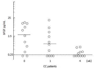
The concentrations of bFGF in perfusion fluid are presented in Table 1. The median values of bFGF concentrations in perfusion fluid were at wk 0, 1 and 4, were 2.7 pg/mL (range: 0.2-14.0), 0.9 pg/mL (range: 0.2-18.3) and 0.2 pg/mL (range: 0.2-0.7), respectively. The decline of bFGF perfusate concentrations over time was significant when analyzed by the Friedman’s ANOVA (P < 0.01) (Figure 1). A correlation (P < 0.05, r = 0.702) that existed between bFGF and ECP perfusate concentrations at wk 0 disappeared at wk 1 and 4. A correlation between bFGF and albumin in perfusates remained significant throughout the study period (P < 0.05 and r = 0.709, r = 0.805, r = - 0.670 at wk 0, 1 and 4 respectively). The median values of bFGF concentrations in serum were 9.1 ng/L (range: 1.7-44), 2.9 ng/L (range: 0.9-17.6) and 3.6 ng/L (range: 0.3-20.9) at wk 0, 1 and 4, respectively, but the decline over time was not significant.
Figure 1.

The individual values of basic fibroblast growth factor (bFGF) concentrations in perfusion fluids from rectum of collagenous colitis (CC) patients before and after one and four weeks of steroid treatment. The bars indicate median values. The dotted line indicates the detection limit for the assay (bFGF: 0.25 pg/mL).
Analysis of VEGF
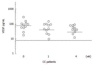
Table 1 shows all the individual concentrations of VEGF in perfusion fluid. The median values of VEGF concentrations in perfusates of these individuals at wk 0, 1 and 4 were 86.1 pg/mL (range: 17.6-299.7), 48.2 pg/mL (range: 9.6-146.7), and 41.0 pg/mL (range: 12.3-90.3), respectively. When the Friedman’s ANOVA was used to analyze the decline of VEGF concentrations, a significant difference (P < 0.01) was seen (Figure 2). Furthermore, there was a significant correlation between VEGF and ECP at wk 0 and 1 (P < 0.05; r = 0.627 and 0.625 at wk 0 and 1 respectively) but not at wk 4. The correlation between VEGF and albumin in perfusate was significant only at wk 1 (P < 0.05; r = 0.666). Similar significant correlations were present between VEGF and bFGF concentrations at wk 0 and 1 (P < 0.01; r = 0.680 and P < 0.05; r = 0.850) but not at wk 4. The median values of VEGF serum concentrations at wk 0, 1 and 4 were 842 ng/L (range: 622-1517), 775 ng/L (range: 385-1215) and 811 ng/L (range: 574-1114), respectively. The decline in serum concentration of VEGF between wk 0 and 4 was significant (P < 0.05) but the over time change was not significant.
Figure 2.

The individual values of vascular endothelial growth factor (VEGF) concentrations in perfusion fluids from rectum of collagenous colitis (CC) patients before and after one and four weeks of steroid treatment. The bars indicate median values. The dotted line indicates the detection limit for the assay (VEGF: 9 pg/mL).
DISCUSSION
The immediate decrease in frequency of bowel movements and the improvement of the general well-being of these patients under steroid medication were similar to previous reports in IBD, including CC[25,26]. After four weeks of treatment with the oral steroid prednisolone, the concentrations of ECP, bFGF, and VEGF in rectal perfusion fluids were reduced significantly. There were correlations between ECP, bFGF, VEGF and albumin concentrations initially, but these correlations disappeared at wk 4.
Local infiltration of the colonic mucosa by inflammatory cells is one of the histopathological characteristics of CC, and its composition includes eosinophils. The activation of eosinophils is supported by perfusion studies on UC and CC patients, where significantly increased ECP concentrations were found in these patients compared to controls[23,27,28]. Other studies have confirmed that eosinophils are activated and participate in the inflammatory process of CC[29,30]. The pro-inflammatory and profibrotic actions of eosinophils can be down-regulated by different medications including glucocorticoids[19-21]. In the present study, six, one and none out of 11 patients had ECP concentrations above the detections level at wk 0, 1 and 4, respectively. This decrease of ECP perfusate concentrations, according to Wilcoxon signed rank test between wk 1 and 4, suggests that glucocorticoids significantly reduce the secretion of ECP in the colonic mucosa of CC patients. The absence of significant difference when the over time changes in ECP concentrations according to Friedman’s ANOVA did not contradict the finding because ECP median values were equal at wk 1 and 4. This happened because all the perfusate ECP values at wk 1 and 4 were below the detection level, with the exception of one case at wk 1. The anti-inflammatory actions of corticosteroids are numerous, including enhancement of eosinophilic apoptosis, increase of tissue eosinophilic clearance, deactivation of eosinophils and diminished recruitment of eosinophils as mentioned above[19-21]. The reduced perfusate concentrations of ECP found in this study, suggests that corticosteroids diminish the mucosal availability of ECP in CC. Corticosteroid treatment reduces the number of eosinophils in the colonic mucosa of CC, as shown in a small previous study[31]. Furthermore, our observations in CC resemble airway eosinophilia in asthma, which is quickly reduced by steroid treatment[32,33]. In a similarly designed therapeutic intervention study on ulcerative proctitis, the local release of ECP, EPO and MPO dropped significantly under steroid medication[34]. Contrary to CC, the numbers of neutrophils in the inflammatory infiltrate of UC is very high and this is well reflected by the high MPO concentrations found in the perfusion fluids of UC patients[35,36]. The similarity in design and the difference in results between these studies suggest that the role of neutrophils in the pathogenesis of CC is probably of little importance, except in cases where acute exacerbation of the disease cannot be excluded[37].
The bFGF concentrations in the rectal perfusates decreased 3 times from wk 0 to 1 and 11 times from wk 0 to 4. The significant downfall of intraluminal bFGF concentrations and the disappearance of the correlation between bFGF and ECP, imply that steroids markedly decrease mucosal bFGF secretion. Based on our previous findings[16] that local bFGF secretion is increased in CC patients, we suggested that bFGF enhances collagen synthesis in CC by stimulating pericryptal myofibroblasts. Furthermore, in vitro experiments showed that bFGF stimulated fibroblasts/myofibroblasts, collected from IBD patients, to produce more collagen, compared to corresponding cells from controls[12]. These observations and the findings in the present study suggest pericryptal myofibroblasts are synthetically active in CC[38,39] , are de-activated by corticosteroids and this may be the reason behind the diminished collagen layer observed in corticosteroid-treated CC[30]and asthma patients[31]. The presence of correlations between bFGF and ECP perfusate concentrations in this study, suggests that bFGF, separately or in relation with other proteins, perpetuates chronic inflammation and triggers tissue remodeling in CC.
VEGF concentrations in the perfusion fluid were halved at wk 4 after the start of steroid treatment in CC patients. The amount of VEGF concentrations decreased significantly, as was the case for ECP, bFGF and albumin. Correlations existed between VEGF and ECP, bFGF and albumin. The correlation between VEGF and ECP disappeared at the end of this study, indicating a down-regulating effect of corticosteroids on eosinophils. VEGF and ECP are produced by eosinophils[7,29] and less activated eosinophils entail reduction of released cytokines. There is evidence that the synergetic relationship between VEGF and bFGF depends on the bFGF property to upregulate the expression of VEGF receptors (flt-1 and KDR)[40]. The correlations found between these mediators in this study may have reflected this synergism. The reduction of VEGF secretion may not only depend on steroids and de-activation of eosinophils, but also on deactivation of other VEGF producing cells, such as fibroblasts and other mesenchymal cells. This is in line with previous studies[24], which concluded that VEGF participates not only in the inflammatory process of UC and Crohn´s disease[15,41,42], but also in CC.
The clinical improvement during corticosteroid medication occurred simultaneously with a reduction in mediator concentrations, but no statistically significant correlation was found in relation to the decline in frequency of bowel movements (data not shown). In the present study, the patients themselves were the controls because the first perfusion (control) took place before starting steroid treatment.
The luminal release of albumin decreased, as in the case of ECP, significantly from wk 0 to 4 while the corresponding changes over time analyzed by the Friedman’s ANOVA, did not show any significant difference. This may be due to the small number of patients included in this study. This finding, together with the correlation of VEGF and albumin, supports the hypothesis that VEGF enhances mucosal permeability in CC as previously suggested[24]. The disappearance of this correlation at wk 4 suggests that corticosteroids normalize permeability in CC. The correlation between bFGF and albumin indicates that bFGF might play a role in the increased colonic permeability. The appearance of a negative correlation at wk 4 is most probably caused by the numbers of bFGF values under detection level which have the same value, increased between wk 0 and 4 from two to six (Figure 1). Because the bFGF values under the detection levels are in the majority, the relevance of this correlation is questionable. The VEGF and ECP serum concentrations declined after introduction of corticosteroid treatment according to the Wilcoxon signed rank test but not according to Friedman’s ANOVA. This may reflect a corticosteroid effect on blood eosinophils, as we did not find any difference in previous studies between the serum concentration of VEGF and ECP in CC patients, compared with controls[23,24]. This finding once again stresses the relevance of methods such as perfusion technique in studying predominantly locally restricted mucosal inflammatory processes, especially in CC.
In summary, our study confirms that the secretion of ECP, bFGF and VEGF is increased in CC and we suggest that corticosteroids relieve symptoms by decreasing the availability, release and functions of these inflammatory mediators in colonic mucosa of CC patients.
ACKNOWLEDGMENTS
The technical assistance of Ingrid Stolt and Saba Haile is gratefully acknowledged.
Footnotes
Supported by grants from the Swedish Medical Research Council, the Swedish Society of Medicine, the Medical Faculty of Uppsala University and Department of Medicine, Karolinska Institutet at Karolinska University Hospital Huddinge
S- Editor Wang GP L- Editor Ma JY E- Editor Ma WH
References
- 1.Jawhari A, Talbot IC. Microscopic, lymphocytic and collagenous colitis. Histopathology. 1996;29:101–110. doi: 10.1046/j.1365-2559.1996.d01-498.x. [DOI] [PubMed] [Google Scholar]
- 2.Bogomoletz WV. Collagenous, microscopic and lymphocytic colitis. An evolving concept. Virchows Arch. 1994;424:573–579. doi: 10.1007/BF00195769. [DOI] [PubMed] [Google Scholar]
- 3.Jessurun J, Yardley JH, Giardiello FM, Hamilton SR, Bayless TM. Chronic colitis with thickening of the subepithelial collagen layer (collagenous colitis): histopathologic findings in 15 patients. Hum Pathol. 1987;18:839–848. doi: 10.1016/s0046-8177(87)80059-x. [DOI] [PubMed] [Google Scholar]
- 4.Aigner T, Neureiter D, Müller S, Küspert G, Belke J, Kirchner T. Extracellular matrix composition and gene expression in collagenous colitis. Gastroenterology. 1997;113:136–143. doi: 10.1016/s0016-5085(97)70088-x. [DOI] [PubMed] [Google Scholar]
- 5.Tagkalidis P, Bhathal P, Gibson P. Microscopic colitis. J Gastroenterol Hepatol. 2002;17:236–248. doi: 10.1046/j.1440-1746.2002.02640.x. [DOI] [PubMed] [Google Scholar]
- 6.Moqbel R, Levi-Schaffer F, Kay AB. Cytokine generation by eosinophils. J Allergy Clin Immunol. 1994;94:1183–1188. doi: 10.1016/0091-6749(94)90330-1. [DOI] [PubMed] [Google Scholar]
- 7.Horiuchi T, Weller PF. Expression of vascular endothelial growth factor by human eosinophils: upregulation by granulocyte macrophage colony-stimulating factor and interleukin-5. Am J Respir Cell Mol Biol. 1997;17:70–77. doi: 10.1165/ajrcmb.17.1.2796. [DOI] [PubMed] [Google Scholar]
- 8.Weller PF. The immunobiology of eosinophils. N Engl J Med. 1991;324:1110–1118. doi: 10.1056/NEJM199104183241607. [DOI] [PubMed] [Google Scholar]
- 9.Metwali A, Blum AM, Ferraris L, Klein JS, Fiocchi C, Weinstock JV. Eosinophils within the healthy or inflamed human intestine produce substance P and vasoactive intestinal peptide. J Neuroimmunol. 1994;52:69–78. doi: 10.1016/0165-5728(94)90164-3. [DOI] [PubMed] [Google Scholar]
- 10.Williams TJ. The eosinophil enigma. J Clin Invest. 2004;113:507–509. doi: 10.1172/JCI21073. [DOI] [PMC free article] [PubMed] [Google Scholar]
- 11.Powell DW, Mifflin RC, Valentich JD, Crowe SE, Saada JI, West AB. Myofibroblasts. II. Intestinal subepithelial myofibroblasts. Am J Physiol. 1999;277:C183–C201. doi: 10.1152/ajpcell.1999.277.2.C183. [DOI] [PubMed] [Google Scholar]
- 12.Lawrance IC, Maxwell L, Doe W. Altered response of intestinal mucosal fibroblasts to profibrogenic cytokines in inflammatory bowel disease. Inflamm Bowel Dis. 2001;7:226–236. doi: 10.1097/00054725-200108000-00008. [DOI] [PubMed] [Google Scholar]
- 13.Salas A, Fernández-Bañares F, Casalots J, González C, Tarroch X, Forcada P, González G. Subepithelial myofibroblasts and tenascin expression in microscopic colitis. Histopathology. 2003;43:48–54. doi: 10.1046/j.1365-2559.2003.01650.x. [DOI] [PubMed] [Google Scholar]
- 14.Redington AE, Roche WR, Madden J, Frew AJ, Djukanovic R, Holgate ST, Howarth PH. Basic fibroblast growth factor in asthma: measurement in bronchoalveolar lavage fluid basally and following allergen challenge. J Allergy Clin Immunol. 2001;107:384–387. doi: 10.1067/mai.2001.112268. [DOI] [PubMed] [Google Scholar]
- 15.Thörn M, Raab Y, Larsson A, Gerdin B, Hällgren R. Intestinal mucosal secretion of basic fibroblast growth factor in patients with ulcerative colitis. Scand J Gastroenterol. 2000;35:408–412. doi: 10.1080/003655200750023985. [DOI] [PubMed] [Google Scholar]
- 16.Taha Y, Raab Y, Larsson A, Carlson M, Lööf L, Gerdin B, Thörn M. Mucosal secretion and expression of basic fibroblast growth factor in patients with collagenous colitis. Am J Gastroenterol. 2003;98:2011–2017. doi: 10.1111/j.1572-0241.2003.07634.x. [DOI] [PubMed] [Google Scholar]
- 17.Suzuma K, Naruse K, Suzuma I, Takahara N, Ueki K, Aiello LP, King GL. Vascular endothelial growth factor induces expression of connective tissue growth factor via KDR, Flt1, and phosphatidylinositol 3-kinase-akt-dependent pathways in retinal vascular cells. J Biol Chem. 2000;275:40725–40731. doi: 10.1074/jbc.M006509200. [DOI] [PubMed] [Google Scholar]
- 18.Griga T, Tromm A, Schmiegel W, Pfisterer O, Müller KM, Brasch F. Collagenous colitis: implications for the role of vascular endothelial growth factor in repair mechanisms. Eur J Gastroenterol Hepatol. 2004;16:397–402. doi: 10.1097/00042737-200404000-00005. [DOI] [PubMed] [Google Scholar]
- 19.Chiappara G, Gagliardo R, Siena A, Bonsignore MR, Bousquet J, Bonsignore G, Vignola AM. Airway remodelling in the pathogenesis of asthma. Curr Opin Allergy Clin Immunol. 2001;1:85–93. doi: 10.1097/01.all.0000010990.97765.a1. [DOI] [PubMed] [Google Scholar]
- 20.Beckett PA, Howarth PH. Pharmacotherapy and airway remodelling in asthma. Thorax. 2003;58:163–174. doi: 10.1136/thorax.58.2.163. [DOI] [PMC free article] [PubMed] [Google Scholar]
- 21.Walsh GM, Sexton DW, Blaylock MG. Corticosteroids, eosinophils and bronchial epithelial cells: new insights into the resolution of inflammation in asthma. J Endocrinol. 2003;178:37–43. doi: 10.1677/joe.0.1780037. [DOI] [PubMed] [Google Scholar]
- 22.Silvestri M, Sabatini F, Scarso L, Cordone A, Dasic G, Rossi GA. Fluticasone propionate downregulates nasal fibroblast functions involved in airway inflammation and remodeling. Int Arch Allergy Immunol. 2002;128:51–58. doi: 10.1159/000058003. [DOI] [PubMed] [Google Scholar]
- 23.Taha Y, Carlson M, Thorn M, Loof L, Raab Y. Evidence of local eosinophil activation and altered mucosal permeability in collagenous colitis. Dig Dis Sci. 2001;46:888–897. doi: 10.1023/a:1010781224158. [DOI] [PubMed] [Google Scholar]
- 24.Taha Y, Raab Y, Larsson A, Carlson M, Lööf L, Gerdin B, Thörn M. Vascular endothelial growth factor (VEGF)--a possible mediator of inflammation and mucosal permeability in patients with collagenous colitis. Dig Dis Sci. 2004;49:109–115. doi: 10.1023/b:ddas.0000011611.92440.f2. [DOI] [PubMed] [Google Scholar]
- 25.Chande N, McDonald JW, Macdonald JK. Interventions for treating collagenous colitis. Cochrane Database Syst Rev. 2005;(4):CD003575. doi: 10.1002/14651858.CD003575.pub3. [DOI] [PubMed] [Google Scholar]
- 26.Pardi DS. Microscopic colitis. Mayo Clin Proc. 2003;78:614–616; quiz 616-617. doi: 10.4065/78.5.614. [DOI] [PubMed] [Google Scholar]
- 27.Raab Y, Fredens K, Gerdin B, Hällgren R. Eosinophil activation in ulcerative colitis: studies on mucosal release and localization of eosinophil granule constituents. Dig Dis Sci. 1998;43:1061–1070. doi: 10.1023/a:1018843104511. [DOI] [PubMed] [Google Scholar]
- 28.Carlson M, Raab Y, Peterson C, Hällgren R, Venge P. Increased intraluminal release of eosinophil granule proteins EPO, ECP, EPX, and cytokines in ulcerative colitis and proctitis in segmental perfusion. Am J Gastroenterol. 1999;94:1876–1883. doi: 10.1111/j.1572-0241.1999.01223.x. [DOI] [PubMed] [Google Scholar]
- 29.Levy AM, Yamazaki K, Van Keulen VP, Burgart LJ, Sandborn WJ, Phillips SF, Kephart GM, Gleich GJ, Leiferman KM. Increased eosinophil infiltration and degranulation in colonic tissue from patients with collagenous colitis. Am J Gastroenterol. 2001;96:1522–1528. doi: 10.1111/j.1572-0241.2001.03750.x. [DOI] [PubMed] [Google Scholar]
- 30.Ståhle-Bäckdahl M, Maim J, Veress B, Benoni C, Bruce K, Egesten A. Increased presence of eosinophilic granulocytes expressing transforming growth factor-beta1 in collagenous colitis. Scand J Gastroenterol. 2000;35:742–746. doi: 10.1080/003655200750023426. [DOI] [PubMed] [Google Scholar]
- 31.Sloth H, Bisgaard C, Grove A. Collagenous colitis: a prospective trial of prednisolone in six patients. J Intern Med. 1991;229:443–446. doi: 10.1111/j.1365-2796.1991.tb00372.x. [DOI] [PubMed] [Google Scholar]
- 32.Belvisi MG. Regulation of inflammatory cell function by corticosteroids. Proc Am Thorac Soc. 2004;1:207–214. doi: 10.1513/pats.200402-002MS. [DOI] [PubMed] [Google Scholar]
- 33.Ward C, Pais M, Bish R, Reid D, Feltis B, Johns D, Walters EH. Airway inflammation, basement membrane thickening and bronchial hyperresponsiveness in asthma. Thorax. 2002;57:309–316. doi: 10.1136/thorax.57.4.309. [DOI] [PMC free article] [PubMed] [Google Scholar]
- 34.Sangfelt P, Carlson M, Thörn M, Lööf L, Raab Y. Neutrophil and eosinophil granule proteins as markers of response to local prednisolone treatment in distal ulcerative colitis and proctitis. Am J Gastroenterol. 2001;96:1085–1090. doi: 10.1111/j.1572-0241.2001.03743.x. [DOI] [PubMed] [Google Scholar]
- 35.Carlson M, Raab Y, Sevéus L, Xu S, Hällgren R, Venge P. Human neutrophil lipocalin is a unique marker of neutrophil inflammation in ulcerative colitis and proctitis. Gut. 2002;50:501–506. doi: 10.1136/gut.50.4.501. [DOI] [PMC free article] [PubMed] [Google Scholar]
- 36.Raab Y, Gerdin B, Ahlstedt S, Hällgren R. Neutrophil mucosal involvement is accompanied by enhanced local production of interleukin-8 in ulcerative colitis. Gut. 1993;34:1203–1206. doi: 10.1136/gut.34.9.1203. [DOI] [PMC free article] [PubMed] [Google Scholar]
- 37.Stampfl DA, Friedman LS. Collagenous colitis: pathophysiologic considerations. Dig Dis Sci. 1991;36:705–711. doi: 10.1007/BF01311225. [DOI] [PubMed] [Google Scholar]
- 38.Hwang WS, Kelly JK, Shaffer EA, Hershfield NB. Collagenous colitis: a disease of pericryptal fibroblast sheath. J Pathol. 1986;149:33–40. doi: 10.1002/path.1711490108. [DOI] [PubMed] [Google Scholar]
- 39.Flejou JF, Grimaud JA, Molas G, Baviera E, Potet F. Collagenous colitis. Ultrastructural study and collagen immunotyping of four cases. Arch Pathol Lab Med. 1984;108:977–982. [PubMed] [Google Scholar]
- 40.Gudbjörnsson B, Christofferson R, Larsson A. Synovial concentrations of the angiogenic peptides bFGF and VEGF do not discriminate rheumatoid arthritis from other forms of inflammatory arthritis. Scand J Clin Lab Invest. 2004;64:9–15. doi: 10.1080/00365510410003732. [DOI] [PubMed] [Google Scholar]
- 41.Kanazawa S, Tsunoda T, Onuma E, Majima T, Kagiyama M, Kikuchi K. VEGF, basic-FGF, and TGF-beta in Crohn's disease and ulcerative colitis: a novel mechanism of chronic intestinal inflammation. Am J Gastroenterol. 2001;96:822–828. doi: 10.1111/j.1572-0241.2001.03527.x. [DOI] [PubMed] [Google Scholar]
- 42.Beddy D, Watson RW, Fitzpatrick JM, O'Connell PR. Increased vascular endothelial growth factor production in fibroblasts isolated from strictures in patients with Crohn's disease. Br J Surg. 2004;91:72–77. doi: 10.1002/bjs.4453. [DOI] [PubMed] [Google Scholar]


