Abstract
AIM: To investigate the correlation between expression of CD58 and severity of hepatitis B.
METHODS: The level of soluble CD58 (sCD58) in serum of patients with hepatitis B was detected by enzyme-linked immunosorbent assay. The level of expression of membrane CD58 molecule in PBMC was detected by direct immunofluorescence. The levels of serumal TBIL, DBIL, IBIL, ALT and AST were detected by the automated biochemistry analyzer as well.
RESULTS: The levels of sCD58 in serum and membrane CD58 molecule in PBMC of patients with hepatitis B were significantly higher than that in normal controls (P < 0.05). Level of CD58 was related to the levels of serumal TBIL, DBIL, IBIL, ALT and AST.
CONCLUSION: The level of CD58 molecule (in both serum and PBMC form) of patients with hepatitis B is related to the degree of liver damage.
Keywords: Hepatitis B, CD58, Liver damage
INTRODUCTION
Hepatitis B is caused by Hepatitis B virus (HBV), but the pathogenesis of hepatitis B is not well understood. HBV infects human liver cells but has no direct cytopathic effect on these cells. Therefore, it is unlikely that direct viral cytotoxicity is the primary cause of pathology in vivo. Several studies have suggested that hepatitis B may be mediated in part by immunopathologic mechanisms. As one of the intercellular adhesion molecules, CD58 provided co-stimulatory signals for the activation of T lymphocyte, it plays an important role in promoting the adhesion of T cells to targeted cells[1-3]. In this study, we used double antibody sandwich ELISA and direct immunofluorescece to analyze the levels of sCD58 in serum and the expression of CD58 on the surface of PBMC of patients with hepatitis B, and compared with those levels of healthy controls to evaluate the role of CD58 in the pathogenesis of hepatitis B.
MATERIALS AND METHODS
Sample collection and processing
Forty-three patients with hepatitis B are selected from outpatients and inpatients of the Department of Infectious Diseases of First Hospital of Xi’an Jiaotong University and Second Hospital of Xi’an Jiaotong University. The patients were divided into four groups, namely mild chronic hepatitis B group (n = 12), moderate chronic hepatitis B group (n = 11), severe chronic hepatitis B group (n = 10) and severe hepatitis B group (n = 10). Eleven healthy persons were taken as normal control group. The diagnostic code for Hepatitis B which edited by 5th Chinese Academic committee of Infection Disease and Parasite in 2000 was used as the classification criteria.
Detection of the sCD58 in serum
The serum samples were diluted 1:50 with assay buffer (5 μL sample + 245 μL assay buffer), and then the levels of sCD58 in the diluted samples were detected by use of double antibody sandwich ELISA following the kit instructions (Australia Science Laboratory company). The standard wells were designated by the instruction as well. The OD value of each well of the samples and the sCD58 standards was read out on an EL311 autoplate reader (BIO-TEK Instruments, American) with a test wavelength of 450 nm and a reference wavelength of 620 nm; hence the optimal concentration of sCD58 could be determined. Blank wells were used as background control.
Detection of membrane CD58 in PBMC
One ml of PBMC sample was mixed with 10 μL of mouse anti-human CD58 antibody marked by fluorescence FITC (American Southern Biotech), incubated in the dark at room temperature, washed twice with PBS, and centrifuged at 2500 r/min before abandoning supernatant. One ml of PBS-formaldehyde was added to fix cells, and CD58 positive cells were dectected by using FACS Calibur flow cytometer.
Detection of the liver function
One mL serum sample was used to test the levels of TBIL, DBIL, IBIL, ALT and AST by the CHEMIX-180 automated biochemistry analyzer (Japanese SYSMEX Corporation).
Statistical Analysis
The significance among different groups was examined by one-way analysis of variance followed by tow-sample Student’s t-test. Differences between groups were considered significant if probability value of P < 0.05 were obtained. The data were presented by means ± SD.
RESULTS
Content of sCD58 in serum
The levels of sCD58 of patient with HBV infection were significantly higher than that of normal control (P < 0.05); and the difference among the groups were significant (P < 0.05) (Figure 1).
Figure 1.
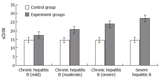
Content of CD58 in serum.
Content of membrane CD58 molecule
The results showed that the level of membrane CD58 molecule in PBMC of patients with HBV infection was significantly higher than that of the normal group and the differences among the groups were significant (P < 0.05). The levels of membrane CD58 molecule increased significantly in an order from light chronic hepatitis B, medium group, severe group and severe hepatitis B. The membrane CD58 molecule in PBMC might relate to the severity of the disease (Figures 2-5, Table 1).
Figure 2.
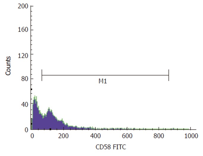
Content of membrane CD58 molecule in mild chronic hepatitis B.
Figure 5.
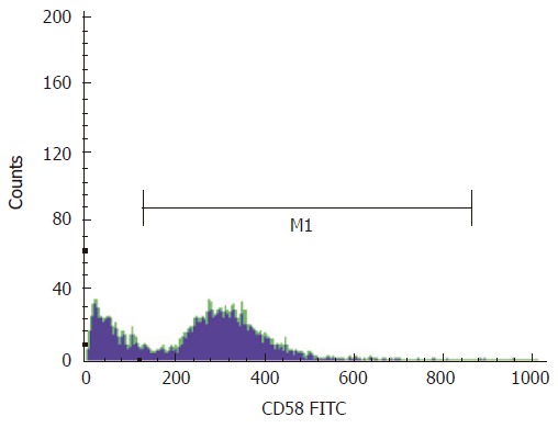
Content of membrane CD58 molecule in severe hepatitis B.
Table 1.
The liver function
| Group | n | TBIL (μmol/L) | DBIL (μmol/L) | IBIL (μmol/L) | ALT (IU/L) | AST (IU/L) |
| Normal control | 11 | 11.25 ± 2.14 | 3.12 ± 1.54 | 6.41 ± 1.85 | 25.19 ± 2.58 | 19.57 ± 3.06 |
| Chronic hepatitis B (mild) | 12 | 15.14 ± 3.26a | 15.92 ± 2.05a | 10.39 ± 2.63a | 63.33 ± 3.68a | 46.67 ± 9.81a |
| Chronic hepatitis B (moderate) | 11 | 39.21 ± 8.73a | 25.21 ± 7.11a | 23.74 ± 3.87a | 98.21 ± 18.90a | 114.43 ± 12.80a |
| Chronic hepatitis B (severe) | 10 | 105.33 ± 17.67a | 49.88 ± 8.62a | 50.86 ± 16.05a | 221.61 ± 18.19a | 157.01 ± 22.54a |
| Severe hepatitis B | 10 | 143.57 ± 23.15a | 75.26 ± 6.56a | 117.35 ± 15.27a | 116.73 ± 28.57 | 94.82 ± 41.49 |
aP < 0.05, compared with control group.
Figure 3.
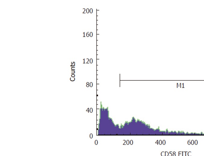
Content of membrane CD58 molecule in moderate chronic hepatitis B.
Figure 4.
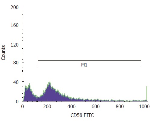
Content of membrane CD58 molecule in severe chronic hepatitis B.
DISCUSSION
CD58 is also called lymphocyte function associated antigen-3 (LFA-3)[4-6], which belongs to the CD2 family. As an important co-stimulating molecule, CD58 plays an important role in promoting the adhesion of T cells to targeted cells, and enhancing the recognition and sensitivity of T lymphocyte to the superantigen[7-9]. CD58 promotes hyperplasia and activation of T cell[1,2], promotes T cells soak inflammatory parts and takes part in signal transmission of T cells. Combined with CD2 molecules on the surface of NK cells, CD58 increases the adhesion between NK cells and target cells, activates NK cells[10,11], and increases the toxin of the cells[12,13]. After integrating with activated T cells, CD58 and CD2 facilitate interferon γ and IL-2 mRNA record and translate, differentiate CD4+T to Th1 and further initiate the immune response of the cell[3,14]. Some researchers proved that integrated with matching cells, CD58 may boost the ability of activated T cells and NK cells[15,16].
Our experiment showed that the levels of sCD58 in serum and membrane CD58 molecule in PBMC of patients with HBV infection were significantly higher than that of the normal group. The levels of CD58 varied from different groups of patients with hepatitis B, correlated to the severity of the disease. The results also showed that the percentage of CD58+ cell of patients with hepatitis B might be related with TBIL, DBIL, IBIL, ALT and AST, which prove the expression of CD58 is closely correlated with the liver damage. According to the results of the research, the increased levels of sCD58 molecule in serum and membrane CD58 molecule in PBMC of patients with hepatitis B could enhance the adhesion of APC and T cells to identify antigen[17,18]. This study proved that the combination of CD58 and CD2 activated T cells was similar to the way of TCR/CD3. This might lead to a series of responses in the cells, including the increase of the concentration of Ca2+, the activation of PKC, several limpid genes in the resting cells such as IL-2 and IFN-γ being recorded and the transformation of T cells from G0 stage to S stage. The T cells activated by integration of CD58 and CD2 would increase the record and translate of IL-2 and IFN-γ mRNA, and then differentiate into Th1 which would enhance the cell immune response. Combined with CD2 molecules on the surface of NK cells, CD58 could activate NK cells and increase the cytotoxic effect[19]. And this would contribute to the organisms to eliminate HBV. The cytotoxic effect of the immune cells might be implemented by releasing PF or inducing apoptosis of CD95 cells[20].
In summary, this study showed that the sCD58 in serum and membrane CD58 molecule in PBMC of the patients with hepatitis B is related to the severity of the disease and the liver damage. CD58 might enhance the elimination of viruses through activating T and NK cells and promoting cell immune response. However, this would also lead to the damage of liver cells.
Footnotes
Supported by a grant from the National Natural Science Foundation of China, No. 30371321
S- Editor Wang J L- Editor Zhao JB E- Editor Ma WH
References
- 1.Oh S, Hodge JW, Ahlers JD, Burke DS, Schlom J, Berzofsky JA. Selective induction of high avidity CTL by altering the balance of signals from APC. J Immunol. 2003;170:2523–2530. doi: 10.4049/jimmunol.170.5.2523. [DOI] [PubMed] [Google Scholar]
- 2.Lopez RD, Waller EK, Lu PH, Negrin RS. CD58/LFA-3 and IL-12 provided by activated monocytes are critical in the in vitro expansion of CD56+ T cells. Cancer Immunol Immunother. 2001;49:629–640. doi: 10.1007/s002620000148. [DOI] [PMC free article] [PubMed] [Google Scholar]
- 3.Le Guiner S, Le Dréan E, Labarrière N, Fonteneau JF, Viret C, Diez E, Jotereau F. LFA-3 co-stimulates cytokine secretion by cytotoxic T lymphocytes by providing a TCR-independent activation signal. Eur J Immunol. 1998;28:1322–1331. doi: 10.1002/(SICI)1521-4141(199804)28:04<1322::AID-IMMU1322>3.0.CO;2-I. [DOI] [PubMed] [Google Scholar]
- 4.Henniker AJ. CD58 (LFA-3) J Biol Regul Homeost Agents. 2001;15:190–192. [PubMed] [Google Scholar]
- 5.Elangbam CS, Qualls CW Jr, Dahlgren RR. Cell adhesion molecules--update. Vet Pathol. 1997;34:61–73. doi: 10.1177/030098589703400113. [DOI] [PubMed] [Google Scholar]
- 6.Hahn WC, Menu E, Bothwell AL, Sims PJ, Bierer BE. Overlapping but nonidentical binding sites on CD2 for CD58 and a second ligand CD59. Science. 1992;256:1805–1807. doi: 10.1126/science.1377404. [DOI] [PubMed] [Google Scholar]
- 7.Geppert TD, Lipsky PE. Immobilized anti-CD3-induced T cell growth: comparison of the frequency of responding cells within various T cell subsets. Cell Immunol. 1991;133:206–218. doi: 10.1016/0008-8749(91)90192-e. [DOI] [PubMed] [Google Scholar]
- 8.Shaw S, Shimizu Y. Two molecular pathways of human T cell adhesion: establishment of receptor-ligand relationship. Curr Opin Immunol. 1988;1:92–97. doi: 10.1016/0952-7915(88)90058-1. [DOI] [PubMed] [Google Scholar]
- 9.Halvorsen R, Leivestad T, Gaudernack G, Thorsby E. Accessory cell-dependent T-cell activation via Ti-CD3. Involvement of CD2-LFA-3 interactions. Scand J Immunol. 1988;28:277–284. doi: 10.1111/j.1365-3083.1988.tb01449.x. [DOI] [PubMed] [Google Scholar]
- 10.Barber DF, Long EO. Coexpression of CD58 or CD48 with intercellular adhesion molecule 1 on target cells enhances adhesion of resting NK cells. J Immunol. 2003;170:294–299. doi: 10.4049/jimmunol.170.1.294. [DOI] [PubMed] [Google Scholar]
- 11.Fletcher JM, Prentice HG, Grundy JE. Natural killer cell lysis of cytomegalovirus (CMV)-infected cells correlates with virally induced changes in cell surface lymphocyte function-associated antigen-3 (LFA-3) expression and not with the CMV-induced down-regulation of cell surface class I HLA. J Immunol. 1998;161:2365–2374. [PubMed] [Google Scholar]
- 12.Grosenbach DW, Schlom J, Gritz L, Gómez Yafal A, Hodge JW. A recombinant vector expressing transgenes for four T-cell costimulatory molecules (OX40L, B7-1, ICAM-1, LFA-3) induces sustained CD4+ and CD8+ T-cell activation, protection from apoptosis, and enhanced cytokine production. Cell Immunol. 2003;222:45–57. doi: 10.1016/s0008-8749(03)00080-7. [DOI] [PubMed] [Google Scholar]
- 13.Gollob JA, Ritz J. CD2-CD58 interaction and the control of T-cell interleukin-12 responsiveness. Adhesion molecules link innate and acquired immunity. Ann N Y Acad Sci. 1996;795:71–81. doi: 10.1111/j.1749-6632.1996.tb52656.x. [DOI] [PubMed] [Google Scholar]
- 14.Gollob JA, Li J, Kawasaki H, Daley JF, Groves C, Reinherz EL, Ritz J. Molecular interaction between CD58 and CD2 counter-receptors mediates the ability of monocytes to augment T cell activation by IL-12. J Immunol. 1996;157:1886–1893. [PubMed] [Google Scholar]
- 15.Gollob JA, Li J, Reinherz EL, Ritz J. CD2 regulates responsiveness of activated T cells to interleukin 12. J Exp Med. 1995;182:721–731. doi: 10.1084/jem.182.3.721. [DOI] [PMC free article] [PubMed] [Google Scholar]
- 16.Chen CM, Li SC, Lin YL, Hsu CY, Shieh MJ, Liu JF. Consumption of purple sweet potato leaves modulates human immune response: T-lymphocyte functions, lytic activity of natural killer cell and antibody production. World J Gastroenterol. 2005;11:5777–5781. doi: 10.3748/wjg.v11.i37.5777. [DOI] [PMC free article] [PubMed] [Google Scholar]
- 17.Polese L, Angriman I, Giuseppe DF, Cecchetto A, Sturniolo GC, Renata D, Scarpa M, Ruffolo C, Norberto L, Frego M, et al. Persistence of high CD40 and CD40L expression after restorative proctocolectomy for ulcerative colitis. World J Gastroenterol. 2005;11:5303–5308. doi: 10.3748/wjg.v11.i34.5303. [DOI] [PMC free article] [PubMed] [Google Scholar]
- 18.Qiu WH, Zhou BS, Chu PG, Chen WG, Chung C, Shih J, Hwu P, Yeh C, Lopez R, Yen Y. Over-expression of fibroblast growth factor receptor 3 in human hepatocellular carcinoma. World J Gastroenterol. 2005;11:5266–5272. doi: 10.3748/wjg.v11.i34.5266. [DOI] [PMC free article] [PubMed] [Google Scholar]
- 19.Han YN, Yang JL, Zheng SG, Tang Q, Zhu W. Relationship of human leukocyte antigen class II genes with the susceptibility to hepatitis B virus infection and the response to interferon in HBV-infected patients. World J Gastroenterol. 2005;11:5721–5724. doi: 10.3748/wjg.v11.i36.5721. [DOI] [PMC free article] [PubMed] [Google Scholar]
- 20.Thompson CB. Apoptosis in the pathogenesis and treatment of disease. Science. 1995;267:1456–1462. doi: 10.1126/science.7878464. [DOI] [PubMed] [Google Scholar]


