Abstract
AIM: To investigate the hepatoprotective effect of manual acupuncture at Yanglingquan (GB34) on CCl4-induced chronic liver damage in rats.
METHODS: Rats were injected intraperitoneally with CCl4 (1 mL/kg) and treated with manual acupuncture using reinforcing manipulation techniques at left GB34 (Yanglingquan) 3 times a week for 10 wk. A non-acupoint in left gluteal area was selected as a sham point. To estimate the hepatoprotective effect of manual acupuncture at GB34, measurement of liver index, biochemical assays including serum ALT, AST, ALP and total cholesterol, histological analysis and blood cell counts were conducted.
RESULTS: Manual acupuncture at GB34 reduced the liver index, serum ALT, AST, ALP and total cholesterol levels as compared with the control group and the sham acupuncture group. It also increased and normalized the populations of WBC and lymphocytes.
CONCLUSION: Manual acupuncture with reinforcing manipulation techniques at left GB34 reduces liver toxicity, protects liver function and liver tissue, and normalizes immune activity in CCl4-intoxicated rats.
Keywords: Manual acupuncture, Yanglingquan (GB34), CCl4-induced liver damage, Hepatoprotective effect
INTRODUCTION
Herbal medicine and acupuncture are the two main methods to treat disease in oriental medicine. Because of chemical residue contamination, there is recent gaining suspicion that herbs may be harmful to the liver. Accordingly, acupuncture is getting more interest these days for the treatment of liver diseases in oriental medical clinics. In the present study, we tried to investigate the effects of manual acupuncture on long-term liver damage. To investigate the effects of manual acupuncture at GB34 on liver damage, we used CCl4-intoxicated rat model and chose GB34 as an acupoint to protect liver and treat liver damage induced by CCl4 administration.
GB34 is an acupoint located on the gall bladder meridian. In oriental medical theory, the liver and gall bladder corresponds to each other and their meridians are also closely related with each other in the ‘interior-exterior relationship’[1,2]. Gall bladder meridian pertains to the gall bladder organ and connects with the liver organ. The liver meridian pertains to the liver organ and connects with the gall bladder organ. Therefore, the acupoints on the liver meridian are used to treat gall bladder organ diseases as well as liver organ diseases. Consequently, the acupoints on the gall bladder meridian are used to treat liver organ diseases as well as gall bladder organ diseases. Hence, GB34 is closely related with the liver as well as the gall bladder. It explains why GB34 is used to treat liver disease so often. Moreover, GB34 is the He (meaning “sea”) point of gall bladder. In oriental medical theory, a “sea He” point is considered to be the entrance of the meridian energy to the corresponding organ[3]. Therefore, GB34 influences the liver and gall bladder more strongly than other acupoints. The functions of this point are regulating and tonifying the liver, regulating the gallbladder, spreading liver Qi (oriental medical term for “vital energy”), subduing liver Yang, draining liver pathogens, etc. GB34 has been clinically used for hypochondriac pain, jaundice, hepatitis, acute biliary tract diseases, cirrhosis of the liver and hypertension due to liver Yang excess, etc[1,4].
We presume that neuronal activity is involved in the transmission of acupuncture stimulation, so the animals were not anesthetized during the acupuncture administration. To keep the animals from moving during the acupuncture administration, they were put in cages with five holes for tail and four limbs. To estimate and exclude the effect of stress from restriction within the cage, the rats in the control group were also kept in the cages in the same manner as the acupuncture group.
The action of acupuncture could be influenced by acupuncture techniques as well as point selection. There are two main categories of acupuncture techniques: reinforcing technique and reducing technique. Clockwise needle rotation, scraping downward of needles, and odd number of manipulating operations are considered as reinforcing techniques. On the other hand, counterclockwise needle rotation, scraping upward of needles, and even number of manipulating operations are considered as reducing techniques. Reinforcing acupuncture manipulation techniques are used for chronic and deficient syndrome, while reducing techniques are used for acute and excess syndrome[1]. Since the animals in the present study were injected with CCl4 for a long period, we considered it as a chronic and deficient condition, and therefore, we administered acupuncture with reinforcing manipulation techniques.
MATERIALS AND METHODS
Animals
Sprague-Dawley male rats (200-250 g) were purchased from Deahan Biorink Co. The animals were adapted to the environment of 22 ± 2°C room temperature, 12-h light/dark cycle for 2 wk and had free access to water and food. Our animal experiment has been conducted in accordance with the Use of Laboratory Animals as adopted and promulgated by the U.S. National Institutes of Health.
Experimental design
Experimental animals were randomly divided into four groups: normal; control (CCl4); Sham (CCl4 + manual acupuncture at sham point); and GB34 (CCl4 + manual acupuncture at left GB34). Each group consisted of 7 rats. Liver injury was induced by intraperitoneal injections with 500 mL/L CCl4 (Sigma, USA) solution in olive oil
(1 mL/kg), twice a week for 10 wk. Manual acupuncture was administered 3 times a week during the same period[5].
Acupuncture
Cages with five holes for the tail and four limbs were manufactured for this study. During acupuncture administration, the animals were kept in the cage with left hind limb fastened to the wall of the cage with tape. Sterilized disposable stainless steel needles (0.25 mm × 30 mm, Dongbang Acupuncture Inc., Korea) were inserted perpendicularly as deep as 2-3 mm at left GB34 or sham point. Rat GB34 was determined according to human GB34 which locates on gall bladder meridian, in the depression anterior and inferior to the head of the fibula[1]. A non-acupoint in the gluteal region was selected as a sham point (Figure 1). After the needles were inserted, they were rotated clockwise with amplitude of 90° nine times, and scraped downward nine times. Nine rotations and nine scrapes constituted one manipulation unit, and three manipulation units constituted one treatment session. After each manipulation unit, there was a hold for about 1 s. The full manipulation took about 30 s. The rats in the control group were kept in a cage for 30 s with the left hind limb tied without acupuncture treatment (Figure 2). All acupuncture was administered by a trained and experienced oriental medical doctor who was unknown about the research protocol except the needle manipulating methods.
Figure 1.
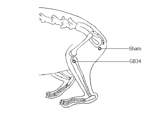
GB34 and sham point for acupuncture.
Figure 2.
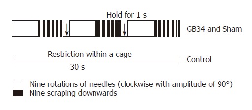
Acupuncture scheme.
Liver index measurement
Rat’s body weight was measured before the animals were sacrificed. Rat liver was removed and weighed right after the animal was sacrificed and the liver index (% of liver weight/body weight)[6,7] was estimated.
Biochemical analysis and blood counts
Forty-eight hours after the last administration with CCl4, the rats were anesthetized with ethyl ether and blood samples were taken from the heart. Blood was centrifuged at 3000 r/min for 15 min and serum was taken. Alanine aminotransferase (ALT), aspartate aminotransferase (AST), alkaline phosphotase (ALP), total cholesterol in serum and the populations of RBC, WBC, lymphocytes in plasma were detected.
Histological analysis
The rats were sacrificed and the liver tissues were obtained individually from each group and fixed in 40 g/L formaldehyde. After decalcification in 50 mL/L formic acid, the specimens were processed for paraffin embedding. Tissue sections were obtained and stained with hematoxylin and eosin (HE) or masson’s trichrome (MT). Tissue destruction and fatty changes of liver were observed at 400 × magnification.
Statistical analysis
Data were obtained from the rats which survived to the end of the experiment. All in normal group, 3 in control group, 3 in sham group and 5 in GB34 group survived to the end of the experiment. Data were expressed as mean ± SD. Statistical significance of difference between groups was determined using ANOVA, followed by t-test. P < 0.05 was considered statistically significant.
RESULTS
Liver index
CCl4 injection induced a significant increase in liver index. On the other hand, manual acupuncture at GB34 lowered it similar to the normal value (Figure 3).
Figure 3.
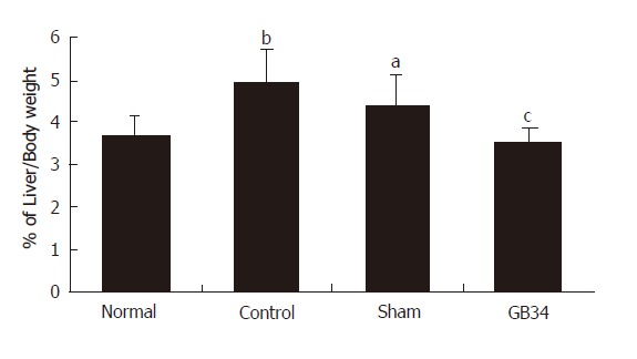
Effect of manual acupuncture at BG34 on liver index of CCl4-intoxicated rats. aP < 0.05, bP < 0.01 vs normal group; cP < 0.05 vs control group.
Serum ALT, AST, ALP and total cholesterol
ALT, AST, ALP and total cholesterol in serum were increased remarkably by long-term CCl4 administration, indicating damage to the liver. Manual acupuncture at GB34 significantly reduced serum ALT, AST and total cholesterol in comparison with the control group. Serum ALP was also reduced by manual acupuncture at GB34 but no statistical significance was found (Figure 4).
Figure 4.
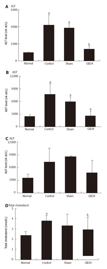
Effect of manual acupuncture at GB34 on serum ALT, AST, ALP and total cholesterol of CCl4-intoxicated rats. aP < 0.01 vs normal group; bP < 0.05 vs control group
Blood cell counts
The number of RBC was slightly reduced by CCl4 intoxication and restored by manual acupuncture at GB34, though statistical significance was not observed. The number of WBC and the percentage of lymphocytes out of WBC was significantly reduced by CCl4 intoxication and restored by manual acupuncture at GB34 close to the normal level (Figure 5).
Figure 5.

Effect of manual acupuncture at GB34 on RBC, WBC and lymphocytes in blood. aP < 0.05, bP < 0.01 vs normal group; cP < 0.05, dP < 0.001 vs control group.
Liver histology
Histological analysis using HE stain showed that necrosis of liver tissue and fatty changes were viciously induced by CCl4 administration. The group treated with manual acupuncture at GB34 showed reduced feature of hepatocyte necrosis and fatty change compared to the control group and the sham group. In addition, MT staining showed lower accumulation of extracellular matrix in the GB34 group compared to the control group and sham group (Figure 6).
Figure 6.
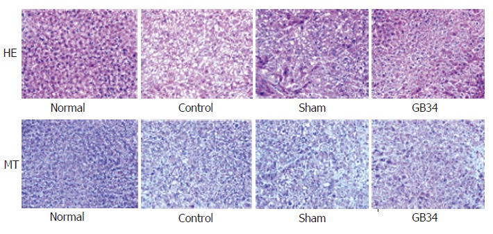
Histological results of liver tissue. Necrosis and fatty change could be found in the control group (top). GB34 group revealed lower accumulation of extracellular matrix compared to the control group and sham group (bottom) (×400 magnification)
DISCUSSION
CCl4 has been widely used to induce experimental hepatic damage[8,9]. It induces liver cell necrosis and apoptosis, and can be used to induce hepatic fibrosis or cirrhosis by repetitive administration[10-12]. Liu et al[13] investigated the effect of manual acupuncture at ST36 and LR3 on CCl4-induced acute liver damage. In the present study, we investigated the effect of manual acupuncture at GB34 on chronic liver damage induced by long-term CCl4 administration.
ALT and AST are the specific markers to assess hepatocellular damage leading to liver cell necrosis[14]. Slight to moderate increases in ALP (1-2 times normal) occurred in liver disorders[15]. Serum cholesterol is one of the general indications of the synthetic and general metabolic capacity of the liver[16]. In the present study, CCl4 injection significantly increased serum ALT, AST, ALP and cholesterol levels, indicating induction of hepatic damage. Manual acupuncture at GB34 inhibited the increases of these parameters, indicating that manual acupuncture at GB34 protected liver and reduced liver toxicity. Histological analysis also showed that the acupuncture at GB34 protected liver tissue against CCl4-intoxication. Our histological analysis in this study was only qualitative. Quantitative assessment of anti-fibrotic effect of acupuncture, such as determination of hepatic hydroxyproline
content, would be meaningful in the next study.
In the present study, long-term CCl4 administration reduced the population of leucocytes and lymphocytes in blood. We infer that this reduction was due to the decline of immune activity by long-term liver damage. Manual acupuncture at GB34 recovered the population of leucocytes and lymphocytes in blood. Therefore, we presume that manual acupuncture at GB34 restored the immune activity, which is also in agreement with Hau’s report on the white cell increase by acupuncture[17]. However, the effect of acupuncture on inflammation in liver is not clear yet. It is known that inflammatory cells are infiltrated in CCl4-intoxicated liver, and there exists an abnormality of cytokines in liver tissue. For instance, TNF-α and IL-10 are increased, and IL-2 and IFN-γ are reduced in CCl4-intoxicated liver tissue[18,19]. More profound studies are required on how acupuncture influences the chronic inflammatory conditions of the liver.
Based on the results of the present study, we speculate that manual acupuncture at GB34 is beneficial to protect liver function and tissue, reduce hepatic toxicity and normalize immune activity against CCl4-intoxication in rats. The hepatoprotective effect of manual acupuncture at GB34 in this study may be related to the immune reinforcing effect of acupuncture or neuro-immune interaction on the pathway of the transmission of acupuncture stimulation.
Footnotes
S- Editors Pan BR and Wang J L- Editor Kumar M E- Editor Bai SH
References
- 1.Deng L, Gan Y, He S, Ji X, Li Y, Wang R, Wang W, Wang X, Xu H, Xue X, et al. Chinese acupuncture and moxibustion. Beijing: Foreign Languages Press; 1997. pp. 54, 71–74, 208, 325-327. [Google Scholar]
- 2.Captchuk TJ. The web that has no weaver: understanding Chinese medicine. NY: Contemporary books; 2000. p. 8. [Google Scholar]
- 3.Choe YT, Yu CH, Kim CH, Kim DH. Oriental medicine series. Vol 2; Acupuncture & moxibustion. Seoul: Res Inst of Oriental Med Inc; 1987. p. 160. [Google Scholar]
- 4.Lade A. Acupuncture points images & functions. Washington: Eastland Press; 1998. p. 230. [Google Scholar]
- 5.Ulicná O, Greksák M, Vancová O, Zlatos L, Galbavý S, Bozek P, Nakano M. Hepatoprotective effect of rooibos tea (Aspalathus linearis) on CCl4-induced liver damage in rats. Physiol Res. 2003;52:461–466. [PubMed] [Google Scholar]
- 6.Yang Q, Xie RJ, Geng XX, Luo XH, Han B, Cheng ML. Effect of Danshao Huaxian capsule on expression of matrix metalloproteinase-1 and tissue inhibitor of metalloproteinase-1 in fibrotic liver of rats. World J Gastroenterol. 2005;11:4953–4956. doi: 10.3748/wjg.v11.i32.4953. [DOI] [PMC free article] [PubMed] [Google Scholar]
- 7.Lin WC, Lin WL. Ameliorative effect of Ganoderma lucidum on carbon tetrachloride-induced liver fibrosis in rats. World J Gastroenterol. 2006;12:265–270. doi: 10.3748/wjg.v12.i2.265. [DOI] [PMC free article] [PubMed] [Google Scholar]
- 8.Tsukamoto H, Matsuoka M, French SW. Experimental models of hepatic fibrosis: a review. Semin Liver Dis. 1990;10:56–65. doi: 10.1055/s-2008-1040457. [DOI] [PubMed] [Google Scholar]
- 9.Yao XX, Jiang SL, Tang YW, Yao DM, Yao X. Efficacy of Chinese medicine Yi-gan-kang granule in prophylaxis and treatment of liver fibrosis in rats. World J Gastroenterol. 2005;11:2583–2590. doi: 10.3748/wjg.v11.i17.2583. [DOI] [PMC free article] [PubMed] [Google Scholar]
- 10.Constandinou C, Henderson N, Iredale JP. Modeling liver fibrosis in rodents. Methods Mol Med. 2005;117:237–250. doi: 10.1385/1-59259-940-0:237. [DOI] [PubMed] [Google Scholar]
- 11.Chang ML, Yeh CT, Chang PY, Chen JC. Comparison of murine cirrhosis models induced by hepatotoxin administration and common bile duct ligation. World J Gastroenterol. 2005;11:4167–4172. doi: 10.3748/wjg.v11.i27.4167. [DOI] [PMC free article] [PubMed] [Google Scholar]
- 12.Shi GF, Li Q. Effects of oxymatrine on experimental hepatic fibrosis and its mechanism in vivo. World J Gastroenterol. 2005;11:268–271. doi: 10.3748/wjg.v11.i2.268. [DOI] [PMC free article] [PubMed] [Google Scholar]
- 13.Liu HJ, Hsu SF, Hsieh CC, Ho TY, Hsieh CL, Tsai CC, Lin JG. The effectiveness of Tsu-San-Li (St-36) and Tai-Chung (Li-3) acupoints for treatment of acute liver damage in rats. Am J Chin Med. 2001;29:221–226. doi: 10.1142/S0192415X01000253. [DOI] [PubMed] [Google Scholar]
- 14.Amacher DE. Serum transaminase elevations as indicators of hepatic injury following the administration of drugs. Regul Toxicol Pharmacol. 1998;27:119–130. doi: 10.1006/rtph.1998.1201. [DOI] [PubMed] [Google Scholar]
- 15.Isselbacher KJ, Braunwald E, Wilson JD, Martin JB, Fauci AS, Kasper DL. Harrison's principles of internal medicine, 13th ed. NY: McGraw-Hill; 1994. pp. 1444–1446. [Google Scholar]
- 16.Fregia A, Jensen DM. Evaluation of abnormal liver tests. Compr Ther. 1994;20:50–54. [PubMed] [Google Scholar]
- 17.Hau DM. Effects of electroacupuncture on leukocytes and plasma protein in the X-irradiated rats. Am J Chin Med. 1984;12:106–114. doi: 10.1142/S0192415X84000118. [DOI] [PubMed] [Google Scholar]
- 18.Jeong DH, Lee GP, Jeong WI, Do SH, Yang HJ, Yuan DW, Park HY, Kim KJ, Jeong KS. Alterations of mast cells and TGF-beta1 on the silymarin treatment for CCl(4)-induced hepatic fibrosis. World J Gastroenterol. 2005;11:1141–1148. doi: 10.3748/wjg.v11.i8.1141. [DOI] [PMC free article] [PubMed] [Google Scholar]
- 19.Yu XH, Zhu JS, Yu HF, Zhu L. Immunomodulatory effect of oxymatrine on induced CCl4-hepatic fibrosis in rats. Chin Med J (Engl) 2004;117:1856–1858. [PubMed] [Google Scholar]


