Abstract
AIM: To identify the variants in UDP-glucuronosyltransfe-rase 1 (UGT1A1) gene in Gilbert’s syndrome (GS) and to estimate the association between homozygosity for TA insertion and GS in India, as well as the frequency of TA insertion and its impact among normal controls in India.
METHODS: Ninety-five GS cases and 95 normal controls were selected. Liver function and other tests were done. The promoter and all 5 exons of UGT1A1 gene were resequenced. Functional assessment of a novel trinucleotide insertion was done by in silico analysis and by estimating UGT1A1 promoter activity carried out by luciferase reporter assay of appropriate constructs in Hep G2 cell line.
RESULTS: Among the GS patients, 80% were homozygous for the TA insertion, which was several-fold higher than reports from other ethnic groups. The mean UCB level was elevated among individuals with only one copy of this insertion, which was not significantly different from those with two copies. Many new DNA variants in UGT1A1 gene were discovered, including a trinucleotide (CAT) insertion in the promoter found in a subset (10%) of GS patients, but not among normal controls. In-silico analysis showed marked changes in the DNA-folding of the promoter and functional analysis showed a 20-fold reduction in transcription efficiency of UGT1A1 gene resulting from this insertion, thereby significantly elevating the UCB level.
CONCLUSION: The genetic epidemiology of GS is variable across ethnic groups and the epistatic interactions among UGT1A1 promoter variants modulate bilirubin glucuronidation.
Keywords: Unconjugated hyperbilirubinemia, UGT1A1 gene, DNA resequencing, Luciferase reporter assay
INTRODUCTION
Gilbert’s syndrome (GS) is generally considered to be an autosomal recessive disorder (OMIM #143500) characterized by mild, chronic, non-hemolytic unconjugated hyperbilirubinemia in the absence of liver disease. Although it has long been perceived to be an innocuous clinical entity with a benign course, recent data suggest that affected individuals may be predisposed to development of liver injury following treatment with various drugs and xenobiotics and that the genetic defect in GS may influence the outcome of liver transplantation and other clinical conditions. The disorder due to a deficiency in bilirubin glucuronidation, is commonly caused by dinucleotide (TA) insertions and [TA (TA)7TAA] alleles in the promoter region of the bilirubin UDP-glucuronosyltransferase 1 (UGT1A1) gene. This dinucleotide insertion reduces the efficiency of transcription of the gene and decreases hepatic UDP-glucuronosyltransferase activity to about 30% of normal levels in homozygous subjects[6-9]. The prevalence of GS, the frequency of the (TA)7TAA allele in the UGT1A1 promoter and the proportion of GS patients who are homozygous for the (TA)7TAA allele vary widely across populations[10-19]. Recent data have also shown that variants in UGT1A1 gene other than the classical dinucleotide insertion in its promoter, can contribute to hyperbilirubinemia, including the milder form as in GS, although their effects appear to be variable across populations[5,20-24]. Thus, as recently emphasized[20], detailed investigations into genetic polymorphisms of UGT1A1 gene in Asian populations may provide a better understanding of unconjugated hyperbilirubinemia.
The objectives of our case-control study were to estimate the association between homozygosity for the (TA)7TAA allele and GS in India, to identify and study the impact of this and other polymorphisms in the promoter and exons of the UGT1A1 gene on serum bilirubin levels, and to estimate the frequency of the (TA)7TAA homozygous genotype in the general population in India.
MATERIALS AND METHODS
Participant recruitment and clinical biochemistry analysis
This study was conducted in 95 consecutive patients presenting to the Liver Clinic at the Department of Gastroenterology of the Institute of Postgraduate Medical Education & Research, Kolkata, India, for evaluation of persistent unconjugated hyperbilirubinemia (serum bilirubin greater than 1.2 mg/dL) documented at least twice over a period of one month. Serum total bilirubin and its unconjugated fraction were estimated in fasting condition. Each of these individuals had a normal finding on physical examination, normal hepatobiliary ultrasound, normal liver function test excluding hyperbilirubinemia and a normal reticulocyte count. A total of 95 adult healthy volunteers from the same ethnic population as the GS patients were included as controls. Each control also underwent the same set of investigations as the patients. Their fasting serum bilirubin level estimated twice at intervals of fifteen days was consistently less than 1.0 mg/dL. Patients were excluded if they were under any medication during the past one month or consumed alcohol regularly or had present or past history of hepatic/hematological disease. Additionally, quantitative estimation of hemoglobin fractions (A0, A2 and F) was carried out on each patient and control by cation-exchange HPLC using the VariantTM Hemoglobin Testing System Beta Thalassemia Short Program (Bio-Rad Diagnostics, Hercules, USA). A 5 mL blood sample was collected from each patient by venipuncture and DNA was isolated using a standard protocol[25]. Data and samples were collected with written informed consent of the patients and controls, after the approval was obtained from the Human Research Ethics Committee of the Institute of Postgraduate Medical Education & Research, Kolkata.
DNA re-sequencing and identification of variant alleles
DNA re-sequencing of the promoter and exon 1*1 encoding the substrate-specific region of bilirubin-UGT1 gene as well as the 4 common exons (exons 2-5) was carried out using an automated DNA sequencer (ABI-3100; Applied Bio-systems, Foster City, USA.). DNA samples were first amplified by the polymerase chain reaction technique using an ABI-9700 thermal cycler. PCR products were then cleaned using exonuclease-I (USB. Corporation, Cleveland, USA) and shrimp alkaline phosphatase (Amersham, Freiburg, Germany), and sequencing reactions were carried out. DNA resequencing was carried out in both forward and reverse directions. Raw DNA sequences were analyzed as previously described[26], and variant genotypes were identified.
Functional analysis of a novel trinucleotide insertion by luciferase reporter assay
UGT1A1 promoter fragments, 331-336 bp in length depending on the number of TA repeats and CAT insertion, were amplified using primers (F: 5’-tgtagatcttcctctctggtaacact-3’) and (R: 5’-atgaagctttgctcctgccagaggttc-3’) from genomic DNA of individuals homozygous for six and seven TA repeats, and CAT insertion on (TA)7TAA background. DNA amplification was carried out with polymerase chain reaction (PCR) using FastStart Taq DNA polymerase (Roche, Mannheim, Germany) with an initial denaturation at 95 °C for 10 min, followed by 35 cycles at 94 °C for 1 min, at 56 °C for 45 s and at 72 °C for 30 s on an ABI-9 700 (Applied Bio-systems, Foster City, USA.) thermal cycler. To facilitate subcloning of the PCR products in the reporter gene construct, oligonucleotides F and R were designed with a Bgl II and a Hind III restriction enzyme site at the 5’ end, respectively. PCR products were doubly digested with Bgl II and Hind III, purified by Qiagen gel purification kit (Qiagen, Hilden, Germany) and subcloned into pGL3-basic vector (Promega, Madison, USA). The integrity of the resulting plasmids was confirmed by restriction mapping and sequencing analyses. Promoter activity of each construct was measured in HepG2 cell line. Cells were grown in MEM medium supplemented with 2 mmol/L L-glutamine, 0.1 mmol/L non-essential amino acids, 1 mmol/L sodium pyruvate and 10% fetal calf serum (GIBCO -BRL, Grand Island, USA) at 37 °C with 50 mL/L CO2. Exponentially growing cells were trypsinized, seeded at 2.5×10⁵ cells, and incubated overnight prior to transfection. Transfection was carried out by lipofectamin (Invitrogen, Carlsbad, USA) using 2 μg of each of the constructs (namely (TA)6 TAA, (TA)7 TAA and CAT insertion on (TA)7 TAA background), as well as the pGL3-basic and pGL3-control as positive controls (TA)7TAA, the luciferase gene was under the control of SV40 promoter and enhancer. After 48 h of transfection, the cells were lysed and centrifuged in cell culture lysis buffer (Promega, Madison, USA). Luciferase activity was assayed in the cell lysate by measuring the photoluminescence in a Monolight 2010 single channel luminometer and the total cellular protein content was measured by the standard Bradford’s method. Luciferase activity was normalized to total cell protein concentration. Normalized luciferase activity of each construct was expressed as a ratio to that of the pGL3-basic vector. Three independent experiments were performed for each construct and all measurements were determined in duplicate.
Statistical analysis
Equality of proportions was statistically tested by the standard normal test procedure[27]. Equalities of mean values of various hematological parameters for individuals belonging to different genotypes at various polymorphic loci were statistically tested using the Student t-test or the analysis of variance (ANOVA) procedure, as appropriate. Regression analysis was performed to test the significance of dependence of un-conjugated serum bilirubin level on some relevant variables. Allele frequencies were estimated using the gene-counting method.
RESULTS
Characteristics of patients and controls
No statistically significant (P > 0.05) differences in the proportions of males and females were observed between the patients (87 males, 8 females) and controls (77 males, 18 females). The mean ages of male and female patients (30.1 ± 1.1 years of males, 26.8 ± 5.1 years of females) and controls (30.2 ± 1.1 years of males, 31.1 ± 2.9 years of females) were not significantly different (P > 0.05). Since elevated HbA2 levels could potentially increase the unconjugated bilirubin level, we tested the significance of the regression coefficient of HbA2 level on unconjugated bilirubin, separately for patients and controls. In both sets, there was no statistically significant impact of HbA2 (P-values for patients and controls were 0.638 and 0.106, respectively). The difference in the mean values of HbA2 between patients and controls was not significantly different (t = 1.76, d.f. = 128, P > 0.05). The 8 GS patients, but none of the normal controls, who were habitual smokers were asked to refrain from smoking for 24 h prior to blood collection. We tested whether the inclusion of these 8 GS patients had a significant impact on our findings. We therefore, examined whether the mean value of un-conjugated bilirubin among patients who were smokers (n = 8) was significantly higher than that among patients who were not smokers (n = 87). No significant difference was found (mean for smoker patients=2.67 ± 0.33, mean for non-smoker patients = 3.38 ± 0.30, t = 0.696, d.f. = 93, P = 0.488). We have performed all analyses that were reported below with and without the inclusion of the 8 GS patients who were habitual smokers. No differences in inferences were found (results not shown). Therefore, we presented all results including these 8 GS subjects.
UGT1A1 sequence variants and their relation with bilirubin level
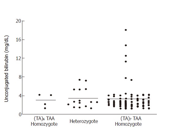
Eleven sequence variants in UGT1A1 gene was observed, of which 3 each were in the promoter, 3 in exon 1, 2 in exon 2, 1 in exon 3 and 2 in exon 4. No variant was found in exon 5. The genotype and variant allele frequencies at these positions in patients and controls are given in Table 1. Of these 11 variant sites, 2 were non-polymorphic (frequency of the rarer allele < 1%), while the remaining 9 sites were polymorphic either among patients or among controls or in both. One of the polymorphic sites was the TA insertion [(TA)7TAA allele] in the TATA box of the UGT1A1 promoter[2,3]. The frequencies of the (TA)7TAA allele (0.879) and the 7/7 genotype (80%) among the patients were significantly higher (P < 0.005) than those among the controls (0.384 and 10%, respectively). The un-conjugated serum bilirubin values for GS patients belonging to the 6/6, 7/6 and 7/7 genotypes are presented in Figure 1. The mean values of un-conjugated bilirubin for GS patients and normal controls were 2.5 ± 0.25 mg/dL and 0.61 ± 0.09 mg/dL respectively. The mean+SE value of un-conjugated bilirubin among patients with the 7/6 genotype was 3.46 ± 0.54 mg/dL, which was not significantly different (P = 0.05) from patients with the 7/7 genotype (3.31 ± 0.33 mg/dL). The mean un-conjugated serum bilirubin values for these genotypes among controls were only 0.59 ± 0.10 mg/dL and 0.64 ± 0.15 mg/dL, respectively.
Table 1.
DNA sequence variations observed among Gilbert’s syndrome patients and normal controls
| Location of variant and nucleotide position (np) | Description of variant1 | Genotype/Allele frequency (p) | Patients (%) | Control (%) |
| UGT1*1 promoter | CAT insertion | Insertion/Insertion | 3 | 0 |
| nps -85 to -83 | Insertion/non-insertion | 6 | 0 | |
| Non-insertion/non-insertion | 86 | 95 | ||
| p(Insertion) | 0.063 | 0.000 | ||
| UGT1*1 promoter | G→C | GG | 93 | 95 |
| np -63 | GC | 2 | 0 | |
| p(C) | 0.011 | 0.000 | ||
| UGT1*1 promoter | (TA)6 TAA→ | (TA)6 TAA /(TA)6 TAA | 4 | 32 |
| nps -53 to -38 | (TA)7 TAA | (TA)7 TAA /(TA)6 TAA | 15 | 53 |
| (TA)7 TAA/(TA)7 TAA | 76 | 10 | ||
| p[(TA)7 TAA] | 0.879 | 0.384 | ||
| Exon 1 | G→A | GG | 85 | 90 |
| np +211 | (G71R) | GA | 9 | 5 |
| AA | 1 | 0 | ||
| p(A) | 0.058 | 0.026 | ||
| Exon 1 | T→C | TT | 93 | 94 |
| np +476 | (I159T) | TC | 2 | 1 |
| p(C) | 0.011 | 0.005 | ||
| Exon 1 | T→C | TT | 94 | 95 |
| np +625 | (R209W) | TC | 1 | 0 |
| p(C) | 0.005 | 0.000 | ||
| Exon 2 | C→G | CC | 95 | 66 |
| np +6 844 | (A321G) | CG | 0 | 29 |
| p(G) | 0.000 | 0.152 | ||
| Exon 2 | A→G | AA | 87 | 89 |
| np +6 846 | (I322V) | AG | 7 | 6 |
| GG | 1 | 0 | ||
| p(G) | 0.042 | 0.032 | ||
| Exon 3 | G→A | GG | 94 | 95 |
| np +7 640 | (D359N) | GA | 1 | 0 |
| p(A) | 0.005 | 0.000 | ||
| Exon 4 | C→T | CC | 93 | 95 |
| np +7 939 | (P364L) | CT | 2 | 0 |
| p(T) | 0.011 | 0.000 | ||
| Exon 4 | A→G | AA | 92 | 95 |
| np +7 975 | (H376R) | AG | 3 | 0 |
| p(G) | 0.016 | 0.000 |
Amino acid changes resulting from nucleotide changes in the exons are indicated in parentheses.
Figure 1.

Distributions of un-conjugated serum bilirubin values among Gilbert’s syndrome patients classified by the genotype of the common TA insertion in the TATA box of the UGT1A1 promoter.
No significant differences (P = 0.05) were found in the mean bilirubin levels among individuals belonging to the various genotypes at the remaining single nucleotide polymorphic loci. With respect to the C6844G polymorphism resulting in a non-synonymous amino acid change (A321G), all patients were CC homozygotes, while about 30% of the controls were CG heterozygotes (Table 1). We did not find any significant effect of the G71R polymorphism on un-conjugated bilirubin level. This polymorphism also showed no significant interaction with the TATA box insertion polymorphism (results not shown). These findings are discordant with those reported among the Japanese[19].
A novel human-specific trinucleotide insertion in Gilbert’s syndrome patients and its impact on bilirubin level
Among the polymorphic variants described in Table 1, aside from the familiar TA insertion, the most striking was the CAT insertion (nucleotide positions -85 to -83) in the CAAT box of UGT1*1 promoter. Normally, there is one copy of the CAT trinucleotide present in human Genbank (http://www.ncbi.nlm.nih.gov/Genbank/). We found two copies of this trinucleotide in some GS patients. To confirm that this was an insertion we searched the chimpanzee (gi|6456543|gb|AF135463.1|AF135463) and gorilla (gi|6456545|gb|AF135464.1|AF135464) databases. In both species there is only copy, confirming that the single copy is the ancestral state. This CAT insertion was found only among nine (10%) GS patients, who were all homozygous for the TA insertion. We found that GS patients with the CAT-insertion had a significantly (P < 0.001) elevated mean level (6.13 ± 1.61 mg/dL) compared to those without the insertion (2.93 ± 0.28 mg/dL). Individuals who possessed the CAT insertion did not consistently possess a variant allele at any of the other sites. One individual who was heterozygous for the CAT insertion was also heterozygous for the I322V variant, and other three CAT-insertion heterozygotes were also heterozygotes for the H376R variant.
Functional analysis of CAT insertion
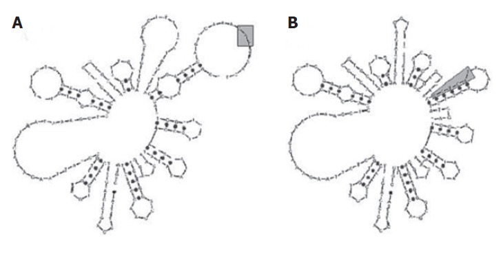
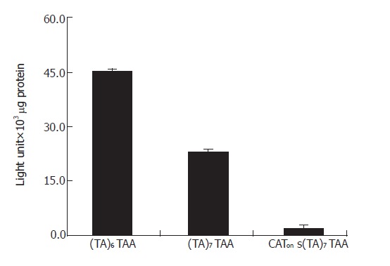
Since about 10% of the (TA)7TAA homozygotes carried the CAT insertion and the insertion significantly elevated the bilirubin level, we postulated that the insertion had a functional impact. To examine this, we studied the change in DNA-folding of the promoter region caused by this insertion. The most stable structures, namely those with lowest free energy (dG) values, are given in Figure 2 for the UGT1A1 promoter region without (dG = -14.4) and with (dG = -14.8) the additional CAT insertion. A striking structural change was found around the region of the insertion, the loops were converted to stems. It is well known that formation of stems in the promoter could reduce transcription of the gene. A detailed functional analysis of the UGT1A1 promoter by luciferase reporter assay confirmed that there was a two-fold decrease in transcription efficiency for the variant (TA)7TAA promoter allele, and a 20-fold decrease when there was a CAT insertion in the promoter on the background of the (TA)7TAA allele compared to the normal (TA)6TAA promoter allele (Figure 3).
Figure 2.

Folded DNA structures of the UGT1A1 promoter region with one copy of the CAT trinucleotide (shaded region) (A) and two copies of the CAT trinucleotide (CAT insertion allele, shaded region) (B).
Figure 3.

Transcriptional activities of the (TA) 6TAA, (TA)7 TAA and CAT alleles as assessed by a luciferase reporter assay.
DISCUSSION
Genetically determined un-conjugated hyperbilirubinemia constitutes a spectrum of clinical entities characterized by incremental serum bilirubin values related to graded reduction of UGT1A1 enzyme activity. The most novel finding of our study is that a trinucleotide (CAT) insertion in the nucleotide positions -85 to -83 of the UGT1A1 promoter was present in GS patients who were homozygous for the causal TA insertion, but not in other GS patients or in controls. This insertion significantly (P < 0.001) elevated the un-conjugated bilirubin level (mean = 6.13 mg/dL) in the patients to the range usually seen in Crigler-Najjar (CN) II syndrome. We have demonstrated that this insertion in the background of the (TA)7 allele classically described in GS, diminishes UGT1A1 transcriptional activity to about 5% (2.4 light units/103 μg protein) of the normal (44.9 light units/103 μg protein), much lower than the 50% reduction (22.9 light units/103 μg protein) observed for (TA)7 alone. Thus the combined biological effect of the CAT and TA insertions is a further elevation in the un-conjugated bilirubin level compared to that of the TA insertion alone, which is exactly what we have observed in the GS patients carrying the CAT insertion. This initial description of the interactive influence of a novel trinucleotide insertion and the GS-type abnormality in the promoter region alone, in the absence of a consistent exonic mutation, to quantitatively elevate un-conjugated bilirubin to levels observed in Crigler-Najjar II (CN II) provides further evidence of genetic heterogeneity and overlap of the clinical syndromes of un-conjugated hyperbilirubinemia. Thus, Gilbert’s, Crigler-Najjar I (CN I) and CN II syndromes may not be mutually exclusive clinical-genetic entitities but are different windows of the quantititative spectrum of elevated serum unconjugated bilirubin levels.
The association (80%) between homozygosity for the (TA)7TAA allele and GS is much stronger in India than reported earlier from most other ethnic groups[14,15,16,20]. Graded reduction of UGT1 activity has been demonstrated with increasing length of the TA repeats in a recent study[28]. It was demonstrated that 7/7 homozygotes and 7/6 heterozygotes have, respectively, a 52% and a 37% reduction of UDP glucuronyl transferase activity in liver tissue homogenates. In our study, serum un-conjugated bilirubin values among 7/6 heterozygote GS patients were high but not significantly different from the 7/7 homozygotes. The finding of increased serum bilirubin values among the 7/6 heterozygote GS patients to the same extent as those homozygous for the classically described 7/7 genotype, even in the absence of any other consistent change in the UGT1A1 gene, is intriguing. We could not explain this finding, but speculate the role of other interacting non-UGT1A1 genetic variants.
Although the insertion of additional TA repeats in the (TA)6 TAA promoter, sequence of the UGT1A1 gene is the most common variation associated with Gilbert’s syndrome, (TA)5 and (TA)8 sequences in the TATAA box have also been found[4,14]. We did not find (TA)5 and (TA)8 alleles in our study participants. Moreover, several mutations in the exons have been found in other populations, particularly from Asia, in association with GS and hyperbilirubinemia, either as the only abnormality or in addition to the more common promoter defect[20-22,29-31]. We have identified several promoter and coding region (all non-synonymous) variants of the gene (Table 1). The G71R variant has been reported earlier[19,22-24], and the P364L variant has recently been reported in one Japanese GS patient[19]. Contrary to the finding among the Japanese[32], the G71R change had no impact on un-conjugated bilirubin level in our samples, nor did it interact with the dinucleotide (TA) insertion in the TATA box of the UGT1A1 gene. Many variants discovered in this study have not been reported earlier and many variants reported earlier were not found in the present sample. Although all the observed variations in exons could result in amino acid substitutions, none of these significantly altered the mean bilirubin level. Thus, the UGT1A1 gene appears to tolerate a large number of mutations without any significant deleterious effect. None of these was strongly associated with the (TA)7TAA allele in our study. However, the amino acid change from isoleucine to valine at the amino acid position 322 was only found among the controls and not among the GS patients. Further studies are necessary to test whether this change helps bilirubin glucuronidation by the UGT1A1 gene.
The UGT1A1 polymorphisms have recently acquired significance because they predispose individuals to altered metabolism and enhanced toxicity of several drugs like paracetamol, propofol, irinotecan, indinavir, etc, which are substrates for glucoronidation by UGT1A1[33-36]. Exons 2-5 are shared by other UGT1A transcripts and isozymes that mediate metabolism of xenobiotics apart from A1 involved in bilirubin glucuronidation[37]. The new variants in these exons may be relevant to drug metabolism, but we have not tested this. Our findings reveal that it is crucial to carry out detailed surveys on genetic variations in and around the UGT1A1 gene and functional studies on these variants for a deeper understanding of quantitative anomalies of bilirubin.
ACKNOWLEDGMENTS
The authors thank Dr. S. Roy Choudhury for permitting us to carry out the cell-biology work in his laboratory at the Indian Institute of Chemical Biology, Kolkata.
Footnotes
Supported by grants from the Department of Biotechnology, Government of India (to PPM) and the Department of Science & Technology, Government of West Bengal (to AC)
Co-correspondents: Partha P Majumder
S- Editor Wang J L- Editor Wang XL E- Editor Bi L
References
- 1.Powell LW, Hemingway E, Billing BH, Sherlock S. Idiopathic unconjugated hyperbilirubinemia (Gilbert's syndrome). A study of 42 families. N Engl J Med. 1967;277:1108–1112. doi: 10.1056/NEJM196711232772102. [DOI] [PubMed] [Google Scholar]
- 2.Bosma PJ. Inherited disorders of bilirubin metabolism. J Hepatol. 2003;38:107–117. doi: 10.1016/s0168-8278(02)00359-8. [DOI] [PubMed] [Google Scholar]
- 3.Sampietro M, Lupica L, Perrero L, Comino A, Martinez di Montemuros F, Cappellini MD, Fiorelli G. The expression of uridine diphosphate glucuronosyltransferase gene is a major determinant of bilirubin level in heterozygous beta-thalassaemia and in glucose-6-phosphate dehydrogenase deficiency. Br J Haematol. 1997;99:437–439. doi: 10.1046/j.1365-2141.1997.4113228.x. [DOI] [PubMed] [Google Scholar]
- 4.Doyama H, Okada T, Kobayashi T, Suzuki A, Takeda Y, Mabuchi H. Effect of bilirubin UDP glucuronosyltransferase 1 gene TATA box genotypes on serum bilirubin concentrations in chronic liver injuries. Hepatology. 2000;32:563–568. doi: 10.1053/jhep.2000.16331. [DOI] [PubMed] [Google Scholar]
- 5.Jansen PL, Bosma PJ, Bakker C, Lems SP, Slooff MJ, Haagsma EB. Persistent unconjugated hyperbilirubinemia after liver transplantation due to an abnormal bilirubin UDP-glucuronosyltransferase gene promoter sequence in the donor. J Hepatol. 1997;27:1–5. doi: 10.1016/s0168-8278(97)80272-3. [DOI] [PubMed] [Google Scholar]
- 6.Bosma PJ, Chowdhury JR, Bakker C, Gantla S, de Boer A, Oostra BA, Lindhout D, Tytgat GN, Jansen PL, Oude Elferink RP. The genetic basis of the reduced expression of bilirubin UDP-glucuronosyltransferase 1 in Gilbert's syndrome. N Engl J Med. 1995;333:1171–1175. doi: 10.1056/NEJM199511023331802. [DOI] [PubMed] [Google Scholar]
- 7.Koiwai O, Nishizawa M, Hasada K, Aono S, Adachi Y, Mamiya N, Sato H. Gilbert's syndrome is caused by a heterozygous missense mutation in the gene for bilirubin UDP-glucuronosyltransferase. Hum Mol Genet. 1995;4:1183–1186. doi: 10.1093/hmg/4.7.1183. [DOI] [PubMed] [Google Scholar]
- 8.Burchell B, Hume R. Molecular genetic basis of Gilbert's syndrome. J Gastroenterol Hepatol. 1999;14:960–966. doi: 10.1046/j.1440-1746.1999.01984.x. [DOI] [PubMed] [Google Scholar]
- 9.Clarke DJ, Moghrabi N, Monaghan G, Cassidy A, Boxer M, Hume R, Burchell B. Genetic defects of the UDP-glucuronosyltransferase-1 (UGT1) gene that cause familial non-haemolytic unconjugated hyperbilirubinaemias. Clin Chim Acta. 1997;266:63–74. doi: 10.1016/s0009-8981(97)00167-8. [DOI] [PubMed] [Google Scholar]
- 10.de Morais SM, Uetrecht JP, Wells PG. Decreased glucuronidation and increased bioactivation of acetaminophen in Gilbert's syndrome. Gastroenterology. 1992;102:577–586. doi: 10.1016/0016-5085(92)90106-9. [DOI] [PubMed] [Google Scholar]
- 11.McGurk KA, Brierley CH, Burchell B. Drug glucuronidation by human renal UDP-glucuronosyltransferases. Biochem Pharmacol. 1998;55:1005–1012. doi: 10.1016/s0006-2952(97)00534-0. [DOI] [PubMed] [Google Scholar]
- 12.Owens D, Evans J. Population studies on Gilbert's syndrome. J Med Genet. 1975;12:152–156. doi: 10.1136/jmg.12.2.152. [DOI] [PMC free article] [PubMed] [Google Scholar]
- 13.Sieg A, Arab L, Schlierf G, Stiehl A, Kommerell B. [Prevalence of Gilbert's syndrome in Germany] Dtsch Med Wochenschr. 1987;112:1206–1208. doi: 10.1055/s-2008-1068222. [DOI] [PubMed] [Google Scholar]
- 14.Beutler E, Gelbart T, Demina A. Racial variability in the UDP-glucuronosyltransferase 1 (UGT1A1) promoter: a balanced polymorphism for regulation of bilirubin metabolism. Proc Natl Acad Sci U S A. 1998;95:8170–8174. doi: 10.1073/pnas.95.14.8170. [DOI] [PMC free article] [PubMed] [Google Scholar]
- 15.Monaghan G, Ryan M, Seddon R, Hume R, Burchell B. Genetic variation in bilirubin UPD-glucuronosyltransferase gene promoter and Gilbert's syndrome. Lancet. 1996;347:578–581. doi: 10.1016/s0140-6736(96)91273-8. [DOI] [PubMed] [Google Scholar]
- 16.Lampe JW, Bigler J, Horner NK, Potter JD. UDP-glucuronosyltransferase (UGT1A1*28 and UGT1A6*2) polymorphisms in Caucasians and Asians: relationships to serum bilirubin concentrations. Pharmacogenetics. 1999;9:341–349. doi: 10.1097/00008571-199906000-00009. [DOI] [PubMed] [Google Scholar]
- 17.Ando Y, Chida M, Nakayama K, Saka H, Kamataki T. The UGT1A1*28 allele is relatively rare in a Japanese population. Pharmacogenetics. 1998;8:357–360. doi: 10.1097/00008571-199808000-00010. [DOI] [PubMed] [Google Scholar]
- 18.Biondi ML, Turri O, Dilillo D, Stival G, Guagnellini E. Contribution of the TATA-box genotype (Gilbert syndrome) to serum bilirubin concentrations in the Italian population. Clin Chem. 1999;45:897–898. [PubMed] [Google Scholar]
- 19.Takeuchi K, Kobayashi Y, Tamaki S, Ishihara T, Maruo Y, Araki J, Mifuji R, Itani T, Kuroda M, Sato H, et al. Genetic polymorphisms of bilirubin uridine diphosphate-glucuronosyltransferase gene in Japanese patients with Crigler-Najjar syndrome or Gilbert's syndrome as well as in healthy Japanese subjects. J Gastroenterol Hepatol. 2004;19:1023–1028. doi: 10.1111/j.1440-1746.2004.03370.x. [DOI] [PubMed] [Google Scholar]
- 20.Kamisako T. What is Gilbert's syndrome Lesson from genetic polymorphisms of UGT1A1 in Gilbert's syndrome from Asia. J Gastroenterol Hepatol. 2004;19:955–957. doi: 10.1111/j.1440-1746.2004.03524.x. [DOI] [PubMed] [Google Scholar]
- 21.Kaplan M, Hammerman C, Rubaltelli FF, Vilei MT, Levy-Lahad E, Renbaum P, Vreman HJ, Stevenson DK, Muraca M. Hemolysis and bilirubin conjugation in association with UDP-glucuronosyltransferase 1A1 promoter polymorphism. Hepatology. 2002;35:905–911. doi: 10.1053/jhep.2002.32526. [DOI] [PubMed] [Google Scholar]
- 22.Soeda Y, Yamamoto K, Adachi Y, Hori T, Aono S, Koiwai O, Sato H. Predicted homozygous mis-sense mutation in Gilbert's syndrome. Lancet. 1995;346:1494. doi: 10.1016/s0140-6736(95)92514-7. [DOI] [PubMed] [Google Scholar]
- 23.Sato H, Adachi Y, Koiwai O. The genetic basis of Gilbert's syndrome. Lancet. 1996;347:557–558. doi: 10.1016/s0140-6736(96)91266-0. [DOI] [PubMed] [Google Scholar]
- 24.Aono S, Adachi Y, Uyama E, Yamada Y, Keino H, Nanno T, Koiwai O, Sato H. Analysis of genes for bilirubin UDP-glucuronosyltransferase in Gilbert's syndrome. Lancet. 1995;345:958–959. doi: 10.1016/s0140-6736(95)90702-5. [DOI] [PubMed] [Google Scholar]
- 25.Miller SA, Dykes DD, Polesky HF. A simple salting out procedure for extracting DNA from human nucleated cells. Nucleic Acids Res. 1988;16:1215. doi: 10.1093/nar/16.3.1215. [DOI] [PMC free article] [PubMed] [Google Scholar]
- 26.Gordon D, Abajian C, Green P. Consed: a graphical tool for sequence finishing. Genome Res. 1998;8:195–202. doi: 10.1101/gr.8.3.195. [DOI] [PubMed] [Google Scholar]
- 27.Snedecor GW, Cochran WG. Statistical Methods. 8th Edition. Ames: Iowa State University Press; 1989. [Google Scholar]
- 28.Raijmakers MT, Jansen PL, Steegers EA, Peters WH. Association of human liver bilirubin UDP-glucuronyltransferase activity with a polymorphism in the promoter region of the UGT1A1 gene. J Hepatol. 2000;33:348–351. doi: 10.1016/s0168-8278(00)80268-8. [DOI] [PubMed] [Google Scholar]
- 29.Kadakol A, Sappal BS, Ghosh SS, Lowenheim M, Chowdhury A, Chowdhury S, Santra A, Arias IM, Chowdhury JR, Chowdhury NR. Interaction of coding region mutations and the Gilbert-type promoter abnormality of the UGT1A1 gene causes moderate degrees of unconjugated hyperbilirubinaemia and may lead to neonatal kernicterus. J Med Genet. 2001;38:244–249. doi: 10.1136/jmg.38.4.244. [DOI] [PMC free article] [PubMed] [Google Scholar]
- 30.Parvez MK, Goyal A, Kazim N, Hasnain SE, Sarin SK. TA insertion mutation in bilirubin UDP- glucoronyl transferase gene (UGT1A1) promoter in Indian patients with Gilbert's syndrome. J Hepatol. 2002;36(S1):159–160. [Google Scholar]
- 31.Borlak J, Thum T, Landt O, Erb K, Hermann R. Molecular diagnosis of a familial nonhemolytic hyperbilirubinemia (Gilbert's syndrome) in healthy subjects. Hepatology. 2000;32:792–795. doi: 10.1053/jhep.2000.18193. [DOI] [PubMed] [Google Scholar]
- 32.Sugatani J, Yamakawa K, Yoshinari K, Machida T, Takagi H, Mori M, Kakizaki S, Sueyoshi T, Negishi M, Miwa M. Identification of a defect in the UGT1A1 gene promoter and its association with hyperbilirubinemia. Biochem Biophys Res Commun. 2002;292:492–497. doi: 10.1006/bbrc.2002.6683. [DOI] [PubMed] [Google Scholar]
- 33.Ostrow JD, Tiribelli C. New concepts in bilirubin neurotoxicity and the need for studies at clinically relevant bilirubin concentrations. J Hepatol. 2001;34:467–470. doi: 10.1016/s0168-8278(00)00051-9. [DOI] [PubMed] [Google Scholar]
- 34.Esteban A, Pérez-Mateo M. Heterogeneity of paracetamol metabolism in Gilbert's syndrome. Eur J Drug Metab Pharmacokinet. 1999;24:9–13. doi: 10.1007/BF03190005. [DOI] [PubMed] [Google Scholar]
- 35.Le Guellec C, Lacarelle B, Villard PH, Point H, Catalin J, Durand A. Glucuronidation of propofol in microsomal fractions from various tissues and species including humans: effect of different drugs. Anesth Analg. 1995;81:855–861. doi: 10.1097/00000539-199510000-00034. [DOI] [PubMed] [Google Scholar]
- 36.Ando Y, Saka H, Ando M, Sawa T, Muro K, Ueoka H, Yokoyama A, Saitoh S, Shimokata K, Hasegawa Y. Polymorphisms of UDP-glucuronosyltransferase gene and irinotecan toxicity: a pharmacogenetic analysis. Cancer Res. 2000;60:6921–6926. [PubMed] [Google Scholar]
- 37.Zucker SD, Qin X, Rouster SD, Yu F, Green RM, Keshavan P, Feinberg J, Sherman KE. Mechanism of indinavir-induced hyperbilirubinemia. Proc Natl Acad Sci U S A. 2001;98:12671–12676. doi: 10.1073/pnas.231140698. [DOI] [PMC free article] [PubMed] [Google Scholar]


