Abstract
Cavernous hemangiomatosis of the colon and liver in a 38-year-old woman presenting with a history of cramp like abdominal pain and a mass in the right iliac fossa are presented. Abdominal ultrasonography and computed tomography demonstrated multiple liver hemangiomas as well as a noncystic lesion in the right iliac fossa. Operative findings were suggestive of diffuse hemangiomatosis of the right colon and an extensive right hemicolectomy was performed. A review of the literature is presented, considering current diagnostic and therapeutic methods.
Keywords: Hemangiomatosis, Right Colon, Liver
INTRODUCTION
Hemangiomas constitute 7% of all benign vascular tumors and are characterized by increased numbers of normal or abnormal vessels filled with blood and are usually localized; however, when they involve a large number of organs in the body the situation is called angiomatosis or hemangiomatosis[1]. Although a variety of histological and clinical types of hemangiomas exist, capillary and cavernous subtypes are most frequently encountered. Cavernous hemangiomatosis of the colon is uncommon. However, the case reported in this study presents the hemangiomatosis of the colon and the liver, a situation rarely reported.
CASE REPORT
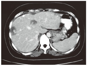
A 38-year-old female patient presented with a 2-mo history of cramp like abdominal pain and radiating ipsilateral lumbar pain, without any rectal passage of blood. The physical examination did not reveal any specific signs. The abdominal ultrasound revealed multiple liver lesions measuring 10 mm × 23 mm each and the computed tomography demonstrated a large non-cystic lesion in the right iliac fossa, as well as multiple small liver lesions with heterogeneous enhancement (Figure 1). Operative findings were suggestive of right colon diffuse hemangiomatosis, innumerable mesenteric and multiple small liver hemangiomas. An extensive right hemicolectomy and end-to-end ileo-transverse anastomosis was carried out.
Figure 1.

Abdominal computed tomography demonstrating liver hemangiomas.
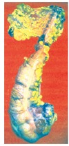
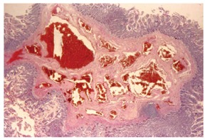
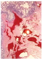
A specimen of a right hemicolectomy was sent to the Pathology Department, presenting grossly as a reddish-hemorrhagic tumor at the ileo-colic valve level, measuring 4 cm × 5 cm (Figure 2). The tumor had a sponge-like consistency and infiltrated the entire wall thickness extending in to the surrounding fatty tissue. The mesocolic and mesenteric fat, as well as the greater omentum, presented multiple hemorrhagic nodules measuring 0.6-1 cm in greater diameter. The specimen was subjected to routine procedures and multiple histological sections, stained with hematoxylin-eosin, were studied microscopically (Figures 3 and 4). The diagnosis of diffuse cavernous hemangiomatosis of the colonic and enteric wall with concomitant lymphangiectatic lesions of the submucosa and infiltration of the mesocolic and mesenteric fat and the greater omentum was decided.
Figure 2.

Macroscopic appearance of the surgical specimen, showing the colon hemangiomatosis.
Figure 3.

Histological section showing enteric wall mucosa and angiomatous lesions through the muscular layer of the colonic wall (HE x 25).
Figure 4.

Histological section of colonic wall showing multiple hyperemic angiomatous spaces in the muscular layer (HE x 25).
DISCUSSION
Hemangiomatosis of the colon is a rare benign disease arising from the submucosal vascular network. According to the literature these lesions originate from embryonic sequestrations of mesodermal tissue[2]. Histologically, hemangiomas are distinct from telangiectasias and angiodysplasias and approximately 80% of colonic hemangiomas are of the cavernous subtype. Capillary hemangiomas are usually solitary and produce no symptoms, while cavernous hemangiomas composed of large thin-walled vascular channels, have no capsule, involve the rectosigmoid region in up to 70% of the cases and present with rectal bleeding (60%-90%), anemia (43%), obstruction (17%) and rarely with platelet sequestration, although approximately 10% of patients remain asymptomatic[3]. A rare case of intussusception in a 39-year old male due to a capillary hemangioma of the right colon has been reported in the literature as a rare complication of colon hemangiomas[4]. The average age of the patient at presentation is 12 years, equally affecting males and females[2]. A case report of a 3-mo old infant presenting with ascites, anemia, minimal rectal bleeding and thrombocytopenia due to several hundred small cavernous hemangiomas of the colon and peritoneal surfaces who underwent subtotal colectomy[5] and a 9-mo old child with an isolated colonic cavernous hemangioma of the hepatic flexure[6], as well as a 3-wk old boy with diffuse hemangiomatosis of the small bowel[7] are some of the youngest cases reported from the review of the literature. Up to 80% of patients undergo an unnecessary surgical procedure before diagnosis is made[8], with an average delay time of 19 years elapsed between initial symptoms and diagnosis. Hemangiomatosis affecting the right colon is rather a rare case and from the review of the English literature fewer than 10 cases have been reported since 1950[4,6,9-12]. Liver and bladder involvement seems to be unusual as well[13,14]. The presence of phleboliths on plain abdominal radiographs is an important diagnostic clue, seen in 26%-50% of adult patients, indicating a sequential process of thrombosis in the tumor caused by perivascular inflammation and stasis of blood flow[15]. In our patient no pleboliths were demonstrated on plain abdominal radiographs. Barium enema reveals non specific polypoid or multilobular masses that collapse with air insufflation. Endoscopically, a bluish, soft, submucosal polypoid mass consistent with widened varices usually indicates the presence of a hemangioma. Mesenteric angiography shows hypervascularity and delayed venous pooling associated with cavernous hemangiomas. Abdominal CT scans can give useful information about the size of the lesion and the extension to adjacent organs. Recently, three dimensional CT colonography was used to gain a more complete knowledge of the characteristics and distribution of lesions throughout the colon[9]. Complete surgical resection is the definitive therapy, although (1) a pedunculated polypoid hemangioma (2) less or equal to 2.5 cm, with (3) an endoscopic ultrasonography demonstrating that the depth is limited in the submucosal layer are the three major criteria for endoscopic polypectomy[16]. In conclusion, a high suspicion index is required when a young patient presents with abdominal pain, painless rectal bleeding, phleboliths in plain radiographs or heterogeneous masses in CT, in order to approach a definite diagnosis before planning any therapeutic intervention.
Footnotes
S- Editor Wang J L- Editor Alpini GD E- Editor Bai SH
References
- 1.Schoen FJ, Cotran RS. Robbins Basic Pathology. 7th ed. Philadelphia: Saunders; 2003. pp. 355–357. [Google Scholar]
- 2.Lyon DT, Mantia AG. Large-bowel hemangiomas. Dis Colon Rectum. 1984;27:404–414. doi: 10.1007/BF02553013. [DOI] [PubMed] [Google Scholar]
- 3.Hsu RM, Horton KM, Fishman EK. Diffuse cavernous hemangiomatosis of the colon: findings on three-dimensional CT colonography. AJR Am J Roentgenol. 2002;179:1042–1044. doi: 10.2214/ajr.179.4.1791042. [DOI] [PubMed] [Google Scholar]
- 4.Gesslein M, Koscheck T, Kusch B. [Capillary hemangioma of the colon as a rare cause of intussusception in adults] Dtsch Med Wochenschr. 2004;129:1970–1972. doi: 10.1055/s-2004-831834. [DOI] [PubMed] [Google Scholar]
- 5.Ibarguen E, Sharp HL, Snyder CL, Ferrell KL, Leonard AS. Hemangiomatosis of the colon and peritoneum: case report and management discussion. Clin Pediatr (Phila) 1988;27:425–430. doi: 10.1177/000992288802700903. [DOI] [PubMed] [Google Scholar]
- 6.Masterson J, Woods D, Lau G, Dobranowski J. Isolated colonic hemangioma in a child. Can Assoc Radiol J. 1991;42:431–434. [PubMed] [Google Scholar]
- 7.Scafidi DE, McLeary MS, Young LW. Diffuse neonatal gastrointestinal hemangiomatosis: CT findings. Pediatr Radiol. 1998;28:512–514. doi: 10.1007/s002470050397. [DOI] [PubMed] [Google Scholar]
- 8.Pérez C, Andreu J, Llauger J, Valls J. Hemangioma of the rectum: CT appearance. Gastrointest Radiol. 1987;12:347–349. doi: 10.1007/BF01885179. [DOI] [PubMed] [Google Scholar]
- 9.Scopinaro F, Signori C, Massa R, Strollo F, Pippa G, Balestrieri P. Right colonic hemangioma diagnosed by scintigraphy with 99-mTc sucralfate. A case report. Ital J Surg Sci. 1989;19:89–92. [PubMed] [Google Scholar]
- 10.Rogers HG, Adler F. Hemangiomas of the cecum. Colonoscopic diagnosis and therapy. Gastroenterology. 1976;71:1079–1082. [PubMed] [Google Scholar]
- 11.Larsen S, Kronborg O. Acute hemorrhage from right-colonic hemangiomas: report of a case. Dis Colon Rectum. 1976;19:363–365. doi: 10.1007/BF02590937. [DOI] [PubMed] [Google Scholar]
- 12.Paris J, Goudemand M, Leduc M, Delmas-Marsalet Y. [Isolated angioma of the right colon with repeated gastrointestinal hemorrhage for 15 years] Lille Med. 1967;12:592–594. [PubMed] [Google Scholar]
- 13.Mohanty R, Gondal SC. Diffuse cavernous haemangioma of the rectosigmoid with bladder involvement--a case report. Indian J Pathol Microbiol. 1999;42:487–489. [PubMed] [Google Scholar]
- 14.Mills CS, Lloyd TV, Van Aman ME, Lucas J. Diffuse hemangiomatosis of the colon. J Clin Gastroenterol. 1985;7:416–421. doi: 10.1097/00004836-198510000-00009. [DOI] [PubMed] [Google Scholar]
- 15.Oner Z, Altaca G. Diffuse cavernous rectal hemangioma--clinical appearance, diagnostic modalities and sphincter saving approach to therapy: report of 2 and a collective review of 79 cases. Acta Chir Belg. 1993;93:173–176. [PubMed] [Google Scholar]
- 16.Takayoshi K, Massanobu T. Polypoid cavernous hemangioma removed under colonoscopy. Digestive Endoscopy. 2003;15:338–340. [Google Scholar]


