Abstract
The current technologies that trend in digital radiology (DR) are toward systems using portable smart mobile as patient-centered care. We aimed to develop a mini-mobile DR system by using smart devices for wireless connection into medical information systems. We developed a mini-mobile DR system consisting of an X-ray source and a Complementary Metal–Oxide Semiconductor (CMOS) sensor based on a flat panel detector for small-field diagnostics in patients. It is used instead of the systems that are difficult to perform with a fixed traditional device. We also designed a method for embedded systems in the development of portable DR systems. The external interface used the fast and stable IEEE 802.11n wireless protocol, and we adapted the device for connections with Picture Archiving and Communication System (PACS) and smart devices. The smart device could display images on an external monitor other than the monitor in the DR system. The communication modules, main control board, and external interface supporting smart devices were implemented. Further, a smart viewer based on the external interface was developed to display image files on various smart devices. In addition, the advantage of operators is to reduce radiation dose when using remote smart devices. It is integrated with smart devices that can provide X-ray imaging services anywhere. With this technology, it can permit image observation on a smart device from a remote location by connecting to the external interface. We evaluated the response time of the mini-mobile DR system to compare to mobile PACS. The experimental results show that our system outperforms conventional mobile PACS in this regard.
Keywords: Digital radiography, Web technology, Medical devices, Portable DR, Smart device, Wireless communication
Introduction
Digital radiography (DR) systems have been replacing traditional analog or screen-film (SF) systems over the last 3 decades [1]. They have been used in combination with X-ray detectors to instantly record X-ray exposure data and output this data in a computer-readable electronic format. DR technologies have gained popularly in many medical and industrial applications because they possess a number of useful capabilities such as electronic archiving, real-time acquisition and post-processing, etc [2, 3].
Radiography facilities have been burdened with the positioning challenges that characterize DR detectors at the present. Taking up a great deal of space, weighing more than 100 lb, the present digital X-ray devices and “rooms” that are primarily designed to install in a radiology suite or for in-hospital use in fixed locations, they cannot easily be moved once installed. To solve this problem, many DR systems have been developed with mini-mobile DR systems [4–6]. They are gaining importance in radiographic acquisition because they can meet the positioning requirements of nearly all commonly encountered radiography examinations and environments. In order to solve positioning requirements, most suppliers offer wireless DR systems in a variety of configurations, ranging from basic to fully automated systems. These DR systems are easy to move if comparing to older generation large-room X-ray machines and systems; however, they are not yet portable. It still requires multiple separate devices, power cables, and data communication in order to provide similar functionality.
Advances in DR system technology have shrunk the size and weight of X-ray generators and associated with digital X-ray components to the point where “mobile” or “portable” X-ray devices on wheels or in multiple component configurations are now possible. Moreover, newer systems have tended toward using flat panel detectors (FPDs) because they offer better imaging performance at a lower radiation dose [7, 8].
In this paper, we describe a mini-mobile DR system that uses wireless smart devices. It includes a touch screen interface and embedded computer system coupled with an X-ray generator. It also includes a communication module that can connect with smart devices. Our suggested DR system focuses on diagnostics at outpatient clinics, in operating rooms, and emergency rooms. We evaluated the response time performance of the mini-mobile DR system compared to mobile Picture Archiving and Communication System (PACS) which is general PACS system supporting mobile devices in hospital. The experimental results show that the mini-mobile DR system outperforms conventional mobile PACS in this regard.
Materials and Methods
Design of the Mini-Mobile DR System
Most of the DR system receives X-ray signals that have passed through a part of the patient and have been measured by an X-ray detector and directly converts these signals to digital signals, which are then directly sent to the image processing and PACS computer systems [9–14]. However, in the case of portable DR, all components cannot be split. Therefore, we designed a method for embedded systems while developing the portable DR system.
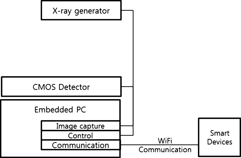
We suggested a system tailored to bring all the separate components together. The structure of the mini-mobile DR system with smart devices is shown in Fig. 1. The mini-mobile DR system consists of an X-ray generator and a FPD based on a type of Complementary Metal Oxide Semiconductor (CMOS image sensor [15]) and the embedded PC (in “The Internal Processing and Imaging Service”). The embedded PC provides the control services for image processing and communication services supporting smart devices using a WiFi communication interface module as an external interface.
Fig. 1.
An illustration of the structure of the mini-mobile DR system and smart devices
We developed a mini-mobile DR system consisting of an X-ray source (40–80 kVp, 0.3 mA) and a CMOS sensor-based FPD (120 × 150 mm, pixel size 75 μm) for small-scale field diagnostics in patients. Moreover, its weight is 25 kg less, and the external interface is designed for portable use. We used procedures established by the American Association of Physicists in Medicine Task Group 116, formed to establish radiation guidelines. The system operates under standardized radiation exposure conditions.
The difference between other products and ours is that we included an X-ray generator and detector in an integrated system. However, our system does not have an image view function or the ability to display images. We used smart devices, such as smartphones, table PC, and Pads to display images instead. The embedded PC that handles image processing is based on Windows OS. This system was designed not only to provide radiology imaging services using the DICOM 3.0 standard but also to convert the raw data to JPEG image files. For smart device capability, we use the JPEG image file for image service better than DICOM Web standard because fast to view the result and easy to access. It is trend of mobile image service. Most of the image data, such as the DICOM files, are transferred to the PACS database. However, our proposed system provides image processing services in an embedded PC and can be quickly checked using smart devices without access to a PACS system that is why we consider that without interaction with the PACS we can connect with mini-mobile DR system directly. The external interface also provides comprehensive and complete image services. Medical staff can use the viewer application via Android-based applications to view images wherever a wireless environment is available. In addition, some functions in the Android application are accessible even the user is off-line. Those functions include displaying images, pan/zoom, and obtaining region of interest (ROI) and distance measurements.
The Internal Processing and Imaging Service
The embedded PC is Windows 7 OS-based; we used an Exynos 4210 with a 1.2 GHz CPU that supports interaction with smart devices. For radiography image processing, we used a correction algorithm that was performed in two steps. In the first step, we found the position of bad pixels with a 3 × 3 differentiation operator. If the differentiation at the given pixel was greater than a threshold value, the pixel was classified as a bad pixel. In the second step, the intensity value of the bad pixel was determined from linear interpolation of nearby pixel values [16]. Then, the next process involves generating the DICOM files. After obtaining the DICOM files, the image data were stored in a specific directory on the embedded computer.
To provide an imaging service, the embedded PC includes the functions of a Web server (Apache). The Web server can refer to either the hardware or the software to help deliver content that can be accessed through the Internet. The primary function of the Web server is to deliver DICOM files or converted JPEG image files requested by smart devices and communication modules. The communication module is designed to handle multi-device communication through a Web server to avoid loss of information from multiple users viewing images at the same time. In order to provide DICOM imaging services in the embedded PC, we decided to use a compressed version of the DICOM image in JPEG format. This DICOM-compatible version is generated using the Dcm2Jpg function from the DCM4CHE library [17]. Then, image related data is stored in MySQL database. It includes patient characteristics (e.g., name, age, sex, etc.) for image search.
Smart Viewer Based on an External Interface
The external interface provides a viewer for the patient anatomy and remote the shooting as condition in smart device setting condition. The interface of the smart device (Android OS-based) uses the fast and stable IEEE 802.11n (2.4 GHz) wireless protocol, which was adapted in this device for connection with medical information systems (PACS) and smartphones using a communication module. IEEE 802.11n wireless local area networks (WLANs) have a greater coverage radius (up to 100 m) and support higher data rates (up to 11 Mbps), making it more suitable than other protocols for the transfer of large files such as images. With the rapid development of information technology infrastructure, WLANs are commonly used in hospitals and outdoor environments. In the field of DR, ubiquitous access to patient records and images using WLANs is becoming a functional necessity. The smart viewer includes an image viewer as well as functionality to control the mini-mobile DR system. As a part of the view functions have the following features:
An image contrast adjustment, image zoom in/out functionality
An image rotation feature for image analysis (at 90°, 180°, and 270°)
An image angle measurement, an ROI settings and interests of the area analysis function, and measure the length
Search of image by attributes (name, id, age, or date)
Evaluation of Mini-Mobile DR System Efficacy
To evaluate the performance of the mini-mobile DR system image service, we used a client viewer program for the image service on a smart device. The client viewer programs were run on a quad-core Exynos 4412-based smartphone (1.4 GHz) running Android 4.0. The time stamps are measured on the smartphone by using the System.currentTimeMillis() function from MIDP 2.1 – CLDC 1.1 that returns 10-ms precision time stamps. Therefore, we measured the response time for the image service on a smartphone in 2 different ways for the mini-mobile DR system and mobile PACS.
Results and Discussion
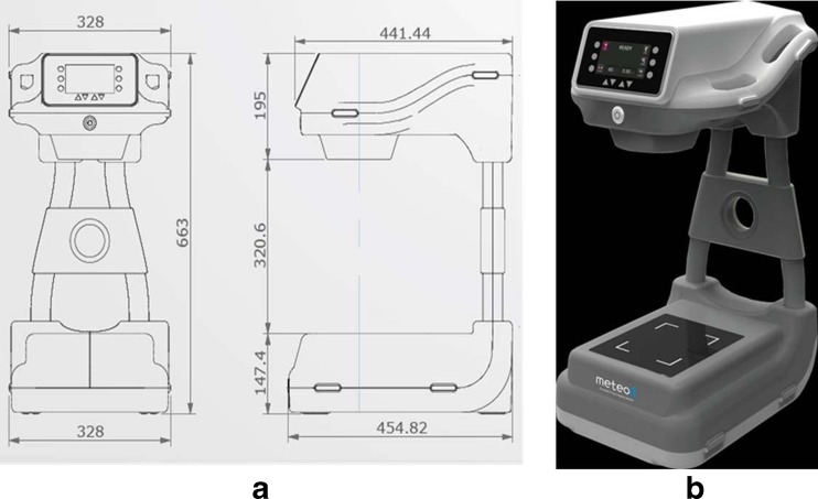
The mini-mobile DR system and smart devices are shown in Fig. 2. The dimensions of the suggested systems are 328 (width) × 455 (depth) × 663 mm (height). This is smaller than the other similar products.
Fig. 2.
The design of the structure (a) and mini-mobile DR system (b) are shown
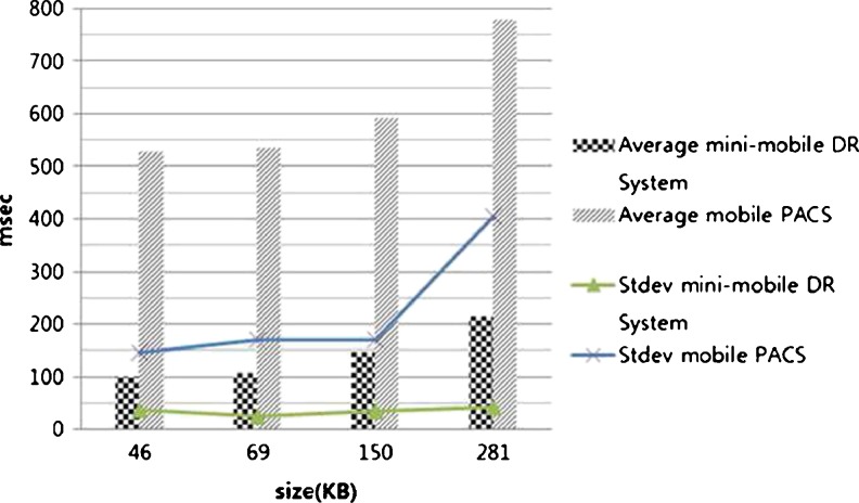
We analyzed the smart device response time from image storage on the embedded PC and database storage on the mobile PACS. Our results show the advantage of using the mini-mobile DR system. Figure 3 shows the response time in milliseconds for imaging services according to file size. The response time of the mini-mobile DR system is less than for the mobile PACS. The gap is very large between the response times of the mini-mobile DR system and mobile PACS.
Fig. 3.
A chart showing the mean response time of the image service on a smart device. The response time of the mini-mobile DR system was about 400 ms faster than mobile PACS
Smaller image sizes and faster response times mean less processing and transmission time required on the smart device, which leads to lower power consumption and a faster imaging service. This satisfies the physical constraints of smart devices and achieves our goal. These results support that the mini-mobile DR system is effective with smart devices. The mini-mobile DR system offers a perfectly good solution for the majority of implementations with greater flexibility and lower overhead compared to mobile PACS.
Then, our system, the size is small that it is possible to move if comparing to the existing DR systems and even fracture patients also can shoot digital image and provide image service easily.
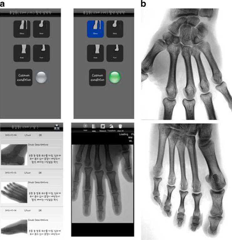
We evaluated the smart viewer function execution of our mini-mobile DR system with smart devices. The smart device could display images on an external monitor apart from the monitor in the DR system, as shown in Fig. 4.
Fig. 4.
The main screen and image results shown on a mobile device. The user interface on a smartphone device (a) shows hand and foot images (b) generated from the mini-mobile DR system
Figure 4 is the smartphone-based user interface. X-ray detector and X-ray generating device according to the patient’s preset value of each part to shoot was to use; smart devices on the affected part of the interface is provided with individual buttons.
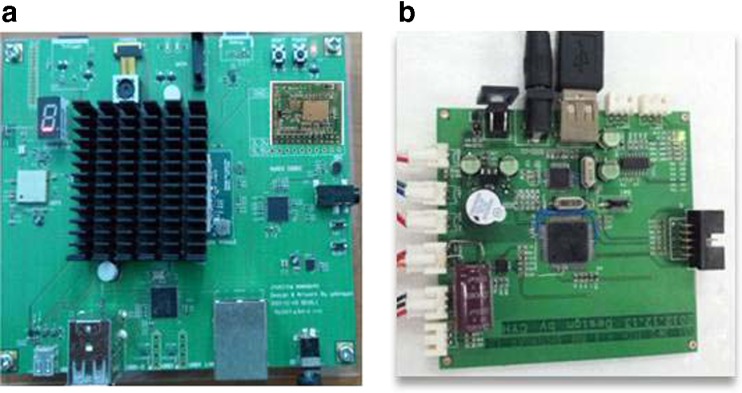
The communication modules (WiFi and the RS-232C protocol) and board (Ubuntu Linux OS-based) supporting smart devices were used, and a smart viewer (an Android application) based on the external interface of the mobile DR system was developed (Fig. 5). The system could display image files on various smart devices. In addition, it has the advantage that it can reduce the radiation exposure of an operator by using a smart device as a remote control.
Fig. 5.
The smart device board and communication module of the mobile DR system. The smart device board (a) and communication module (b) are shown
The major concern over our suggested DR system is portable and mobility technology using smart devices. It was improved environment limitation problems developed to small size DR device and using smart devices. Our system will provides diagnostics at outpatient clinics, in operating rooms, and emergency rooms. The current study showed that, with the use of a mini-mobile DR system, the response time in image service to the smart device is markedly reduced. We obtained 400 ms faster image service when we used mini-mobile DR system.
Conclusion
We developed a mini-mobile DR system by using smart devices that can provide X-ray imaging services anywhere. This system consists of an X-ray source (40–80 kVp, 0.3 mA) and a CMOS sensor-based FPD (120 × 150 mm, pixel size, 75 μm) and allows high mobility because of its low weight (25 kg less than other similar products). Moreover, our system can permit image observation on a smart device from a remote location that is connected to the external interface through the communication module. The experimental results show that the mini-mobile DR system outperforms conventional mobile PACS. We expect the mini-mobile DR system in this study to be useful for clinical radiology services.
Our future work includes applying the clinical evaluation and diagnostic test of the suggested mini-mobile DR system. We will also extend our research to the area of minimization of the delivery of radiation dose to patient and we will promotion of clinical efficiency and continuous image quality improvement.
Acknowledgments
This study was supported by the grant of the Korean Health Technology R&D Project, Ministry of Health & Welfare, Republic of Korea (A120152).
Contributor Information
Chang-Won Jeong, Email: mediblue@wku.ac.kr.
Su-Chong Joo, Email: scjoo@wku.ac.kr.
Jong-Hyun Ryu, Email: jhryu@wku.ac.kr.
Jinseok Lee, Email: gonasago@wku.ac.kr.
Kyong-Woo Kim, Email: kwkim@nfr.kr.
Kwon-Ha Yoon, Phone: +82-10-81011677, FAX: +82-63-8591009, Email: khy1646@wku.ac.kr.
References
- 1.Cho HM, Kim HJ, Lee CL, Nam S, Jung JY. Imaging characteristics of the direct and mobile indirect digital radiographic systems. IEEE Nucl Sci Symp Conf Rec. 2007;5:3840–3846. [Google Scholar]
- 2.Lanc¸a L, Silva A. Digital radiography detectors- a technical overview part 1. Radiogr Elsevier. 2009;15(1):9–19. [Google Scholar]
- 3.Jeong MH, Kim HS, Lee BS, Han BS, Lee SC, Kim EK. Development of a portable digital radiographic system based on FOP-coupled CMOS image sensor and its performance evaluation. IEEE Trans Nucl Sci. 2005;52(5):1604–1609. [Google Scholar]
- 4.Cowen AR, Kengyelics SM, Davies AG. Solid-state, flat-panel, digital radiography detectors and their physical imaging characteristics. Clin Radiol. 2008;64:487–498. doi: 10.1016/j.crad.2007.10.014. [DOI] [PubMed] [Google Scholar]
- 5.Williams MB, Krupinski EA, Strauss KJ, Breeden WK, Rzeszotarski MS, Applegate K, Wyattg M, Bjork S, Seibert JA. Digital radiography image quality: image acquisition. Am Coll Radiol. 2007;4(6):371–388. doi: 10.1016/j.jacr.2007.02.002. [DOI] [PubMed] [Google Scholar]
- 6.Gauntt DM, Barnes GT. An automatic and accurate x-ray tube focal spot/grid alignment system for mobile radiography: system description and alignment accuracy. Med Phys. 2010;37(12):6402–6410. doi: 10.1118/1.3518085. [DOI] [PMC free article] [PubMed] [Google Scholar]
- 7.Lehnert T, Naguib NN, Ackermann H, Schomerus C, Jacobi V, Balzer JO, Vogl TJ. Novel, portable, cassette-sized, and wireless flat-panel digital radiography system: initial workflow results versus computed radiography. Am J Roentgenol. 2011;196(6):1368–1371. doi: 10.2214/AJR.10.5867. [DOI] [PubMed] [Google Scholar]
- 8.Tzeng W-S, Kuo K-M, Liu C-F, Yao H-C, Chen C-Y, Lin H-W. Managing repeat digital radiography image—a systematic approach and improvement. J Med Syst. 2012;36(4):2697–2704. doi: 10.1007/s10916-011-9744-8. [DOI] [PubMed] [Google Scholar]
- 9.Langer SG. A flexible database architecture for mining DICOM objects: the DICOM data warehouse. J Digit Imaging. 2012;25(2):206–212. doi: 10.1007/s10278-011-9434-6. [DOI] [PMC free article] [PubMed] [Google Scholar]
- 10.Lee HJ, Lee KH, Hwang SI, Kim H-C, Seo EH, Kim TG, Ha K-S. The effect of wireless LAN-based PACS device for portable imaging modalities. J Digit Imaging. 2010;23(2):185–191. doi: 10.1007/s10278-008-9174-4. [DOI] [PMC free article] [PubMed] [Google Scholar]
- 11.Hsiao C-H, Shiau C-Y, Liu Y-M, Chao MM, Lien C-Y, Chen C-H, Yen S-H, Tang S-T. Use of a rich internet application solution to present medical images. J Digit Imaging. 2011;24(6):967–978. doi: 10.1007/s10278-011-9374-1. [DOI] [PMC free article] [PubMed] [Google Scholar]
- 12.Sakai S, Yabuuchi H, Matsuo Y, Okafuji T, Kamitani T, Honda H, Yamamoto K, Fujiwara K, Sugiyama N, Doi K. Integration of temporal subtraction and nodule detection system for digital chest radiographs into picture archiving and communication system (PACS): four-year experience. J Digit Imaging. 2008;21(1):91–98. doi: 10.1007/s10278-007-9014-y. [DOI] [PMC free article] [PubMed] [Google Scholar]
- 13.Kim DK, Kim EY, Yang KH, Lee CK, Yoo SK. A mobile tele-radiology imaging system with JPEG2000 for an emergency care. J Digit Imaging. 2011;24(4):709–718. doi: 10.1007/s10278-010-9335-0. [DOI] [PMC free article] [PubMed] [Google Scholar]
- 14.Bui AAT, Morioka C, Dionisio JDN, Johnson DB, Sinha U, Ardekani S, Taira RK, Aberle DR, El-Saden S, Kangarloo H. openSourcePACS: an extensible infrastructure for medical image management. IEEE Trans Inf Technol Biomed. 2007;11(1):94–109. doi: 10.1109/TITB.2006.879595. [DOI] [PubMed] [Google Scholar]
- 15.Spyropoulou VA, Fountos GP, Kalyvas NI, Valais IG, Kandarakis IS, Panayiotakis GS. Experimental and theoretical evaluation of a high resolution CMOS based detector under x-ray imaging conditions. IEEE Trans Nucl Sci. 2011;58(1):314–322. doi: 10.1109/TNS.2010.2094206. [DOI] [Google Scholar]
- 16.Rinkel J, Gerfault L, Est’eve F, Dinten J-M. A new method for x-ray scatter correction: first assessment on a cone-beam CT experimental setup. Phys Med Biol. 2007;52(15):4633–4653. doi: 10.1088/0031-9155/52/15/018. [DOI] [PubMed] [Google Scholar]
- 17.Open Source Clinical Image and Object Management. Available at http://www.dcm4che.org/. Accessed 1 October 2012


