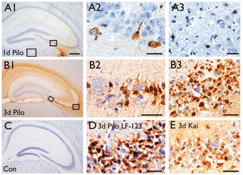
Figure 1.
Osteopontin expression in the hippocampus 1–3 days after SE. Osteopontin immunohistochemistry using 2A1 (A–C and E) and LF-123 (D) antibodies are shown for brain sections 1 (A1–A3) and 3 days after pilocarpine-(B1–B3, D) and 3 days after kainate-(E) induced SE in comparison to a control CF1 mouse (C). One day after pilocarpine-induced SE there is little staining in the hippocampus restricted to some cells with neuronal morphology (A2) and some small cells in the thalamus (A3). (B) Three days after pilocarpine-induced SE neurons with healthy morphology are not stained (B2), while smaller cells with the morphology of pyknotic neurons are stained in the pyramidal cell layers (B3, D). Note that the immunolabelings with the 2A1 (B3) and LF-123 (D) antibodies in the CA3b area are similar. D shows less robust but similar labeling in the CA3b area 3 days after kainate-induced SE. The scale bar is 500 μm for A1, B1, and C. All other scale bars = 50 μm
Epilepsia © ILAE

