INTRODUCTION
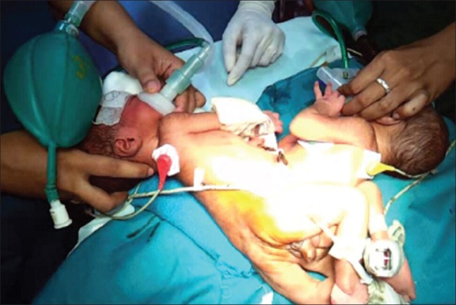
The ischiopagus variety of conjoined-twins is the most complicated form of twinning, contributing only 6% to all with significant organ sharing[1] [Figure 1]. The type of the organ shared and the degree of cross-circulation add to the complexity and determine the success rate of the separation surgery, if undertaken and the same factors also determine the success rate of any palliative surgery. As for unknown reasons the foetuses who survive to the point of birth are females and still-borns are usually males, they follow a female predominance in the order of 3:1.[2] The incidence is 1:50,000-1:2,00,000 in general and is further higher in Indian and African race, that is 1:14,000-25,000 live-births.[3] We share our experience of the anaesthetic management for colostomy in tetrapus variety of ischiopagus with cross-circulation, on their third postnatal day with an aim of thorough discussion of the anaesthetic considerations and implications of such anatomical abnormalities.
Figure 1.

Conjoined-twins fused from their front pelvis
CASE REPORT
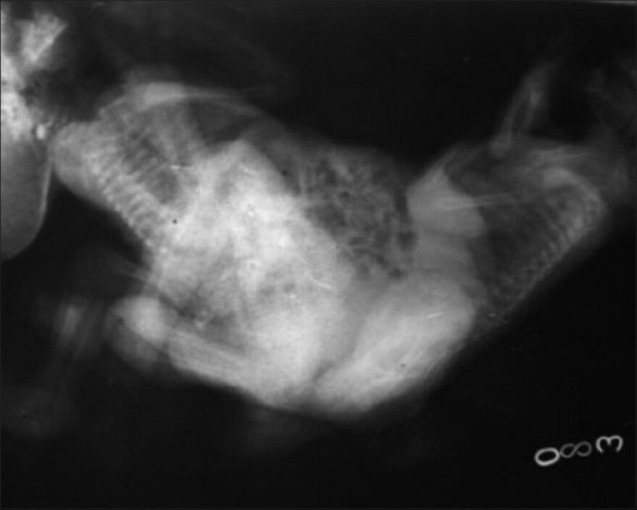
Three day-old male twins joined from their front pelvis were admitted to our hospital for colostomy. They were actively moving limbs with a conjoined-weight of 4.0 kg. The routine investigations were within normal limits and the clinical examination revealed common male genitalia and no external anal opening. X-ray showed two separate spines and pelvic bones, which were apparently conjoined at ischii [Figure 2]. Plain and contrast enhanced computed tomography of the abdomen and pelvis suggested that both lung fields and mediastinal shadows were normal and the twins were fused along anterior abdominal wall as well as pelvis and perineum.[4] Right sided liver parenchyma could be seen separately in both twins, but left parenchyma were fused across the midline with contiguous intrahepatic channels. Multiple small bowel loops in continuation across the fused abdomen suggested their communication. Ureters were not visualised with single urinary bladder and normally appearing separate kidneys in both twins. Spinal columns appeared unfused with two separate pelvic bones. The twins in reference were referred to our centre from a peripheral hospital. General anaesthesia was planned and they were labelled as twin-A and B. The intravenous (i/v) lines as well as all the monitoring cables for electrocardiography, arterial oxygen saturation and blood pressure (BP) were attached and labelled as A and B, respectively. The pre-operative values of heart rate (HR), BP and oxygen saturation (SpO2) for them were 160/min, 90/60 mmHg, 98%, and 140/min, 86/58 mmHg and 96%, respectively. Twin-A was administered injection glycopyrrolate 0.02 mg i/v, which increased its HR to 180/min with a concomitant increase in HR of twin-B to 160/min. Before induction, all relevant equipments were checked and two separate anaesthesia machines were arranged for two conjoined individuals. The twins were pre-oxygenated with 100% oxygen for 5 min using Jackson-Rees circuits. The drugs were calculated as for the combined body weight and halved for each individual. Twin-A was induced first in sequence with sevoflurane (4-8%) and injection ketamine 4 mg i/v and endotracheal intubation was facilitated with an endotracheal tube, size- 3.0 using injection succinylcholine 4.0 mg i/v. As soon as twin-A was induced, twin-B showed signs of respiratory depression demanding its immediate inductions, which was done in a similar way. The heads and limbs of both the twins were covered with cotton. Anaesthesia was maintained on O2:N2O (50%:50%), sevofluorane 0.25% and injection atracurium 1.0 mg i/v for each of the twins. Two repeated doses of atracurium of 0.5 mg each were given to twin-B at the interval of 10 min. The neuromuscular blockade was reversed with injection neostigmine 0.1 mg i/v and injection glycopyrrolate 0.02 mg i/v for each of the twins separately at the end of an hour surgery. Each twin had received injection paracetamol 30 mg i/v. They were shifted on T-piece with SpO2 of 95-96% (twin-A) and 98-99% (twin-B) and were extubated next day in paediatric intensive care unit. Each twin was individually administered 16 ml of warm paediatric maintenance solution for initial two- third duration of surgery followed by 10 ml of hydroxyl-ethyl starch - 3% over rest one-third. Intraoral temperature monitoring was done perioperatively.
Figure 2.

X-ray baby gram of ischiopagus
DISCUSSION
Although the incidence of conjoined- twins is 1 in 50,000-1 in 2,00,000,[5] the reports of separation surgeries are few and those of palliative procedures are much fewer. Any palliative procedure in such a case has to be carried out within 1 or 2 postnatal days.
Anaesthetic-management at such an early stage of life not supported with any investigations to assess the extent of organ sharing and cross-circulation in most of the settings further adds to the complexity.
The ischiopagii, considered the most complicated form of conjoined twinning have potential involvement of pelvis, liver, intestine and genitourinary tract. The primary and most important step is the pre-operative assessment of the twins to evaluate the extent of cross-circulation and organ sharing. Presence or absence of cross-circulation plays an important role in planning induction as well as maintenance of anaesthesia, haemodynamics during surgery and the post-operative outcome. For evaluation of cross-circulation, Toyoshima et al., injected a bolus of indigo-carmine and the pigment appeared in the urine of the other twin. They also undertook radioimmune angiography, which showed that radionucleotides in one twin were similar to those in other after 5-10 min.[6] In our set of twins also the portal venous channels were contiguous along the fused left liver parenchyma leading to admixture of blood to a variable degree as supported by the facts that twin-B showed signs of muscle-activity and needed two repeated doses of atracurium and also that the recovery of twin-A was delayed with poor respiratory efforts in comparison to twin-B, possibly explaining the residual effects of drug too.
Szmuk et al., have reported anaesthetic management of 14 month-old thoracopagus twins with cyanotic heart disease for cardiac assessment, where 1 h after induction of anaesthesia, while twin-A was fully relaxed, twin-B showed signs of muscular activity and required three repeated doses of rocuronium over 15 min to achieve relaxation, indicating a considerable amount of cross-circulation from twin-B to A. This is similar to the present case, where twin-B required two repeated doses of atracurium of 0.5 mg each, although it was induced second in the sequence. To confirm this, they administered 20 mg of propofol to twin-B when bispectral index (BIS) value was 70 in both twins. After 2 min, the BIS value in twin-A decreased to 45 while it remained 68-70 in twin-B. After another 10 min, the BIS value in twin-A returned to 70.[7]
Seefelder et al., have successfully administered awake caudal anaesthesia for inguinal surgery in one conjoined-twin (omphalocele) for placement of a surgically tunnelled femoral central line.[8] In the present case also caudal anaesthesia could have replaced general anaesthesia if proper positions were possible.
CONCLUSION
Although use of regional anaesthesia eliminates the need for unwanted general anaesthesia in the presence of cross-circulation, general anaesthesia can still be managed uneventfully with meticulous pre-operative assessment, comprehensive intraoperative management, and intensive post-operative care with a good inter-disciplinary team work.
REFERENCES
- 1.Khan YA. Ischiopagus tripus conjoined twins. APSP J Case Rep. 2011;2:5. [PMC free article] [PubMed] [Google Scholar]
- 2.Chalam KS. Anaesthetic management of conjoined twins‘ separation surgery. Indian J Anaesth. 2009;53:294–301. [PMC free article] [PubMed] [Google Scholar]
- 3.Ezike HA, Ajuzieogu VO, Amucheazi AO, Ekenze SO. General anesthesia for repair of omphalocele in a pair of conjoined twins in Enugu, Nigeria. Saudi J Anaesth. 2010;4:202–4. doi: 10.4103/1658-354X.71579. [DOI] [PMC free article] [PubMed] [Google Scholar]
- 4.Kingston CA, McHugh K, Kumaradevan J, Kiely EM, Spitz L. Imaging in the preoperative assessment of conjoined twins. Radiographics. 2001;21:1187–208. doi: 10.1148/radiographics.21.5.g01se011187. [DOI] [PubMed] [Google Scholar]
- 5.Hansen J. Incidence of conjoined twins. Lancet. 1975;306:1257. doi: 10.1016/s0140-6736(75)92092-9. [DOI] [PubMed] [Google Scholar]
- 6.Toyoshima M, Fujihara T, Hiroki K, Namatame R, Ka K, Ooe K. Evaluation of cross circulation in conjoined twins. Masui. 1993;42:1347–50. [PubMed] [Google Scholar]
- 7.Szmuk P, Rabb MF, Curry B, Smith KJ, Lantin-Hermoso MR, Ezri T. Anaesthetic management of thoracopagus twins with complex cyanotic heart disease for cardiac assessment: Special considerations related to ventilation and cross-circulation. Br J Anaesth. 2006;96:341–5. doi: 10.1093/bja/aei313. [DOI] [PubMed] [Google Scholar]
- 8.Seefelder C, Hill DR, Shamberger RC, Holzman RS. Awake caudal anesthesia for inguinal surgery in one conjoined twin. Anesth Analg. 2003;96:412–3. doi: 10.1097/00000539-200302000-00021. [DOI] [PubMed] [Google Scholar]


