Abstract
Knee replacement is an effective treatment for pain and functional impairment secondary to degenerative joint conditions. The number of knee replacements performed continues to rise. Periprosthetic fractures around total knee arthroplasties are a relatively rare complication but are complex injuries that require the treating surgeon to be familiar with and proficient at arthroplasty and trauma reconstructive techniques. An increase in life expectancy and in the functional demands of elderly patients may lead to an increased incidence of periprosthetic fractures. Supracondylar fractures of the femur are the most common type and this review will focus on the incidence, risk factors, classification, investigation, and treatment options for periprosthetic fractures around total knee arthroplasties.
Keywords: Arthroplasty, Replacement, Knee, Femur, Periprosthetic fractures
Introduction
The number of knee replacement procedures performed annually is increasing dramatically. In the United Kingdom, 90,842 knee replacement procedures were performed in 2012, which represents a 7.3 % increase over the previous year [1]. It is, therefore, expected that the incidence of periprosthetic fractures around the knee associated with primary and revision total knee arthroplasty (TKA) will also rise. Treatment of these fractures is complex as well as time and resource consuming. Newer treatment modalities in the form of locking plate fixation and nailing techniques as well as endoprostheses have improved the outcomes in these technically demanding injuries. The purpose of the current article is to review the incidence and prevalence of these fractures, along with the various classification systems in use and review the management of these fractures. The focus will be on femoral fractures as these are by far the commonest with significant morbidity and mortality.
Incidence
The incidence of periprosthetic fractures around the knee in primary TKA has been variably reported to be between 0.1 % and 2.5 % [2–4]. Supracondylar femoral periprosthetic fractures are by far the commonest with an incidence of 0.3 %–2.5 % after primary TKA and 1.6–38 % after revision TKA [3, 5–8, 9•]. Tibial periprosthetic fractures are less common with an incidence of 0.4 % in the primary setting and a higher incidence in revision TKA [10]. Patellar periprosthetic fractures appear to be more common when the patella is resurfaced [11] but their overall incidence is low. This has been reported in literature as around 0.68 % but the true incidence may be considerably higher as majority of these fractures tend to be asymptomatic and therefore undetected [12].
Predictors of fracture
Osteopenia appears to be an important predisposing factor contributing to periprosthetic fractures [3, 13, 14].Other risk factors include old age [9•], chronic use of corticosteroids [15], inflammatory arthropathy including rheumatoid arthritis [3], local osteolysis and stress risers from screw holes, as well as significant deformity or previous surgery [16•]. Rheumatoid arthritis is a risk factor in itself although associated osteopenia and use of corticosteroids may contribute to the increased incidence [17•]. Patients with pre-existing neurologic problems like epilepsy, Parkinson’s disease, and poliomyelitis also appear to be at a higher risk of periprosthetic fractures [18]. Anterior femoral notching is controversial. Although deep anterior notches more than 3 mm have been shown to reduce flexural and torsional strength in biomechanical studies [19, 20], this does not seem to be reflected in the clinical setting [21, 22]. Meek et al reviewed the Scottish database and identified, age above 70 years and female gender to be at a higher risk of periprosthetic fractures [9•].
Singh et al reported an incidence of 1.1 % and 2.5 % in primary and revision TKA, respectively, in their review of more than 17,000 primary and 4000 revision TKA [23•]. Interestingly, in their study, a lower age was associated with a higher incidence of fracture. Patients between the age of 60 and 80 were found to have a 40 % lower risk than those below the age of 60 years. The authors felt that this was attributed to a more active lifestyle of younger patients, and the fact that patients having a TKA at a younger age were more likely to have an underlying condition making them prone for osteoporosis and fractures. There was an increased risk of periprosthetic fractures if the patient had a 3+ score on the Deyo-Charlson index for comorbidities [24]. In revision TKA, an underlying diagnosis of nonunion, infection, or previous surgery increased the risk of periprosthetic fracture.
Classification
Periprosthetic fractures around the knee can be broadly classified into intraoperative fractures and postoperative fractures. They can be further classified by the anatomic location into femoral, tibial, or patellar fractures.
Intraoperative fractures
Intraoperative fractures on the tibial side are more common in revision surgery. Intraoperative femoral fractures can be either metaphyseal or diaphyseal and usually are detected only in the postoperative period. They are more likely to occur in posterior stabilized knees.
Postoperative fractures
A significant proportion of these fractures occur in the supracondylar area of the femur. Various classification systems have been proposed, but the one used most commonly is the Lewis and Rorabeck classification [4, 25], which considers fracture displacement and fixation status of the femoral component. Type 1 fractures are undisplaced, type 2 fractures have greater than 5 mm of displacement or an angulation more than 5° but the prosthesis is stable. Type 3 fractures have a loose component irrespective of fracture displacement. An alternative classification was proposed by Kim et al, which took into consideration the amount of bone in the distal fragment, the position and fixation of the component, and fracture reducibility, thereby guiding management [26]. Type 1 fractures had a stable and well-aligned component. 1A fractures were undisplaced or reducible, and 1B fractures were irreducible necessitating open reduction and internal fixation. Type 2 fractures were reducible with good bone stock but a loose or maligned component, which benefitted from a revision arthroplasty. Type 3 fractures were severely comminuted with poor distal bone stock and a loose and maligned component and warranted the use of a distal femoral replacement.
Tibial periprosthetic fractures can be classified into 4 types based again on anatomic location and component fixation [10]. Type 1 fractures involve the tibial plateau. These are the commonest and are frequently associated with loosening. Type 2 fractures occur around the prosthetic stem and are essentially traumatic in etiology, although osteolysis is a contributing factor. Type 3 fractures occur distal to the component and type 4 involves the tibial tuberosity. These are further subcategorized into those with well-fixed prosthesis, those with a loose prosthesis, and those which are intraoperative.
Patellar periprosthetic fractures are also classified taking into account the component stability, quality of the bone stock, and integrity of the extensor mechanism. Type 1 and 2 fractures have a well-fixed prosthesis and types 3 and 4 have a loose prosthesis. The extensor mechanism is disrupted in type 2 fractures, whereas type 4 fractures are associated with poor bone stock [12].
Investigations
Standard anteroposterior and lateral radiographs of the knee, to include views of the length of the affected bone when a fracture is present is the mainstay of radiological investigation. The fracture is classified according to the chosen classification system as described above. The radiographs are carefully analyzed for any evidence of implant loosening such as migration with reference to bony landmarks of the segment still attached to the prosthesis, radiolucencies at the bone/cement interface, implant/cement interface, or bone/implant interface. If previous radiographs are available, these should also be analyzed for comparison and note should be made of any progression.
If the patient was experiencing pain associated with the affected joint prior to sustaining the fracture, this may suggest pre-existing loosening of the implant. Note should be made of the bone stock available for fixation with particular reference to osteolysis and comminution. If doubt remains, a computerized tomography (CT) scan is a useful adjunct to assess bone stock and the relationship to the implant in situ when planning fixation. If fixation has been planned, the surgeon should have adequate resources available to change to a suitable revision implant intraoperatively if loosening is discovered. Loosening may be secondary to infection. The authors recommend screening with a thorough patient history and serological markers (CRP, ESR) where there is any suggestion of loosening. If these are raised or the suspicion of infection is raised by the history or imaging, preoperative aspiration should be performed with synovial white cell count (WCC), polymorphonuclear (PMN) cell proportion and microbiological analysis. The authors recommend the use of the thresholds described by the International Consensus on Periprosthetic Joint Infection (CRP >10 mg/L, ESR >30 mm/hour, synovial WCC >3000 cells/μL and synovial PMN% >80 %) [27].
Treatment
Intraoperative fractures
These fractures, when detected intraoperatively are relatively easy to manage. They are usually undisplaced and implant stability is not usually compromised. Moreover, they are not associated with soft tissue damage. Metaphyseal femoral fractures can usually be managed with a single transcondylar screw fixation and diaphyseal perforations can be treated with stemmed components, which bypass the perforation by at least 2 cortical dimensions [25]. Tibial intraoperative fracture is more common in the revision scenario and again can usually be treated by either a screw fixation or a stemmed implant or both without significant change in the postoperative rehabilitation protocol. Transverse patellar fractures usually tend to be undisplaced and can be treated conservatively, although occasionally these warrant tension band wiring to minimize the chances of extensor mechanism disruption in the postoperative period. When their fractures are detected in the immediate postoperative period, conservative management is usually satisfactory with a period of protected weight bearing with or without hinged braces, although these cases need to be monitored carefully for loss of alignment.
Postoperative fractures
The treatment of periprosthetic fractures of the distal femur with a well-fixed TKA in situ can be divided into nonoperative and operative intervention. The population that suffers from periprosthetic distal femoral fractures is often elderly with multiple comorbidities; this leads to a high risk of mortality. When the risk of mortality is high, nonoperative intervention may be chosen but is generally limited to those with nondisplaced fractures [28, 29] and nonambulatory patients.
Conservative
Conservative treatment typically requires a prolonged period of immobility with union taking between 2 and 4 months [30]; this period of immobility and its attendant complications may be a greater risk to the patient than that posed by operative intervention. Comparative cohort studies have shown that better functional results are achieved with stable fixation than with conservative treatment, by permitting early motion and avoiding postoperative stiffness [18]. Close observation of those treated conservatively is required. Loss of alignment or displacement may require operative fixation where this is an option for the patient. Although techniques such as the use of intramedullary Rush rods improve stability compared with conservative treatment alone, the time to union and necessary period of protected activity is not improved [31, 32].
Operative fixation
When operative intervention is chosen, the method will be guided by a variety of factors such as how well fixed the implant is, the fracture pattern (including presence or absence of comminution), the presence of infection, other implants proximal or distal to the TKA, periprosthetic bone stock, and bone quality [33••]. When there is no infection present and the implant is well-fixed, the option to retain the implant may be taken. Operative strategies in this context include the use of retrograde intramedullary nailing, open reduction and internal fixation (ORIF) with non-locked or locked plates and the use of external fixation techniques.
The technique selected will in part depend upon the skill-set of the treating surgeon, the soft tissue envelope, bone stock, equipment availability, and implant compatibility [34]. The goals of surgery are to achieve satisfactory fixation in the distal femur and proximally, restoring alignment with respect to flexion/extension, varus/valgus and rotation, minimizing soft tissue stripping, avoiding damage to the indwelling implants and the fixation interface, and preventing intraoperative and postoperative complications. Patients should be medically optimized as far as possible prior to surgical intervention to minimize the associated morbidity and mortality in this high-risk population. When the implants are loose or bone stock insufficient to achieve stability, revision of the TKA with standard revision components or an endoprosthesis is recommended and will be discussed later. Comparison of the different methods is hampered by the design of available studies (predominantly level IV studies), small case numbers, selection and reporting bias.
Intramedullary nailing
Retrograde intramedullary nailing is an attractive option, in that it may minimize any soft tissue stripping required if fracture reduction can be achieved by closed means. It provides a load-sharing construct and when satisfactory fixation is achieved, allows for early weight bearing. It is worth remembering that few patients in this cohort are capable of effective touch or partial weight bearing and, therefore, the authors’ preferred strategy is one of full weight bearing as tolerated as soon as possible. Where this cannot be achieved, patients are realistically non-weight bearing.
In addition to the size and location of the box of the TKA design dictating compatibility with nailing, the presence of a stem on the femoral component, patella baja, and hardware or implants proximal to the knee may preclude the use of a nail. The authors prefer to perform a formal arthrotomy to correctly identify the nail insertion point, confirm that this is satisfactory to achieve femoral alignment, and protect the implants themselves from the nail instrumentation and nail during insertion. Intraoperative fluoroscopy is essential to determine correct alignment. A diaphyseal fit is required to achieve satisfactory stability and as such long, proximally locked nails are required. If a total hip arthroplasty is present proximally, care must be taken to avoid creating a stress riser between the implants. Bridging of a region between proximal and distal implants may be required if this situation arises. Intramedullary nail designs that permit the creation of a fixed angle device are useful augments to the technique.
Plating
Open reduction and internal fixation with plates may be divided into non-locking (standard or conventional) plating and locking plates. Polyaxial screw orientation, which is available in some locking plate designs, allows the surgeon to optimize stability while avoiding damage to the in situ implant or interface [35, 36•]. Although some early reports suggested satisfactory results with non-locking plates [37], these have been unsatisfactory in the treatment of periprosthetic distal femoral fractures where comminution and osteoporosis were present [38]. Cohort studies have demonstrated that the use of locked plate fixation achieves satisfactory rates of union (96 %) but this may take up to 6 months and require restricted weight bearing for up to 3 months [39•]. In this study, the quickest recovery was noted in distal femoral replacements, utilized when implants in situ were unstable. Norrish et al found a mean time to union of 3.7 months with the use of a LISS plate although the follow-up in their series was incomplete [40]. Union was achieved in 11 of 12 cases; follow-up and matching to primary knee replacements in 5 patients revealed no difference in the Oxford Knee Score or Short-Form 12 score. Recent evidence suggests that supplementation of locking plate fixation with cerclage wires may lead to a lower rate of complication, a faster time to union, and a lower revision rate (0 % cf 20 %) [41••], although care must be taken not to excessively strip the soft tissues in the metaphyseal region, which risks devascularization and an increased risk of nonunion.
Hoffmann et al assessed the outcome of 36 periprosthetic distal femoral fractures fixed with locking plates and noted nonunion in 8 cases and hardware failure in 3 cases [42••]. Nonunion was less frequent when less invasive surgical methods were employed but there was no difference in infection rate. Postoperative stiffness was common in this study. Ehlinger et al stated that minimally invasive surgery in conjunction with the use of locked plating may prevent further soft tissue damage [43••]. The method may lead to an increased need for restricted weight bearing in the postoperative period but achieves high rates of consolidation (94 %).
Retrograde intramedullary nailing has been shown to be superior in some respects to ORIF when non-locking plates are used [44]. In this particular study, the only superiority was in operative time and intraoperative blood loss. When the use of retrograde intramedullary nails is compared with non-locked plates, a relative risk reduction of 87 % for developing a nonunion and 70 % for requiring revision surgery is observed [45]. In vitro comparisons have shown that the retrograde intramedullary nail may provide superior stability when compared with locking plate fixation in specimens with no fracture gap or a 10 mm fracture gap [46]. The authors suggest that locked lateral plating may not be appropriate in the setting of significant medial comminution or a fracture gap. Large et al found in vivo that the use of non-locked plates or retrograde intramedullary nails was associated with higher rates of malunion (47 % cf 20 %), nonunions (16 % cf 0 %), and complication rates (42 % cf 12 %) than when locked plates were used. Kiliçoğlu et al demonstrated no difference in the time to union, range of motion, Knee Society Score or sagittal and coronal alignment when retrograde intramedullary nailing was compared with ORIF with locking plates [47•]. Althausen et al compared a small series of patients in whom 4 different methods were used for operative stabilization of periprosthetic femoral fractures (5 LISS plates, 4 Rush rod fixation, 2 standard plate fixations and 1 retrograde supracondylar nail) [48]. Poorer range of motion was seen in Rush rods and standard plating. Adequate correction and maintenance of alignment was only achieved with the use of the LISS.
A recent systematic review demonstrated advantages for retrograde intramedullary nailing and locked plate fixation when compared with nonoperative treatment and standard plating [49••]. When retrograde intramedullary nailing was compared with locked plating, no significant difference was observed for the rate of nonunion (odds ratio = 0.39, 95 % CI 0.13–1.15) or revision surgery (odds ratio 0.65, 95 % CI 0.31–1.35) but a higher malunion rate (odds ratio = 2.37, 95 % CI 1.17–4.81) was seen with nailing. As discussed by the authors of this study, it is worthy of note that only level IV evidence is available for these comparisons and there is a need for robust level I or II evidence to provide robust data to guide treatment.
External fixation
External fixation techniques may provide adequate correction of alignment, stability and allow knee motion during the postoperative phase. Good results have been reported in case series and reports [50–52] where sufficient expertise is available but these reports involve very small numbers and their generalizability is limited.
Revision arthroplasty
Revision TKA is the preferred option especially when the components are loose or malaligned. If the bone stock is adequate, fracture reduction and a stemmed revision arthroplasty is a feasible option. Realistically, this is only feasible when the fracture pattern is simple without any ligamentous deficiency, there is no significant bone stock loss after component removal, but the prosthesis is loose or malaligned [53•]. However, if the bone stock is deficient, the choices are between a distal femoral megaprosthesis and allograft prosthesis composites. Saidi et al compared the results of allograft implant composites, standard revision components, and distal femoral replacement in the management of periprosthetic fractures especially in the elderly [54••]. Although the numbers in this study were small, all 3 groups had similar functional outcomes. Despite popular belief, the distal femoral replacement group did not have an increased complication rate and interestingly the recovery of patients was quicker with a shortened surgical time and decreased blood loss.
Endoprostheses
Distal femoral replacements have been commonly used in tumor surgery and their efficacy has been proven at least in the short term [55]. They are increasingly being used in the treatment of supracondylar periprosthetic fractures with component loosening and poor bone stock (Figs. 1 and 2). They allow early mobilization and weight bearing thereby reducing the complications of recumbency [56••]. They are also useful in the management of nonunions or implant failure following the fixation of periprosthetic fractures [57–59]. Jassim et al reported on the use of a distal femoral replacement in 11 supracondylar periprosthetic fractures with encouraging results although their complication rate remained high. However, none of their patients needed a reoperation [60••]. In contrast Mortazavi et al were more guarded in recommending distal femoral replacements as they had a 50 % complication rate in their series of 20 patients with at least 5 needing a reoperation [61••]. Although they appear to be an attractive option their use should be restricted to selective indications and surgeons familiar with their use (Figs. 3, 4, and 5).
Fig. 1.
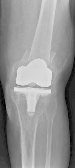
Preoperative AP periprosthetic supracondylar fracture with poor bone stock
Fig. 2.
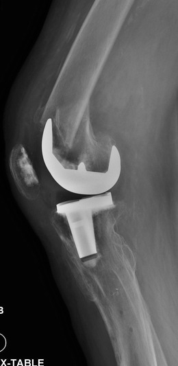
Preoperative Lat periprosthetic supracondylar fracture with poor bone stock
Fig. 3.
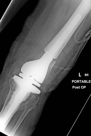
Postoperative AP1 distal femoral replacement
Fig. 4.
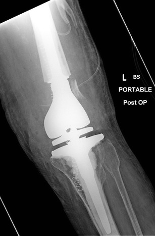
Postoperative AP2 distal femoral replacement
Fig. 5.
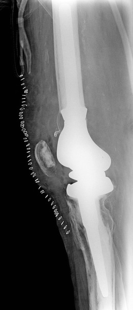
Postoperative Lat distal femoral replacement
Authors’ Preferred treatment
In our unit, these fractures are managed by a team approach with surgeons competent in fracture fixation as well as revision arthroplasty. The initial radiographs are assessed jointly by the 2 groups of surgeons and as discussed previously the patient is medically optimized. Computed tomography scans are obtained if necessary and an infection screen is performed if there is any suspicion. Fracture fixation is the preferred modality of treatment except in elderly patients with a loose or malaligned prosthesis with bone stock loss where revision arthroplasty is considered, although intraoperatively all options are available to the operating surgeon.
Complications
Periprosthetic fractures are associated with a very high morbidity and mortality [9•, 62, 63]. The mortality figures can be up to 17 % at 6 months and 30 % at 1 year [64–66]. These fractures carry a much higher risk of mortality than distal femoral fractures or total knee arthroplasties (TKA) in isolation [67•]. Postoperative mobility of patients with periprosthetic distal femoral fractures is reduced with a significant proportion of patients needing long-term ambulatory aid assistance. Some loss of alignment and loss of movement is common in most cases, and nonunion remains a major concern with reports varying from 0 %–50 % in various studies [42••]. Indirect reduction techniques and submuscular plate insertion techniques are believed to reduce the incidence of nonunion [68].
Other complications include infection, extensor mechanism disruption, implant failure, and dislocation. Most complications are attributable to poor bone quality, fracture location, and patient factors. Complications like pressure ulcers, pulmonary embolism, confusion, urinary tract, and respiratory infections are all consequences of prolonged immobility [42••].
Conclusions
In conclusion, periprosthetic fractures around the knee have devastating consequences for the patient and pose a technical challenge for the surgeon. These fractures frequently occur in frail, elderly patients with significant medical comorbidities and their successful treatment involves a team approach with skills in fixation as well as revision arthroplasty. A significant proportion of these cases can be treated with fixation by an intramedullary device or a locked plating system, but selected cases will benefit from revision surgery or indeed a distal femoral endoprosthesis, especially in patients with poor bone stock. The goal of treatment should be to have a well-aligned and mobile knee joint and an ambulatory patient with a united fracture while minimizing the risk to the patient.
Acknowledgments
Compliance with Ethics Guidelines
ᅟ
Conflict of Interest
MR Whitehouse is employed by the National Institute of Health Research; the NIHR currently funds his post as a Clinical Lecturer. He also receives funding for DePuy for a clinical research fellow post in the department he works in. He previously received the McMinn Scholar, funded by a scholarship from the British Hip Society, and he previously received a traveling fellowship award from the BOA, funded by Zimmer.
S Mehendale declares that he has no conflict of interest.
Human and Animal Rights and Informed Consent
This article does not contain any studies with human or animal subjects performed by any of the authors.
References
Papers of particular interest, published recently, have been highlighted as: • Of importance •• Of major importance
- 1.NJR Steering Committee. National Joint Registry for England, Wales and Northern Ireland: 10th Annual Report, Hemel Hempstead, UK. 2013.
- 2.Aaron RK, Scot R. Supracondylar fracture of the femur after total knee arthroplasty. Clin Orthop Relat Res. 1987;219:136–9. [PubMed] [Google Scholar]
- 3.Merkel KD, Johnson EW. Supracondylar fracture of the femur after total knee arthroplasty. J Bone Joint Surg Am. 1986;68:29–43. [PubMed] [Google Scholar]
- 4.Rorabeck CH, Taylor JW. Classification of periprosthetic fractures complicating total knee arthroplasty. Orthop Clin North Am. 1999;30:209–14. doi: 10.1016/S0030-5898(05)70075-4. [DOI] [PubMed] [Google Scholar]
- 5.Healy WL, Siliski JM, Incavo SJ. Operative treatment of distal femoral fractures proximal to total knee replacements. J Bone Joint Surg Am. 1993;75:27–34. doi: 10.2106/00004623-199301000-00005. [DOI] [PubMed] [Google Scholar]
- 6.Inglis AE, Walker PS. Revision of failed knee replacements using fixed-axis hinges. J Bone Joint Surg (Br) 1991;73:757–61. doi: 10.1302/0301-620X.73B5.1894661. [DOI] [PubMed] [Google Scholar]
- 7.Ritter MA, Faris PM, Keating EM. Anterior femoral notching and ipsilateral supracondylar femur fracture in total knee arthroplasty. J Arthroplasty. 1988;3:185–7. doi: 10.1016/S0883-5403(88)80085-8. [DOI] [PubMed] [Google Scholar]
- 8.Schrøder HM, Berthelsen A, Hassani G, Hansen EB, Solgaard S. Cementless porous-coated total knee arthroplasty: 10-year results in a consecutive series. J Arthroplasty. 2001;16:559–67. doi: 10.1054/arth.2001.23565. [DOI] [PubMed] [Google Scholar]
- 9.•.Meek RMD, Norwood T, Smith R, Brenkel IJ, Howie CR. The risk of peri-prosthetic fracture after primary and revision total hip and knee replacement. J Bone Joint Surg (Br) 2011;93:96–101. doi: 10.1302/0301-620X.93B1.25087. [DOI] [PubMed] [Google Scholar]
- 10.Felix NA, Stuart MJ, Hanssen AD. Periprosthetic fractures of the tibia associated with total knee arthroplasty. Clin Orthop Relat Res. 1997;345:113–24. doi: 10.1097/00003086-199712000-00016. [DOI] [PubMed] [Google Scholar]
- 11.Chalidis BE, Tsiridis E, Tragas AA, Stavrou Z, Giannoudis PV. Management of periprosthetic patellar fractures. A systematic review of literature. Injury. 2007;38:714–24. doi: 10.1016/j.injury.2007.02.054. [DOI] [PubMed] [Google Scholar]
- 12.Ortiguera CJ, Berry DJ. Patellar fracture after total knee arthroplasty. J Bone Joint Surg Am. 2002;84:532–40. doi: 10.2106/00004623-200204000-00004. [DOI] [PubMed] [Google Scholar]
- 13.Engh GA, Ammeen DJ. Periprosthetic fractures adjacent to total knee implants. Treatment and clinical results. J Bone Joint Surg Am. 1997;79:1100–13. [PubMed]
- 14.Beals RK, Tower SS. Periprosthetic fractures of the femur. An analysis of 93 fractures. Clin Orthop Relat Res. 1996;327:238–46. doi: 10.1097/00003086-199606000-00029. [DOI] [PubMed] [Google Scholar]
- 15.Porsch M, Galm R, Hovy L, Starker M, Kerschbaumer F. [Total femur replacement following multiple periprosthetic fractures between ipsilateral hip and knee replacement in chronic rheumatoid arthritis. Case report of 2 patients] Z Orthop Ihre Grenzgeb. 1996;134:16–20. doi: 10.1055/s-2008-1037412. [DOI] [PubMed] [Google Scholar]
- 16.•.Della Rocca G. Periprosthetic fractures about the knee—an overview. J Knee Surg. 2013;26:3–8. doi: 10.1055/s-0033-1333900. [DOI] [PubMed] [Google Scholar]
- 17.•.Platzer P, Schuster R, Aldrian S, Prosquill S, Krumboeck A, Zehetgruber I, et al. Management and outcome of periprosthetic fractures after total knee arthroplasty. J Trauma. 2010;68:1464–70. doi: 10.1097/TA.0b013e3181d53f81. [DOI] [PubMed] [Google Scholar]
- 18.Culp RW, Schmidt RG, Hanks G, Mak A, Esterhai JL, Heppenstall RB. Supracondylar fracture of the femur following prosthetic knee arthroplasty. Clin Orthop Relat Res. 1987;222:212–22. [PubMed] [Google Scholar]
- 19.Lesh ML, Schneider DJ, Deol G, Davis B, Jacobs CR, Pellegrini VD. The consequences of anterior femoral notching in total knee arthroplasty. A biomechanical study. J Bone Joint Surg Am. 2000;82-A:1096–101. doi: 10.2106/00004623-200008000-00005. [DOI] [PubMed] [Google Scholar]
- 20.Zalzal P, Backstein D, Gross AE, Papini M. Notching of the anterior femoral cortex during total knee arthroplasty characteristics that increase local stresses. J Arthroplasty. 2006;21:737–43. doi: 10.1016/j.arth.2005.08.020. [DOI] [PubMed] [Google Scholar]
- 21.Ritter MA, Thong AE, Keating EM, Faris PM, Meding JB, Berend ME, et al. The effect of femoral notching during total knee arthroplasty on the prevalence of postoperative femoral fractures and on clinical outcome. J Bone Joint Surg Am. 2005;87:2411–4. doi: 10.2106/JBJS.D.02468. [DOI] [PubMed] [Google Scholar]
- 22.Gujarathi N, Putti AB, Abboud RJ, MacLean JGB, Espley AJ, Kellett CF. Risk of periprosthetic fracture after anterior femoral notching. Acta Orthop. 2009;80:553–6. doi: 10.3109/17453670903350099. [DOI] [PMC free article] [PubMed] [Google Scholar]
- 23.•.Singh JA, Jensen M, Lewallen D. Predictors of periprosthetic fracture after total knee replacement. Acta Orthop. 2013;84:170–7. doi: 10.3109/17453674.2013.788436. [DOI] [PMC free article] [PubMed] [Google Scholar]
- 24.Charlson ME, Pompei P, Ales KL, MacKenzie CR. A new method of classifying prognostic comorbidity in longitudinal studies: development and validation. J Chronic Dis. 1987;40:373–83. doi: 10.1016/0021-9681(87)90171-8. [DOI] [PubMed] [Google Scholar]
- 25.Engh GA, Rorabeck CH, eds. Revision total knee arthroplasty. Baltimore, Williams & Wilkins, Philadelphia. 1997;275–95.
- 26.Kim K-I, Egol KA, Hozack WJ, Parvizi J. Periprosthetic fractures after total knee arthroplasties. Clin Orthop Relat Res. 2006;446:167–75. doi: 10.1097/01.blo.0000214417.29335.19. [DOI] [PubMed] [Google Scholar]
- 27.Parvizi J, Gehrke T, Chen AF. Proceedings of the international consensus on periprosthetic joint infection. Bone Joint J. 2013;95-B:1450–2. doi: 10.1302/0301-620X.95B11.33135. [DOI] [PubMed] [Google Scholar]
- 28.Garnavos C, Rafiq M, Henry APJ. Treatment of femoral fracture above a knee prosthesis: 18 cases followed 0.5–14 years. Acta Orthop. 1994;65:610–4. doi: 10.3109/17453679408994614. [DOI] [PubMed] [Google Scholar]
- 29.Moran MC, Brick GW, Sledge CB, Dysart SH, Chien EP. Supracondylar femoral fracture following total knee arthroplasty. Clin Orthop Relat Res. 1996;324:196–209. doi: 10.1097/00003086-199603000-00023. [DOI] [PubMed] [Google Scholar]
- 30.Delport PH, Van Audekercke R, Martens M, Muller JC. Conservative treatment of ipsilateral supracondylar femoral fracture after total knee arthroplasty. J Trauma. 1984;24:846–9. doi: 10.1097/00005373-198409000-00013. [DOI] [PubMed] [Google Scholar]
- 31.Ginther JR, Ritter MA. Surgical Techniques in Total Knee Arthroplasty. New York: Springer; 2002. Femoral Periprosthetic Fractures: Rush Rods; pp. 553–7. [Google Scholar]
- 32.Ritter MA, Keating EM, Faris PM, Meding JB. Rush rod fixation of supracondylar fractures above total knee arthroplasties. J Arthroplasty. 1995;10:213–6. doi: 10.1016/S0883-5403(05)80130-5. [DOI] [PubMed] [Google Scholar]
- 33.••.Sarmah SS, Patel S, Reading G, El-Husseiny M, Douglas S, Haddad FS. Periprosthetic fractures around total knee arthroplasty. Ann R Coll Surg Engl. 2013;94:302–7. doi: 10.1308/003588412X13171221592537. [DOI] [PMC free article] [PubMed] [Google Scholar]
- 34.Currall VA, Kulkarni M, Harries WJ. Retrograde nailing for supracondylar fracture around total knee replacement: a compatibility study using the Trigen supracondylar nail. Knee. 2007;14:208–11. doi: 10.1016/j.knee.2006.12.001. [DOI] [PubMed] [Google Scholar]
- 35.Kregor PJ, Hughes JL, Cole PA. Fixation of distal femoral fractures above total knee arthroplasty utilizing the Less Invasive Stabilization System (LISS) Injury. 2001;32(Suppl 3):SC64–75. doi: 10.1016/S0020-1383(01)00185-1. [DOI] [PubMed] [Google Scholar]
- 36.•.Ruchholtz S, Tomás J, Gebhard F, Larsen MS. Periprosthetic fractures around the knee—the best way of treatment. Euro Orthop Traumatol. 2013;4:93–102. doi: 10.1007/s12570-012-0130-x. [DOI] [PMC free article] [PubMed] [Google Scholar]
- 37.Short WH, Hootnick DR, Murray DG. Ipsilateral supracondylar femur fractures following knee arthroplasty. Clin Orthop Relat Res. 1981;158:111–6. [PubMed] [Google Scholar]
- 38.Weber D, Peter RE. Distal femoral fractures after knee arthroplasty. Int Orthop. 1999;23:236–9. doi: 10.1007/s002640050359. [DOI] [PMC free article] [PubMed] [Google Scholar]
- 39.•.Hassan S, Swamy GN, Malhotra R, Badhe NP. Periprosthetic fracture of the distal femur after total knee arthroplasty; prevalence and outcomes following treatment. J Bone Joint Surg (Br) 2012;94-B(Suppl 24):6. [Google Scholar]
- 40.Norrish AR, Jibri ZA, Hopgood P. The LISS plate treatment of supracondylar fractures above a total knee replacement: a case-control study. Acta Orthop Belg. 2009;75:642–8. [PubMed] [Google Scholar]
- 41.••.Ebraheim NA, Sochacki KR, Liu X, Hirschfield G, Liu J. Locking plate fixation of periprosthetic femur fractures with and without cerclage wires. Orthop Surg. 2013;5:183–7. doi: 10.1111/os.12052. [DOI] [PMC free article] [PubMed] [Google Scholar]
- 42.••.Hoffman MF, Jones CB, Sietsema DL, Koenig SJ, Tornetta P. Outcome of periprosthetic distal femoral fractures following knee arthroplasty. Injury. 2012;43:1084–9. doi: 10.1016/j.injury.2012.01.025. [DOI] [PubMed] [Google Scholar]
- 43.••.Ehlinger M, Adam P, Abane L, Rahme M, Moor BK, Arlettaz Y, et al. Treatment of periprosthetic femoral fractures of the knee. Knee Surg Sports Traumatol Arthr. 2011;19:1473–89. doi: 10.1007/s00167-011-1480-6. [DOI] [PubMed] [Google Scholar]
- 44.Bezwada HP, Neubauer P, Baker J, Israelite CL, Johanson NA. Periprosthetic supracondylar femur fractures following total knee arthroplasty. J Arthroplasty. 2004;19:453–8. doi: 10.1016/j.arth.2003.12.078. [DOI] [PubMed] [Google Scholar]
- 45.Herrera DA, Kregor PJ, Cole PA, Levy BA, Jönsson A, Zlowodzki M. Treatment of acute distal femur fractures above a total knee arthroplasty: systematic review of 415 cases (1981-2006) Acta Orthop. 2008;79:22–7. doi: 10.1080/17453670710014716. [DOI] [PubMed] [Google Scholar]
- 46.Bong MR, Egol KA, Koval KJ, Kummer FJ, Su ET, Iesaka K, et al. Comparison of the LISS and a retrograde-inserted supracondylar intramedullary nail for fixation of a periprosthetic distal femur fracture proximal to a total knee arthroplasty. J Arthroplasty. 2002;17:876–81. doi: 10.1054/arth.2002.34817. [DOI] [PubMed] [Google Scholar]
- 47.•.Kilicoglu OI, Akgül T, Sağlam Y, Yazıcıoğlu O. Comparison of locked plating and intramedullary nailing for periprosthetic supracondylar femur fractures after knee arthroplasty. Acta Orthop Belg. 2013;79:417–21. [PubMed] [Google Scholar]
- 48.Althausen PL, Lee MA, Finkemeier CG, Meehan JP, Rodrigo JJ. Operative stabilization of supracondylar femur fractures above total knee arthroplasty: a comparison of four treatment methods. J Arthroplasty. 2003;18:834–9. doi: 10.1016/S0883-5403(03)00339-5. [DOI] [PubMed] [Google Scholar]
- 49.••.Ristevski B, Nauth A, Williams D, Hall J, Whelan D, Bhandari M, et al. Systematic review of the treatment of periprosthetic distal femur fractures. J Orthop Trauma. 2013;1. [Epub ahead of print]. A good systematic review of current evidence regarding treatment with locked plates, conventional plates, conservative treatment and intramedullary nailing.
- 50.Beris AE, Lykissas MG, Sioros V, Mavrodontidis AN, Korompilias AV. Femoral periprosthetic fracture in osteoporotic bone after a total knee replacement: treatment with Ilizarov external fixation. J Arthroplasty. 2010;25:1168.e9–e12. [DOI] [PubMed]
- 51.Simon RG, Brinker MR. Use of Ilizarov external fixation for a periprosthetic supracondylar femur fracture. J Arthroplasty. 1999;14:118–21. doi: 10.1016/S0883-5403(99)90214-0. [DOI] [PubMed] [Google Scholar]
- 52.Hurson C, Synnott K, McCormack D. Above-knee Ilizarov external fixation for early periprosthetic supracondylar femoral fracture—a case report. Knee. 2005;12:145–7. doi: 10.1016/j.knee.2004.06.005. [DOI] [PubMed] [Google Scholar]
- 53.•.Keeney JA. Periprosthetic total knee arthroplasty fractures: revision arthroplasty technique. J Knee Surg. 2013;26:19–26. doi: 10.1055/s-0033-1333903. [DOI] [PubMed] [Google Scholar]
- 54.••.Saidi K, Ben-Lulu O, Tsuji M, Safir O, Gross AE, Backstein D. Supracondylar periprosthetic fractures of the knee in the elderly patients: a comparison of treatment using Allograft-Implant Composites, Standard Revision Components, Distal Femoral Replacement Prosthesis. J Arthroplasty. 2014;29:110–4. doi: 10.1016/j.arth.2013.04.012. [DOI] [PubMed] [Google Scholar]
- 55.Berend KR, Lombardi AV. Distal femoral replacement in nontumor cases with severe bone loss and instability. Clin Orthop Relat Res. 2009;467:485–92. doi: 10.1007/s11999-008-0329-x. [DOI] [PMC free article] [PubMed] [Google Scholar]
- 56.••.Chen AF, Choi LE, Colman MW, Goodman MA, Crossett LS, Tarkin IS, et al. Primary versus secondary distal femoral arthroplasty for treatment of total knee arthroplasty periprosthetic femur fractures. J Arthroplasty. 2013;28:1580–4. doi: 10.1016/j.arth.2013.02.030. [DOI] [PubMed] [Google Scholar]
- 57.Appleton P, Moran M, Houshian S, Robinson CM. Distal femoral fractures treated by hinged total knee replacement in elderly patients. J Bone Joint Surg (Br) 2006;8:1065–70. doi: 10.1302/0301-620X.88B8.17878. [DOI] [PubMed] [Google Scholar]
- 58.Springer BD, Hanssen AD, Sim FH, Lewallen DG. The kinematic rotating hinge prosthesis for complex knee arthroplasty. Clin Orthop Relat Res. 2001;392:283–91. doi: 10.1097/00003086-200111000-00037. [DOI] [PubMed] [Google Scholar]
- 59.Springer BD, Sim FH, Hanssen AD, Lewallen DG. The modular segmental kinematic rotating hinge for non-neoplastic limb salvage. Clin Orthop Relat Res. 2004;421:181–7. doi: 10.1097/01.blo.0000126306.87452.59. [DOI] [PubMed] [Google Scholar]
- 60.••.Jassim SS, McNamara I, Hopgood P. Distal femoral replacement in periprosthetic fracture around total knee arthroplasty. Injury. 2013 doi: 10.1016/j.injury.2013.10.032. [DOI] [PubMed] [Google Scholar]
- 61.••.Mortazavi SMJ, Kurd MF, Bender B, Pos Z, Parvizi J, Purtill JJ. Distal femoral arthroplasty for the treatment of periprosthetic fractures after total knee arthroplasty. J Arthroplasty. 2010;25:775–80. doi: 10.1016/j.arth.2009.05.024. [DOI] [PubMed] [Google Scholar]
- 62.Bhattacharyya T, Chang D, Meigs JA, Estok DM, Malchau H. Mortality after periprosthetic fracture of the femur. J Bone Joint Surg Am. 2007;89:2658–62. doi: 10.2106/JBJS.F.01538. [DOI] [PubMed] [Google Scholar]
- 63.Figgie MP, Goldberg VM, Figgie HE, Sobel M. The results of treatment of supracondylar fracture above total knee arthroplasty. J Arthroplasty. 1990;5:267–76. doi: 10.1016/S0883-5403(08)80082-4. [DOI] [PubMed] [Google Scholar]
- 64.Christodoulou A, Terzidis I, Ploumis A, Metsovitis S, Koukoulidis A, Toptsis C. Supracondylar femoral fractures in elderly patients treated with the dynamic condylar screw and the retrograde intramedullary nail: a comparative study of the two methods. Arch Orthop Trauma Surg. 2005;125:73–9. doi: 10.1007/s00402-004-0771-5. [DOI] [PubMed] [Google Scholar]
- 65.Boyd AD, Wilber JH. Patterns and complications of femur fractures below the hip in patients over 65 years of age. J Orthop Trauma. 1992;6:167–74. doi: 10.1097/00005131-199206000-00006. [DOI] [PubMed] [Google Scholar]
- 66.Dunlop DG, Brenkel IJ. The supracondylar intramedullary nail in elderly patients with distal femoral fractures. Injury. 1999;30:475–84. doi: 10.1016/S0020-1383(99)00136-9. [DOI] [PubMed] [Google Scholar]
- 67.•.Streubel PN, Ricci WM, Wong A, Gardner MJ. Mortality after distal femur fractures in elderly patients. Clin Orthop Relat Res. 2011;469:1188–96. doi: 10.1007/s11999-010-1530-2. [DOI] [PMC free article] [PubMed] [Google Scholar]
- 68.Bolhofner BR, Carmen B, Clifford P. The results of open reduction and internal fixation of distal femur fractures using a biologic (indirect) reduction technique. J Orthop Traum. 1996;10:372–7. doi: 10.1097/00005131-199608000-00002. [DOI] [PubMed] [Google Scholar]


