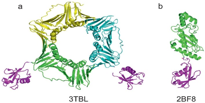
Figure 5.
Summary of the currently available structures of ubiquitinated proteins. (a) Structure of mono-ubiquitinated PCNA (PDB code 3TBL). Note that the last GG moieties on the two Ub molecules do not appear in the PDB structure. K164 of two of the three monomers of the PCNA trimer are showed as sticks; (b) Structure of sumoylated E2-25K (PDB code 2BF8). K14 of E2-25K and G97 of SUMO are reported as sticks to visualise the native isopeptide bond.

