

Abstract
Rheumatoid arthritis (RA) manifests itself in a variety of ways, with its effect being seen in around 90 % of sufferers’ feet. The foot has been found to be the most common reason for incapacity in patients with RA, with the forefoot the most common area. The foot is second, behind only the hand, as the most common place for manifestation of RA. Pain in the foot is commonly the most debilitating condition, which causes the patient to seek specialist help. As well as pain, foot deformities such as hallux valgus and claw toes are common complaints. These symptoms often arise as a result of continued walking on an unstable foot, leading to painful callosities and dislocation of the metatarsophalangeal joints. Other conditions, such as pannus formation and Morton’s neuroma, can be related to RA. This review sets out what we believe to be a successful approach to the rheumatoid forefoot, which aims at the relief of pain and the preservation of ambulation. Key to a successful outcome is appropriate medical control with a multidisciplinary approach that enables close liaison between orthopaedic surgeons, orthotists, and rheumatologists. Combined clinics provide this multidisciplinary care. Those treating RA need to be aware of the high incidence of foot involvement and how early intervention may benefit the patient. The aim of this article is to present current evidence to enable people to develop a treatment algorithm for this condition.
Keywords: Rheumatoid arthritis, Forefoot, Surgery
Introduction
Rheumatoid arthritis (RA) manifests itself in a variety of ways, with its effect being seen in around 90 % of sufferers’ feet [1]. The foot has been found to be the most common reason for incapacity in patients with RA, with the forefoot the most common area [2]. The foot is second, behind only the hand, as the most common place for manifestation of RA [3].
Pain in the foot is commonly the most debilitating condition that causes the patient to seek specialist help. As well as pain, foot deformities such as hallux valgus and claw toes are common complaints. These symptoms often arise as a result of continued walking on an unstable foot, leading to rupture of the collateral ligaments and migration of the plantar fat pads, causing painful callosities and dislocation of the metatarsophalangeal joints. Other conditions, such as pannus formation and Morton’s neuroma, can be related to RA.
A variety of treatment options are available in treating the rheumatoid patients for their forefoot conditions. These range from conservative to surgical intervention. The aim of this article is to present current evidence to enable people to develop a treatment algorithm for this condition.
Pathophysiology
RA is a systematic inflammatory condition that is mediated by the interaction of antigens, antibodies, and complement and immune complexes. The cause of this condition remains unclear. Clinically, it presents with symmetrical erosive synovitis, commonly of the peripheral joints, which leads to progressive destruction and joint deformity. It affects approximately 1 % of the worldwide adult population, and its prevalence increases with age.
The deformities of the foot result from chronic synovitis, which causes capsular distension Classical deformities of the foot include dorsal dislocation of the metatarsophalangeal joints (MTPJ), plantar callosities, and hallux valgus deformity of the great toe (Figs. 1 and 2). These deformities are the result of the inflammatory process, which leads to joint instability by the eventual rupture of the collateral ligaments and the plantar plates following the joint distension. In the MTPJs, the disruption of the soft tissues leads to the progressive instability and deformities seen in the RA foot. This instability is worsened by loading of the MTPJs while mobilizing, causing the plantar displacement of the metatarsal (MT) heads. The classic lesser toe deformities of RA occur due to progressive dorsiflexion of the toes during the gait cycle. Due to dorsal subluxation of the proximal phalanx, the interosseous muscles become ineffective extensors [4]. This leads to a flexor–extensor imbalance, leading to a flexible claw toe deformity that, without treatment, quickly becomes a rigid deformity, leading to the development of painful corns and calluses over the now prominent MT heads. Finally, the plantar plate ruptures at its proximal insertion and moves distally, leading to further plantar callosities [5].
Fig. 1.

X-ray of a rheumatoid forefoot
Fig. 2.
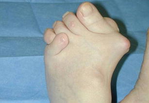
Preoperative picture of severe rheumatoid forefoot
Foot biomechanics
Foot biomechanics and its understanding are important when treating the rheumatoid foot. Studies have shown that RA patients walk slower, have longer double support, and have a change to their ground angle [6]. The gait cycle can be described as three key components: weight acceptance, single limb support, and swing limb advancement. Key to the gait cycle is the three rockers of the foot as described by Perry [7]. The rockers are the heel rocker, ankle rocker, and forefoot rocker and are important in maintaining the propulsive force during the gait cycle.
Clinical evaluation
Despite the foot being a common source of pain and disability in RA, it is often overlooked in clinical assessment of the disease. In recent times, an improvement in medical management may improve the condition, and fewer patients may suffer the severe consequences seen in feet. Evidence of involvement of the forefoot is present on radiological studies in up to a third of patients on presentation with RA [3]. Plain radiographs and magnetic resonance imaging (MRI) has demonstrated this [8, 9]. As RA establishes itself, this can rise to around 90 % [10], and approximately a quarter of all patients with RA will undergo surgery on their forefoot [11].
The DAS 28 is a scoring system used to assess patients with RA [12]. This score is based on assessment of tenderness and swelling over certain joints. Interestingly, this does not include the foot in its assessment. De Jong et al. performed a study where they added a squeeze test across the MTPJs in their assessment using the DAS 28 [13]. They found that this had greater sensitivity for the assessment of disease severity in patients presenting with early RA. A study looked specifically at the use of scoring systems to suggest remission of RA and the presence of foot synovitis [14]. This study compared the 2011 remission criteria agreed upon by the American College of Rheumatologists (ACR) and the European League Against Rheumatism (EULAR) with the DAS 28. They found that remission in the presence of foot joint synovitis was lower in the 2011 criteria, which included assessment of the foot, than with the DAS 28. Van Tuyl et al. reinforced this when they found that up to 40 % of rheumatoid patients had activity in their feet when classified with the DAS 28, as compared with ACR/EULAR 38 joint classification [15]. The authors concluded that this has limited effects on the outcome of the disease in these patients, but we would urge caution on this, since they included only patients with 1-year follow-up.
Radiological studies are often used by clinicians in their investigation of patients with painful joints. Many radiological tests are available to the clinician. The role of the simple radiograph cannot be underestimated as a quick and reliable method of evaluating the rheumatoid forefoot. In the assessment of any deformity, great value can be gained by weight-bearing radiographs. Assessment of alignment of the forefoot to the hindfoot can be made on the lateral weight-bearing radiograph.
MRI scanning has been found to have relatively good specificity and sensitivity in the assessment of patients with undifferentiated RA. Duer-Jensen et al. used the presence of bone oedema in their study and found that its presence in the MTPJ or the wrist joint in a patient who had undifferentiated arthritis was an independent predictor of future RA [16]. The main debate is focused around the use of musculoskeletal ultrasound and MRI of the plantar plates. The plantar plates are often affected in RA, and their rupture can account for large amounts of discomfort. Both ultrasound and MRI are excellent tools for evaluating this structure [17]. The advantage of MRI is that it allows the review of images in the clinic and can often be interpreted by the clinician, as well as the radiologist, while ultrasound requires a skilled radiologist and the images can be difficult for clinicians to read. The advantage of ultrasound is that it allows a clinical distinction to be made between asymptomatic lesions and symptomatic lesions, since the patients can reproduce the pain for the radiologists.
Medical management
Pharmacological control of RA remains the mainstay in the management of the condition. The current standard of care remains the medical management of this condition and is best carried out under the care of a rheumatologist and utilizing the varying medications available. The development of disease-modifying antirheumatic drugs (DMARDs), as well as the development of newer biological agents, has revolutionized the management of this condition.
DMARDs represent drug such as methotrexate and sulfasalazine. These drugs are generally now offered to patients much earlier than previously, and current clinical guidelines recommend the prescription of such drugs ideally within the first 3 months of symptoms [18].
The new biologics represent an advance in the management of this condition. These are antitumour necrosis factors and work by cytokine modulation. These are generally reserved for patients who have not responded well to DMARDs and still have active RA.
The controversy concerning these drugs is whether they should be stopped prior to and after surgery, as well as the length of time for which such stoppages should occur. Most surgeons would allow the DMARDs to be continued during the perioperative and postoperative periods. Evidence exists only for the use of methotrexate, however [19]. It is generally advised to stop the anti-TNF drugs prior to surgery, although recent evidence would suggest that this may not be necessary [20, 21].
Nonoperative
While podiatry has a well-established role in diabetic care, its role in RA is less clear. This was highlighted by an audit of foot care in New Zealand, which found that access to podiatry was either poor or lacking [22]. This was further highlighted in a study in England, where only one third of RA patients received any form of intervention for foot problems [23]. It was found that older RA patients tended to be managed conservatively, while the younger cohort was more likely to be offered surgical intervention. Farrow et al. performed a systematic review of management of foot disease in RA and found that only orthoses had high-quality evidence of benefit [24•].
The effects of RA on the forefoot can lead to changes in plantar plate pressure and altered motion of the foot. Gait analysis has shown that there are marked changes in patients with a painful rheumatoid foot, in particular with increased pressure under the 1st and 4th MTPJs [25]. The suggestion is that by correcting these forces and stabilizing the foot, this may slow or prevent the development of forefoot deformity. Critical reviews have so far not been able to show this to be the case [26, 27].
A Cochrane review was performed to assess the use of custom-made orthotics in foot pain. One of the subgroups that were included in the study was patients with RA. They found that there was some evidence that custom orthoses made a difference in terms of rear foot pain, but the evidence failed to show any difference between supportive footwear and custom-made orthotics for metatarsophalangeal joint pain [28•].
There have been randomized control trials that have assessed the used of orthotics in the management of RA and its forefoot manifestations. The first study looked at the use of custom orthotics over a 3-year period and failed to show a significant difference in the outcome, as compared with thin shoe insoles [29]. This is the longest of the four studies. This differs from the results shown in the other three studies. The issue with these three is that the length of the study was shorter in all three, with lengths of 30 months, 2 months, and 12 weeks [30–32]. These studies showed that there was a significant reduction in reported pain in the group using orthotics, as compared with the standard treatment arm. They suggest that the use of orthotics may have a role in those patients who have mainly a painful forefoot. A problem that undermines the success of orthotics is the low rate of compliance. It has been reported to be as low as 36 % in the rheumatoid population [33].
There is evidence that footwear modifications can improve the mobility, pain, and function of RA patients with manifestation in the forefoot [34]. This result was also seen in a study by Magalhaes [35]. A major criticism of both of these studies is the small study population and short-term follow-up.
The aim of any custom-made orthoses is to reduce vertical pressure and shear forces passing through the foot. It is an essential requirement that the foot be flexible in order to use corrective orthoses. In the presence of a flexible foot deformity, an orthotic needs to incorporate posting, a more rigid material to control and limit the movement, while a rigid deformity needs a softer orthotic for comfort. Outer shoe modifications have also been found to be useful in the management of the painful rheumatoid foot [36].
Surgical management
The aim of any surgical treatment on the foot is to provide relief from pain, to restore ambulation where possible, and to allow for comfortable footwear. Surgical options are many in the rheumatoid forefoot. Numerous procedures have been described with varying degrees of success. The importance in surgical decision making is to understand the deformity and respect the soft tissues, while not forgetting the overarching issues of the disease process itself and its biological sequelae. It is also important to treat the patient bearing in mind the systemic manifestations of the disease, as well as the medication the patient is on. These are key to achieving a good outcome in the surgical management of the rheumatoid forefoot.
One of the first procedures described for the management of the rheumatoid forefoot was a resection arthroplasty of the MTPJs by Hoffman in 1911 [37]. This is still an accepted method of management today. Other types of resection arthroplasty have also been described focusing on the 1st MTPJ by Hueter [38], Mayo [39], and Keller [40]. Resection arthroplasty was first popularized for the management of RA in 1963 by Clayton [41]. In 2001, Fuhrmnn et al. presented their retrospective series on the results of resection arthroplasty of the 1st MTPJ in RA patients [42]. They had used a combination of Keller’s procedures and the Hueter–Mayo technique, with an average of 7.9 years of follow-up in 278 patients. Their overall results are superior in patients treated with the Hueter–Mayo technique than with Keller’s procedure, with a statistical difference between the two. Walking ability was not improved particularly in either group. They conclude that Keller’s procedure should no longer be recommended, since the Hueter–Mayo technique provides superior results. Poor results in terms of recurrence of deformity and pain have been described for these procedures [43, 44]. However, more recent studies using MTPJ resection have found higher rates of satisfaction and pain relief [45, 46]. Despite this, a study by Hattori et al. showed that deformity and deterioration of the hallux occurred following resection arthroplasty in rheumatoid patients [47].
One of the more commonly performed procedures that have been shown to both correct deformity and relieve pain is the 1st MTPJ fusion. This is often used either in isolation or as a combined procedure with lesser MTPJ resection arthroplasty. The advantage of this is that it provides a permanent correction of the deformity, while also providing pain relief of the joint. It has been shown that this method of reconstruction restores weight bearing along the medial border of the foot. Dwyer popularized this method after observing that the effects of lesser MTPJ excision was only short-lived [48]. Mann and Thompson further popularized it by using dorsal incisions and advocating Kirschner wire stabilization for the lesser toe deformities [49]. Coughlin reported his results of using 1st MTPJ fusion with lesser MTPJ excision and found this method to provide good to excellent results in 77 % of the feet operated on [50•]. In one of the few randomized control trials, Grondal et al. found that there was no difference between 1st MTPJ fusion and resection when using the Mayo technique [51]. Kadambande et al. reported the results of 3 years of their method, which involved MTPJ fusion and lesser MTPJ resection, with good results in terms of pain relief, function, and subjective relevant outcome measure [52]. Van der Heide showed that their results for correction of lesser toe deformities were greatly improved when performed with a 1st MTPJ fusion [53].
Another surgical option described in the literature for the management of the RA forefoot is distal-joint-preserving surgery. The Lapidus procedure is one such operation [54]. The Lapidus procedure involves fusion of the first tarsometatarsal joint. Its main use is in patients with a hypermobile 1st ray. Shi et al. described their modified technique for the Lapidus procedure in patients with RA [55]. They achieved good to excellent pain relief in 80 % of patients. These results were echoed by Popleka et al. in their study [56]. They concluded that the Lapidus procedure may be useful as a preventive surgery. This suggestion is one we take with caution, due to the risks of surgery in this patient population and, in addition, the fusion of a seemingly normal joint proximally. Joint-preserving surgery has gained popularity in this patient group. Some feel that with modern DMARDs, there are less erosive changes seen in the RA feet, suggesting that the role of MTPJ fusion and arthroplasty may be appropriate only for the severe cases [57]. Barouk has made this a popular method of treatment for some RA patients [58•]. They proposed the use of a Weil osteotomy, which elevates the metatarsal head, combined with extensor tendon lengthening. The advantage of this osteotomy is that it is biomechanically more stable than other proposed proximal osteotomies [59]. Niki et al. published the results of their combined joint-preserving surgery [60]. The patients in this study had early or intermediate RA. They concluded that this kind of surgical approach to RA forefeet provided better correction of deformity, function, and cosmesis. The disadvantage of these joint-preserving procedures is that they involve technically very demanding surgery. Despite the promising results reported by Barouk [61], these results have not been replicated by others [62].
Joint replacement has been used in the 1st MTPJ with varying degrees of success. Developed in the 1960s primarily as a treatment for either hallux rigidus or hallux valgus, its use has been controversial. Its perceived advantage over fusion is that it allows for a degree of flexibility in the joint. High complications have been reported as a result of implant failure, osteolysis, and cock up toe deformities [62, 63]. Despite this, there are a few series that have shown promising results with this method, reporting high patient satisfaction [64, 65]. The success of this arthroplasty is felt to be due to its acting as a spacer, and the joint is stabilized by a fibrous reaction [66], although the procedure has also been related to high rates of synovitis [63], leading to suggestions that it should be abandoned.
Debridement of plantar callosities has been shown to have some clinical benefit [67]. If the callosity is found to be irritant, then debridement will offer some pain relief. In one of the few randomized control trials, Davys showed that the role of debridement was limited and offered little benefit, as compared with sham procedures [68].
Synovectomy has been found to be a useful surgical option in other areas of the body. Very little has been published on synovectomy in the forefoot. This is often the result of presentation of the patients, once the inflammation has settled, with the resultant deformity. One study has suggested that there may be a role for synovectomy of the MTPJs [69], although others have suggested that synovectomy may not prevent subsequent deformity in the ankle or forefoot [70, 71].
One issue which remains relevant to all surgical methods of treatment of RA feet is the incidence of infection. Kadambande showed a rate of 7 % in their series, which would correspond well with the results of other series on rheumatoid feet [52, 72]. Rate of infection may be dependent on surgical approach, as well as the pharmaceutical agents used to control the RA. It has been reported to be as high as 39 % [73]. We would suggest that longer periods of protected weight bearing to stabilize the wounds may reduce the overall infection rate.
Authors’ Preferred method
The approach to the rheumatoid forefoot depends on the condition of the foot and the problems the patient is reporting. We prefer to follow an algorithm that involves progression of treatment options, leaving surgery as a last resort. We rely on the provision of custom orthotics for the management of early rheumatoid patients. When patients are unable to mobilize without pain and there is evidence of progressive deformity, we favor the use of fusion to deal with issues relating to the 1st MTPJ, and if the lesser toes are involved, we perform excision arthroplasty on them. We prefer the use of a plantar elliptical skin excision approach to excise the lesser metatarsal heads. This is a safe and easy surgical approach, since the metatarsal heads have subluxed into the sole of the foot in most cases. It also helps to excise calloused skin, which, by the process of dermadesis, helps to decrease the severe clawing deformity at the lesser MTP joints. All patients under surgical management are also routinely assessed by their rheumatologist both pre- and postoperatively, if required. We have found that this surgical approach offers excellent pain relief, improvement in the function and mobility of the patients, and improvement in the patient’s subjective relevant outcome measure (Figs. 3 and 4). In a series of 88 feet in 59 rheumatoid patients, we have shown that this method provides excellent results [52]. Complications were low and related mainly to superficial wound infections, which remain an issue in the rheumatoid patient. We currently would give high-risk patients a 3-day course of prophylactic antibiotics and would keep them protected for weight bearing in a heel-bearing shoe for up to 6 weeks following surgery.
Fig. 3.
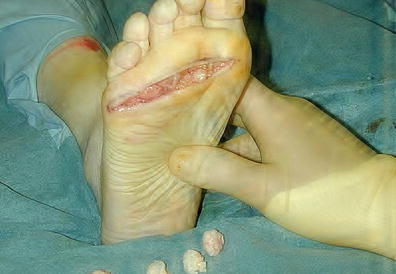
Postexcision, showing the plantar incision
Fig. 4.
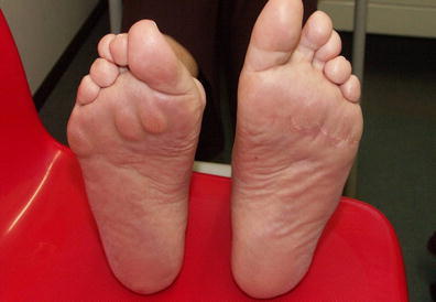
Post-op foot, left, with well-healed plantar scar
Conclusion
The successful approach to management of the rheumatoid forefoot involves a multidisciplinary approach with close liaison between the orthotists and rheumatologists. Combined clinics will offer the patients the ability to have this multidisciplinary care. Those treating RA need to be aware of the high incidence of foot involvement and how early intervention may benefit the patient. It is crucial to be aware of the very significant systemic, pharmacological, and immunological issues when treating these patients, and the multidisciplinary approach with rheumatologists and orthotists is paramount in optimizing the safe surgical management of these patients.
Compliance with Ethics Guidelines
Conflict of Interest
Francis Brooks and Kartik Hariharan declare that they have no conflict of interest.
Human and Animal Rights and Informed Consent
This article does not contain any studies with human or animal subjects performed by any of the authors.
References
Papers of particular interest, published recently, have been highlighted as: • Of importance
- 1.Vainio K. Rheumatoid foot: clinical study with pathological and roentgenological comments. Ann Chir gynaecol Fenniae. 1956;45(supplement):1–107. [PubMed] [Google Scholar]
- 2.Grondal L, Tengstrand B, Nordmark B, Wretenburg P, Stark A. The foot; still the most important reason for walking incapacity in rheumatoid arthritis. Acta Orthopaedica. 2008;79(2):257–261. doi: 10.1080/17453670710015067. [DOI] [PubMed] [Google Scholar]
- 3.Dixey J, Solymossy C, Young A. Is it possible to predict radiological damage in early rheumatoid arthritis? a report on the occurrence, progression and prognostic factors of radiological erosions over the first 3 years in 868 patients from the early RA study (ERAS) J Rheumatol Suppl. 2004;69:48–54. [PubMed] [Google Scholar]
- 4.Stainsby GD. Ptahological anatomy and dynamic effect of the displaced plantar plate and the importance of the plantar plate-deep transverse metatarsal ligament tie-bar. Ann R Coll Surg Engl. 1997;79:58–68. [PMC free article] [PubMed] [Google Scholar]
- 5.Myerson MS. Arthroplasty of the second toe. Semin Arthroplasty. 1992;3:31–38. [PubMed] [Google Scholar]
- 6.Turner DE, Helliwell PS, Siegel KL, Woodburn J. Biomechanics of the foot in rheumatoid arthritis: identifying abnormal function and the factors associated with localised disease ‘impact’. Clin biomechanics. 2008;23(1):93–100. doi: 10.1016/j.clinbiomech.2007.08.009. [DOI] [PubMed] [Google Scholar]
- 7.Perry J, Burnfield JM. Gait analysis: normal and pathological function. 2nd ed. Thorofare: SLACK Incorporated; 2010.
- 8.Pensec VD, Saraux A, Berthelot JM, et al. Ability of foot radiographs to predict rheumatoid arthritis in patients with early arthritis. J Rheumatol. 2004;31:66–70. [PubMed] [Google Scholar]
- 9.Ostendorf B, Scherer A, Modder U, Schneider M. Diagnostic value of magnetic resonance imaging of the forefoot in early rheumatoid arthritis when findings on imaging of the metacarpophalangeal joints of the hands remain normal. Arthritis Rheum. 2004;50:2094–2102. doi: 10.1002/art.20314. [DOI] [PubMed] [Google Scholar]
- 10.Michelson J, Easley M, Wigley FM, Hellmann D. Foot and ankle problems in rheumatoid arthritis. Foot Ankle Int. 1994;15:608–613. doi: 10.1177/107110079401501106. [DOI] [PubMed] [Google Scholar]
- 11.Belt EA, Kaarela K, Lehto MU. Destruction and arthroplasties of the metarsophalangeal joints in seropostiive rheumatoid arthritis: a 20 year follow up study. Scand J Rheumatol. 1998;27:194–196. doi: 10.1080/030097498440804. [DOI] [PubMed] [Google Scholar]
- 12.Van Der Heijde DM, Van’t Hof MA, Van Riel PL, et al. Judging disease activity in clinical practice rheumatoid arthritis: first step in the development of a disease activity score. Ann Rheum Dis. 1990;49:916–20. [DOI] [PMC free article] [PubMed]
- 13.de Jong PH, Weel AE, de Man YA et al. To squeeze or not to squeeze, that is the question!! Optimizing the disease activity score in 28 joints by adding the squeeze test of the metatarsophalangeal joints in early rheumatoid arthritis. Arthritis Rheum. 64(10):3095–3101. [DOI] [PubMed]
- 14.Wechalekar M, Lester S, Proudman S, et al. Active foot synovitis in patients with rheumatoid arthritis: applying clinical criteria for 489 disease activity and remission may result in underestimate of foot 490 joint involvement. Arthritis Rheum. 2012;64(5):1316–22. [DOI] [PubMed]
- 15.Van Tuyl LH, Britsemmer K, Wells GA, et al. Remission in early rheumatoid arthritis defined by 28 joint counts: limited consequences of residual disease activity in the forefeet on outcome. Ann Rheum Dis. 2012;71(1):33–37. [DOI] [PubMed]
- 16.Duer Jensen A, Horslev Petersen K, Hetland ML, et al. Bone edema on magnetic resonance imaging is an independent predictor of rheumatoid arthritis development in patients with early undifferentiated arthritis. Arthritis Rheum. 2011;63(8):2192–2202. [DOI] [PubMed]
- 17.Gregg JM, Silberstein M, Schneider T, et al. Sonography of the plantar plates in cadavers: correlation with MRI and histology. Am J Roentgenol. 2006;186:948–955. doi: 10.2214/AJR.04.1481. [DOI] [PubMed] [Google Scholar]
- 18.National Institute for Health and Clinical Excellence (NICE). Rheumatoid arthritis: the management of rheumatoid arthritis in adults, (NICE clinical guideline; no. 79). National Collaborating Centre for Chronic Conditions. London; 2009. p. 35.
- 19.Bibbo C, Anderson RB, Davis WH, Norton J. The influence of rheumatoid chemotherapy, age, and presence of rheumatoid nodules on postoperative complications in rheumatoid foot and ankle surgery: analysis of 725 procedures in 104 patients. Foot Ankle Int. 2003;24(1):40–44. doi: 10.1177/107110070302400106. [DOI] [PubMed] [Google Scholar]
- 20.den Broeder AA, Creemers MC, Fransen J, et al. Risk factors for surgical site infections and other complications in elective surgery in patients with rheumatoid arthritis with special attention for anti tumor necrosis factor: a large retrospective study. J Rheumatol. 2007;34(4):689–695. [PubMed] [Google Scholar]
- 21.Bibbo C, Goldberg JW. Infectious and healing complication after elective orthopaedic foot and ankle surgery during tumor necrosis factor-alpha inhibitions therapy. Foot Ankle Int. 2004;25(5):331–335. doi: 10.1177/107110070402500510. [DOI] [PubMed] [Google Scholar]
- 22.Rome K, Gow PJ, Dalbeth N, Chapman JM. Clinical audit of foot problems in patients with rheumatoid arthritis treated at counties Manukau district health board, Auckland New Zealand. Foot and Ankle Research. 2009;2:16. doi: 10.1186/1757-1146-2-16. [DOI] [PMC free article] [PubMed] [Google Scholar]
- 23.Backhouse MR, Keenan AM, Hensor EM, et al. Use of conservative and surgical foot care in an inception cohort of patients with rheumatoid arthritis. Rheumatology. 2011;50(9):1586–1595. doi: 10.1093/rheumatology/ker130. [DOI] [PMC free article] [PubMed] [Google Scholar]
- 24.Farrow SJ, Kingsley GH, Scott DL. Interventions for foot disease in rheumatoid arthritis: a systematic review. Arthrtitis and Rheumatism. 2005;53(4):593–602. doi: 10.1002/art.21327. [DOI] [PubMed] [Google Scholar]
- 25.Van Der Leeden M, Steultjens M, Dekker JH, Prins AP, Dekker J. Forefoot joint damage, pain and disability in rheumatoid arthritis patients with foot complaints. The role of plantar pressure and gait characteristics. Rheumatology. 2006;45(4):465–469. doi: 10.1093/rheumatology/kei186. [DOI] [PubMed] [Google Scholar]
- 26.Oldfield V, Felson DT. Exercise therapy and orthotic devices in rheumatoid arthritis: evidence-based review. Current op Rheumatol. 2008;20:353–359. doi: 10.1097/BOR.0b013e3282fd17df. [DOI] [PubMed] [Google Scholar]
- 27.Clark H, Rome K, Plant M, et al. A critical review of foot orthoses in rheumatoid arthritic foot. Rheumatology. 2006;45:139–145. doi: 10.1093/rheumatology/kei177. [DOI] [PubMed] [Google Scholar]
- 28.• Hawke F, Burns J, Radford JA, du Toit V. Custom-made foot orthoses for the treatment of foot pain. Cochrane Database Syst Rev. 2008. doi:10.1002/14651858.CD006801.pub2. A comprehensive review of the use of orthotics in foot pain. [DOI] [PMC free article] [PubMed]
- 29.Conrad KJ, Budiman-Mak E, Roach KE, et al. Impact of foot orthoses on pain and disability in rheumatoid arthritics. J Clin Epidemiol. 1996;49:1–7. doi: 10.1016/0895-4356(96)00534-3. [DOI] [PubMed] [Google Scholar]
- 30.Woodburn J, Barker S, Helliwell PS. A randomized controlled trial of foot orthoses in rheumatoid arthritis. J Rheumatol. 2002;29:1377–1383. [PubMed] [Google Scholar]
- 31.Fransen M, Edmonds J. Off the shelf orthopaedic footwear for people with rheumatoid arthritis. Arthritis Care Res. 1997;10:250–256. doi: 10.1002/art.1790100406. [DOI] [PubMed] [Google Scholar]
- 32.Chalmers AC, Busby C, Goyert J, Porter B, Schulzer M. Metatarsalgia and rheumatoid arthritis: a randomized, single blind, sequential trial comparing 2 types of foot orthoses and supportive shoes. J Rheumatol. 2002;27:1643–1647. [PubMed] [Google Scholar]
- 33.Borman P, Ayhan F, Tuncay F, Sahin M. Foot problem in a group of patients with rheumatoid arthritis: an unmet need for foot care. Open Rheumatology. 2012;6:290–295. doi: 10.2174/1874312901206010290. [DOI] [PMC free article] [PubMed] [Google Scholar]
- 34.Cho NS, Hwang JH, Chang HJ, Koh EM, Park HS. Randomized control trial for clinical effects of varying types of insoles combined with specialized shoes in patients with rheumatoid arthritis of the foot. Clin Rehabil. 2009;23(6):512–521. doi: 10.1177/0269215508101737. [DOI] [PubMed] [Google Scholar]
- 35.de P. Magalhaes E, Davitt M, Filho DJ, Battistela LR, Bertolo MB. The effect of foot orthoses in rheumatoid arthritis. Rheumatology. 2006;45(4):449–453. doi: 10.1093/rheumatology/kei163. [DOI] [PubMed] [Google Scholar]
- 36.Janisses DJ. Prescription footwear for arthritis of the foot and ankle. Clin Orthop. 1998;349:100–107. doi: 10.1097/00003086-199804000-00013. [DOI] [PubMed] [Google Scholar]
- 37.Hoffman P. An operation for severe grades of contracted or clawed toes. Am J Orthop Surg. 1911;9:441–448. doi: 10.1097/00003086-199707000-00002. [DOI] [PubMed] [Google Scholar]
- 38.Hueter C. Klinik der Gelenkkrankheiten mit Einschluß der Orthopädie. Leipzig: Vogel. 1877:10–11.
- 39.Mayo CH. The surgical treatment of bunions. Ann Surg. 1908;48:300–302. doi: 10.1097/00000658-190808000-00018. [DOI] [PMC free article] [PubMed] [Google Scholar]
- 40.Keller WL. The surgical treatment of bunion and hallux valgus. NY Meg J. 1904;80:741. [Google Scholar]
- 41.Fuhrmann RA, Anders JO. The long term results of resection arthroplastie of the first metatarsophalangeal joint in rheumatoid arthrtitis. Int Orthop. 2001;25:312–316. doi: 10.1007/s002640100264. [DOI] [PMC free article] [PubMed] [Google Scholar]
- 42.Vahvanen V, Piirainen H, Kettunen P. Resection arthroplasty of the metarsophalangeal joints in rheumatoid arthritis. A follow up study of 100 patients. Scand J Rheumatol. 1980;9:257–265. doi: 10.3109/03009748009112360. [DOI] [PubMed] [Google Scholar]
- 43.McGarvey SR, Johnson KA. Keller arthroplasty in combination with resection arthroplasty of the lesser metatarsophalangeal joints in rheumatoid arthritis. Foot Ankle. 1988;9:75–80. doi: 10.1177/107110078800900203. [DOI] [PubMed] [Google Scholar]
- 44.Hulse N, Thomas AMC. Metatarsal head resection in the rheumatoid foot: 5-year follow-up with and without resection of the first metatarsal head. Foot Ankle. 2006;45(2):107–112. doi: 10.1053/j.jfas.2005.12.005. [DOI] [PubMed] [Google Scholar]
- 45.Van Loon PJ, Aries RP, Karthans RP, Steenart BJ. Metatarsal head resection in the deformed, symptomatic rheumatic foot. A comparison of two methods. Acta Orthop Belg. 1992;58:11–15. [PubMed] [Google Scholar]
- 46.Broeng L, Jensen C, Torholm C. Resection arthroplasty of the forefoot in rheumatoid arthritis cases. J Foot Ankle Surg. 1995;34:534–536. doi: 10.1016/S1067-2516(09)80073-2. [DOI] [PubMed] [Google Scholar]
- 47.Hattori H, Mibe J, Nohara A, Yamamoto K. Course of damage to the hallux over 5 years after forefoot resection arthroplasty in rheumatoid arthritis patients. Int Orthop. 2007;31:477–481. doi: 10.1007/s00264-006-0221-9. [DOI] [PMC free article] [PubMed] [Google Scholar]
- 48.Dwyer AF. Correction of severe toe deformities. J Bone Joint Surg. 1970;52-B:192. [Google Scholar]
- 49.Mann RA, Thompson FM. Arthrodesis of the first metatarsophalangeal joint for hallux valgus in rheumatoid arthritis. J Bone Joint Surg Am. 1984;66:687–692. [PubMed] [Google Scholar]
- 50.Coughlin MJ. Rheumatoid forefoot reconstruction: a long term follow up study. J Bone Joint Surg. 2000;82(3):322–341. [PubMed] [Google Scholar]
- 51.Grondal L, Brostrom E, Wretenberg P, Stark A. Arthrodesis versus mayo resection: the management of the first metatarsophalangeal joint in reconstruction of the rheumatoid forefoot. J Bone Joint Surg Br. 2006;88:914–919. doi: 10.1302/0301-620X.88B7.17472. [DOI] [PubMed] [Google Scholar]
- 52.Kadambande S, Debnath U, Khurana A, Hemmady M, Hariharan K. Rheumatoid forefoot reconstruction: 1st metatarsophalangeal fusion and excision arthroplasty of lesser metatarsal heads. Acta orthop Belg. 2007;73:8–95. [PubMed] [Google Scholar]
- 53.van der Heide HJL, Louwerens JWK. Reconstructing the rheumatoid forefoot. Foot Ankle. 2010;16:117–121. doi: 10.1016/j.fas.2009.07.001. [DOI] [PubMed] [Google Scholar]
- 54.Lapidus PW. The author’s bunion operation from 1931 to 1959. Clin Orthop. 1960;16:119–1935. [PubMed] [Google Scholar]
- 55.Shi K, Hayashida K, Tomita T, Tanabe M, Ochi T. Surgical treatment of hallux valgus deformity in rheumatoid arthritis: clinical and radiographic evaluation of modified lapidus technique. Foot Ankle. 2000;39(6):376–382. doi: 10.1016/s1067-2516(00)80073-3. [DOI] [PubMed] [Google Scholar]
- 56.Popelka S, Hromadka R, Vavrik P, et al. Hypermobility of the first metatarsal bone in patients with rheumatoid arthritis treated by lapidus procedure. BMC Musculoskelet Disord. 2012;13:148. doi: 10.1186/1471-2474-13-148. [DOI] [PMC free article] [PubMed] [Google Scholar]
- 57.Bibbo C. The assessment and perioperative management of patients with rheumatoid arthritis. Techniques in Foot and ankle surgery. 2004;3(2):126–135. doi: 10.1097/01.btf.0000115112.01657.9d. [DOI] [Google Scholar]
- 58.Barouk LS, Barouk P. Joint preserving surgery in rheumatoid forefoot preliminary study with more than 2 year follow up. Foot Ankle Clin. 2007;12:435–454. doi: 10.1016/j.fcl.2007.05.006. [DOI] [PubMed] [Google Scholar]
- 59.Trnka HJ, Muhlbauher M, Zettle R, et al. Comparison of the results of Weil and helal osteotomies for the treatment of metatarsalgia secondary to dislocation of the lesser metatarsophalangeal joints. Foot Ankle Int. 1999;20:72–79. doi: 10.1177/107110079902000202. [DOI] [PubMed] [Google Scholar]
- 60.Niki H, Hirano T, Okada H, Beppu M. Combination joint-preserving surgery for forefoot deformity in patients with rheumatoid arthritis. J Bone Joint Surg Br. 2010;92(3):380–386. doi: 10.1302/0301-620X.92B3.23186. [DOI] [PubMed] [Google Scholar]
- 61.Thordarson D, Aval S, Krieger L. Failure of hallux MTP preservation surgery for rheumatoid arthritis. Foot Ankle int. 2002;23:486–490. doi: 10.1177/107110070202300603. [DOI] [PubMed] [Google Scholar]
- 62.Granberry WM, Noble PC, Bishop O, Tullos HS. Use of a hinged silicone prosthesis for replacement of the first metatarsophalangeal joint. J Bone Joint Surg. 1991;73-A:1453–1459. [PubMed] [Google Scholar]
- 63.Rahmann H, Fagg PS. Silicone granulomatous reaction after first metatarsophalangeal joint hemiarthroplasty. J Bone Joint Surg. 1993;75-B:637–639. doi: 10.1302/0301-620X.75B4.8331122. [DOI] [PubMed] [Google Scholar]
- 64.Cracchiolo A, Weltmer J, Lian G. Arthroplasty of the first metatarsophalangeal joint with double-stem silicone impalnt. J Bone Joint Surg. 1992;74-A:552–556. [PubMed] [Google Scholar]
- 65.Hanyu T, Yamazaki H, Ishikawa H, et al. Flexible hinge toe implant arthroplasty for rheumatoid arthritis of the first metatarsophalangeal joint: long term results. J Orthop Sci. 2001;6:141–147. doi: 10.1007/s007760100062. [DOI] [PubMed] [Google Scholar]
- 66.Senthil Kumar C, Holt G. Hallux metatarsophalangeal arthroplasty in the rheumatoid forefoot. Foot Ankle Clin N Am. 2007;12:405–416. doi: 10.1016/j.fcl.2007.04.001. [DOI] [PubMed] [Google Scholar]
- 67.Woodburn J, Stableford Z, Helliwell PS. Preliminary investigation of debridement of plantar callosities in rheumatoid arthritis. Rheumatology. 2000;39:652–654. doi: 10.1093/rheumatology/39.6.652. [DOI] [PubMed] [Google Scholar]
- 68.Davys HJ, Turner DE, Helliwell PS, et al. Debridement of plantar callosities in rheumatoid arthritis: a randomized controlled trial. Rheumatology. 2005;44:207–210. doi: 10.1093/rheumatology/keh435. [DOI] [PubMed] [Google Scholar]
- 69.Aho H. Synovectomy of MTP joints in rheumatoid arthritis. J Rheumatol. 1987;11:126–130. [Google Scholar]
- 70.Akagi S, Sugano H, Ogawa R. The long- results of ankle joint synovectomy for rheumatoid arthritis. Clin Rheumatol. 1997;16:284–290. doi: 10.1007/BF02238965. [DOI] [PubMed] [Google Scholar]
- 71.Nakamura H, Tanaka H, Yoshino S. Long term result of multiple synovectomy for patients with refractory rheumatoid arthritis: effects on disease activity and radiologic progression. Clin Exp Rheumatol. 2004;22:151–157. [PubMed] [Google Scholar]
- 72.van der Heijden KW, Rasker JJ, Jacobs JW, Dey K. Kates forefoot arthroplasty in rheumatoid arthritis. A 5 year follow up study. J Rheumatol. 1992;19:154515–154550. [PubMed] [Google Scholar]
- 73.Barton NJ. Arthroplasty of the forefoot in rheumatoid arthritis. J Bone Joint Surg. 1973;55-B:126–133. [PubMed] [Google Scholar]