Abstract
The Drosophila polypyrimidine tract-binding protein (dmPTB or hephaestus) plays an important role during embryogenesis. A loss of function mutation, heph03429, results in varied defects in embryonic developmental processes, leading to embryonic lethality. However, the suite of molecular functions that are disrupted in the mutant remains unknown. We have used an unbiased high throughput sequencing approach to identify transcripts that are misregulated in this mutant. Misregulated transcripts show evidence of significantly altered patterns of splicing (exon skipping, 5′ and 3′ splice site switching), alternative 5′ ends, and mRNA level changes (up and down regulation). These findings are independently supported by reverse-transcription-polymerase chain reaction (RT-PCR) analysis and in situ hybridization. We show that a group of genes, such as Zerknüllt, z600 and screw are among the most upregulated in the mutant and have been functionally linked to dorso-ventral patterning and/or dorsal closure processes. Thus, loss of dmPTB function results in specific misregulated transcripts, including those that provide the missing link between the loss of dmPTB function and observed developmental defects in embryogenesis. This study provides the first comprehensive repertoire of genes affected in vivo in the heph mutant in Drosophila and offers insight into the role of dmPTB during embryonic development.
Introduction
RNA-binding proteins regulate many aspects of post-transcriptional gene expression. The heterogeneous nuclear ribonucleoproteins (hnRNPs) are ubiquitously expressed, associate with nascent transcripts, and play various roles in basic RNA metabolism [1], [2]. One such protein, the polypyrimidine-tract-binding protein (PTB or hnRNP I), binds to pyrimidine-rich sequences containing motifs such as UCUU and UUCU [3]–[7]. It affects mRNA splicing, polyadenylation, translation, mRNA stability/degradation, and mRNA localization (reviewed in [2], [8]–[10]). In humans, of the three PTB genes (PTBP1, PTBP2, and PTBP3), PTBP1 has near-ubiquitious expression, whereas PTBP2 and PTBP3 have tissue-specific expression [11].
Drosophila melanogaster has only one PTB ortholog, hephaestus (heph). Different Drosophila heph mutants have wide-ranging phenotypes, including embryonic lethality, sensory bristle and wing margin abnormalities, and sterility in adult males [6], [12]–[14]. Genetic screens and analysis have implicated the heph gene or dmPTB protein in oskar mRNA translational repression during oogenesis [15], in efficient Grk signaling in the germline [16], and in Notch signaling, for example, to repress Notch activity or pathway following ligand-dependent activation during wing development and embryogenesis [12], [17]–[22]. Very little is known, however, about the role or the downstream targets of dmPTB in these various developmental processes in Drosophila.
We took an unbiased genome-wide approach to study the role of dmPTB in Drosophila embryos through transcriptome profiling. We used RNA-Seq analysis of embryonic mRNAs and show that loss of heph03429 function results in misregulation of numerous transcripts, including those known to be functionally involved in dorso-ventral patterning and dorsal closure during embryogenesis.
Results
High throughput sequence analysis for transcriptome profiling
As noted above, the molecular basis of the developmental defects in the heph03429 mutant remains unknown. The heph03429 is a null allele or an extreme hypomorph (below detectable level) [12], [15]. We therefore used high throughput sequencing as an unbiased approach to identify misregulated mRNAs in the mutant. We performed the RNA-Seq protocol on polyadenylated RNA from the heph 03429 mutant and wild-type control (yw) embryos. We obtained 175,257,719 sequence reads (100-nucleotide length) for the wild-type control and 167,968,932 sequence reads for the heph 03429 mutant. Using TopHat, over 90% of the sequences could be mapped to the Drosophila genome and about 14% corresponded to splice junction reads (Table 1). Multiple sequence reads from the heph 03429 mutant confirmed the insertion of the transposon P element in the heph locus (Figure S1). About 90% of genes were represented. We observed that, of the total 14,794 genes, about 1520 genes showed no sequence reads (1359 for the mutant), about 2652 genes (2799 for the mutant) had <1 FPKM (Fragments per kilobase per million) reads, about 1720 genes (1796 for the mutant) had between 1 and 5 FPKM, about 4414 genes (4202 for the mutant) had between 5 and 25 FPKM and about 4488 genes (4638 for the mutant) had more than 25 FPKM. We conclude that the majority of genes are represented by the sequence reads.
Table 1. Sequencing and mapping statistics for the heph03429 analysis.
| Group | Number of reads | Number of spliced reads | Reads mapped to genome (%) |
| yw embryos (control) replicate 1 | 28,158,603 | 2,331,740 (8%) | 91% |
| heph03429 embryos replicate 1 | 27,820,399 | 2,395,023 (9%) | 92% |
| yw embryos (control) replicate 2 | 175,257,719 | 23,774,538 (14%) | 91% |
| heph03429 embryos replicate 2 | 167,968,932 | 23,383,095 (14%) | 92% |
Transcriptome profiling identifies misregulated isoforms
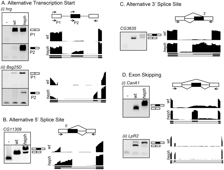
We analyzed the wild-type control and heph03429 transcriptomes for mRNA isoform and expression level differences. With respect to mRNA isoforms, differences could arise from transcription start sites, alternative 5′ splice sites, alternative 3′ splice sites, and/or exon skipping. We manually inspected over 100 candidates from sequence analysis algorithms and selected six of them based on fold difference and isoform type for analysis by RT-PCR. We observed significant isoform differences for each category: Use of alternative 5′ regions from transcription start sites was observed for hrg and Bsg25D genes such that the proximal transcription start site was used only in the heph 03429 mutant (Fig. 1A, i and ii). Alternative 5′ splicing resulted in use of a downstream 5′ splice site and thus a longer transcript for CG11309 in the heph 03429 mutant (Fig. 1B). Switching to an upstream 3′ splice site resulted in a longer transcript for CG3635 in the heph 03429 mutant (Fig. 1C). For the CanA1 transcript, an exon that was skipped in the wild-type control was included in the heph 03429 mutant (Fig. 1D, i). Finally, there was a quantitative switch in the ratio of LpR2 transcripts from the predominantly exon-skipped isoform in the wild-type control to the predominantly exon-included isoform in the heph 03429 mutant (Fig. 1D, ii). Based on our analysis, these are the most significant isoform differences in the heph 03429 mutant. It is possible that further scrutiny may reveal a few more examples of small quantitative differences in alternative transcripts in the heph 03429 mutant. Differences in poly(A) site usage were not analyzed (see the Materials and Methods section). These findings show that the loss of dmPTB function in the heph 03429 mutant resulted in significant differences in mRNA isoforms for specific transcripts.
Figure 1. mRNA isoforms misregulated in the heph 03429 mutant.
RT-PCR analysis for alternative isoform usage between the wild-type control and the heph 03429 mutant. Schematics are shown for each alternative isoform category. A. Alternative transcription start sites, B. Alternative 5′ splice site, C. Alternative 3′ splice site, and D. Exon skipping. Primers used for RT-PCR are indicated. RNA-Seq read pileups across the alternatively expressed section of each gene are shown for the wild-type (yw) control and the heph03429 mutant.
Transcriptome profiling identifies expression level differences
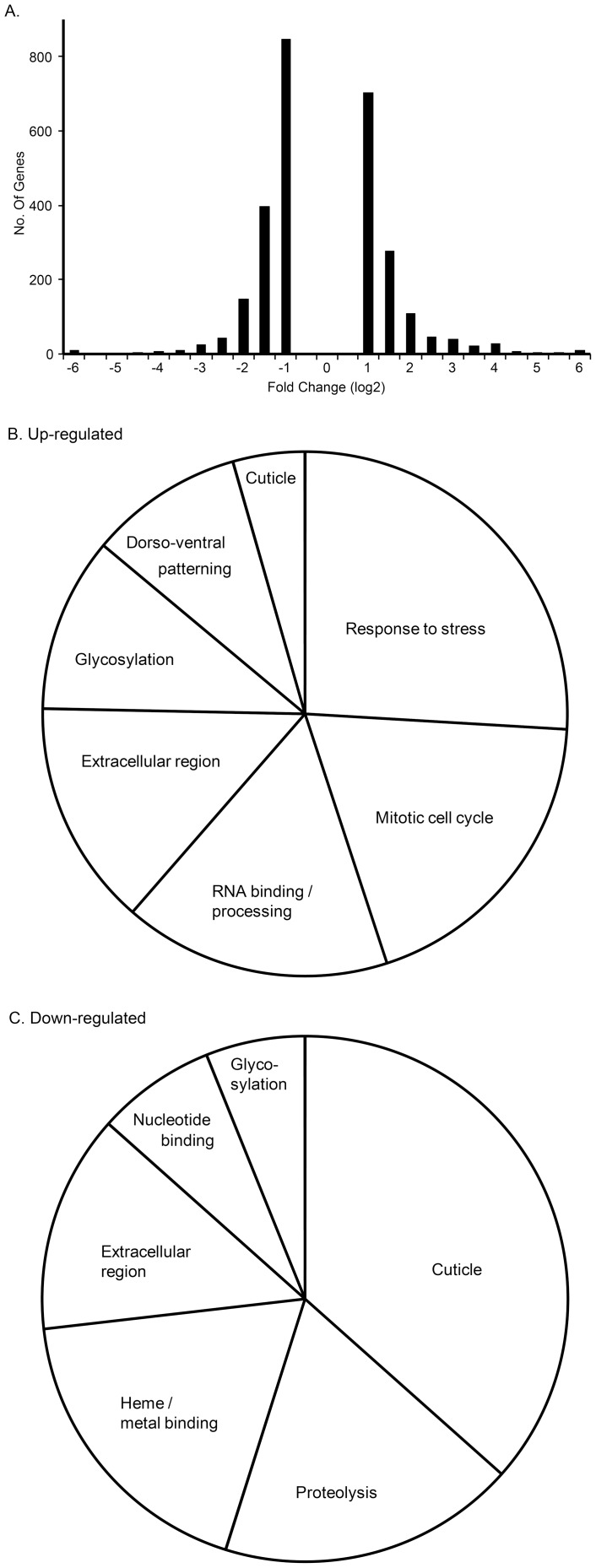
We compared expression levels for each gene between the two samples (Fig. 2A). Of the total 14,794 genes, 1161 genes showed no expression in either sample. 6419 genes were called significant by the algorithm, however, 3709 were below the two-fold cutoff. The remaining 2710 genes contained four snRNA/snoRNA gene categories and five genes had a value of zero (as denominator to preclude fold-change calculation), which were removed from further analysis. Approximately 2217 genes had a difference of 2-3 fold (Fig. 2A). There was over four-fold upregulation for 255 genes and downregulation for 229 genes. Six genes changed by 128-fold or more. These results show significant changes in gene expression of specific genes in the heph 03429 mutant.
Figure 2. Analysis of RNA-Seq differential expression between wild-type control and heph03429 mutant. A.
Histogram depicting fold-change values (above two-fold) of all genes that are significantly changed. B and C. Most overrepresented (four-fold and above up-regulated or down-regulated) gene ontology categories from the DAVID analysis are shown by their percent representation in terms of number of genes. Many genes belong to multiple categories.
Next, we asked if some functional gene ontology categories were over-represented in the differentially expressed genes. We subjected the 463 genes that showed over four-fold difference in expression levels between the wild-type control and the heph 03429 mutant to gene ontology analysis using DAVID; 20 genes were not recognized and thus ignored. We selected the top categories from the output of approximately 240 genes, including 158 in the up-regulated and 82 in the down-regulated set. Our analysis included genes relevant for RNA-binding, cell cycle, stress, signaling, structural components, and axis formation or dorso-ventral patterning (Fig. 2B). Genes relevant for cuticle synthesis were among the most down-regulated (Fig. 2C). These results are consistent with the idea that dmPTB is a potent upstream regulator because it affects numerous cellular functions.
Genes related to dorso-ventral patterning, amnioserosa development or dorsal closure, and cuticle formation are among the most misregulated
We next analyzed components of specific functional categories. Consistent with the previous observations that heph regulates Notch signaling, we found that there was a 75% increase in the Notch transcript level in the heph 03429 mutant. Components of the Notch signaling such as rumi, enhancer of split m4 and Brother of Bearded also showed an increase (Table 2). Genes in other specific functional categories were also severely misregulated in the mutant. For example, several genes that had been functionally linked to dorsal/ventral embryonic axis formation and amnioserosa development such as screw, twisted gastrulation and zerknüllt (zen) showed over 16-fold upregulation (Table 2). Many others such as Protein Z600 and spindle E have also been functionally linked to dorso-ventral patterning and showed a 19-fold and 9-fold increase, respectively [23], [24]. In addition to these up-regulated transcripts, we identified many genes that were considerably down-regulated (up to 165-fold). Many of these genes relate to cuticle development, dorsal closure, proteolysis, signal peptide and the extracellular region (Table 2). We conclude that in addition to the smaller differences observed for Notch signaling-related transcripts, many more transcripts, with much larger differences, are involved in dorso-ventral patterning, amnioserosa development and cuticle formation.
Table 2. List of selected, differentially expressed genes in the heph03429 mutant, relevant for dorso-ventral axis specification, Notch signaling and cuticle formation.
| Gene name | Fold change | Ontology |
| screw | 27 | Axis, AS |
| shrew/CG11582 | 21 | DC, AS |
| zerknüllt | 20 | D/V, AS, CU |
| Protein Z600 | 19 | D/V |
| twisted gastrulation | 16 | Axis, AS |
| spindle E | 9 | D/V |
| nullo | 9 | morph. |
| Cuticular protein 47Eb | 9 | CU |
| gastrulation-defective | 8 | D/V |
| Brother of Bearded A | 6 | Notch |
| krimper | 6 | D/V |
| squash | 5 | D.C. |
| Spook | 5 | D.C., CU |
| rumi | 5 | Notch |
| Oskar | 5 | Axis |
| zerknüllt-related | 5 | D/V, AS |
| swallow | 5 | Axis |
| Shade | 4 | D.C., CU |
| mummy | 3 | D.C. |
| e(spl) m4 (Bearded family) | 2 | Notch |
| Cuticular protein 5C | −5 | CU |
| Cuticular protein 78Cc | −5 | CU |
| Cuticular protein 65Ec | −6 | CU |
| Cuticular protein 49Ae | −10 | CU |
| Adult cuticle protein 1 | −16 | CU |
| Cuticular protein 49Ad | −165 | CU |
Genes are sorted by fold-change. Genes that are related to dorsal/ventral axis formation with less than four-fold change are not shown: tolloid, jumu, wntD, Cg25C, mmy, rl, cactin, dib, Ptp61F, Dif, Src64B, aop, vrt, ImpE1, Abl, zip, Btk29A, ena, and chic. Similarly, genes that are related to Notch signaling with smaller than four-fold change are not shown: O-fut1, bib, sens, Sca, Su(H), CG13465, Ocho, Ebi, nct, Rpd3, Tom, m4, Dl, CG8027, Bre, neur, phyl, Al, elB, malpha, gro, amx, N, Rtf1, Brd, CtBP, dsh, Hs3st-B, HLHgamma, bun, sim, and e(spl). CU = cuticle, D.C. = Dorsal Closure, axis = axis specification, D/V = dorsal/ventral axis specification, morph. = embryonic morphogenesis, AS = amnioserosa.
RT-PCR analysis confirms misregulated transcripts
We randomly selected several candidates from specific gene ontology categories for analysis by RT-PCR for their expression level differences between the wild-type and mutant embryos. Fig. 3 shows that three components of the Notch signaling (BobA, rumi, and m4) and four candidates (scw, tsg, zen, and Z600) relevant to embryonic development (dorsal closure, axis formation, and/or dorso-ventral patterning) all showed upregulation, consistent with the differences observed from high throughput sequencing. Finally, two of the candidates related to cuticle structure or formation (Cpr49Ad and Acp1) recapitulated downregulation observed from the Illumina sequencing reads. We conclude that RT-PCR and high throughput sequencing independently confirm misregulation of specific transcripts in the heph 03429 mutant.
Figure 3. Analysis of differentially expressed genes in the heph03429 mutant using RT-PCR.

Candidates relating to Notch signaling, dorso-ventral axis specification and cuticle formation were randomly picked for analysis. Three of the primer pairs tested did not work.
Misregulation of zen and its link to dorso-ventral patterning defects
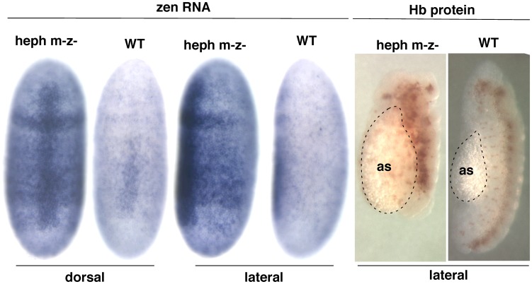
Several components of the dorso-ventral patterning, axis formation, or proper development of amnioserosa are among the highest up-regulated genes. Proper embryonic developmental process involves establishment of Toll and Dpp signaling gradient slopes in opposite directions along the dorso-ventral axis, leading to cell fate specification along this axis [25], [26]. In this context, both temporal and spatial expression patterns are the key to proper development. This important feature of the spatio-temporal expression pattern is lost during RNA isolation from homogenized embryos. Therefore, to investigate the misregulation of zen, which is functionally linked to dorso-ventral patterning [27], we analyzed the expression pattern of zen in the wild-type control and the heph 03429 mutant embryos using in situ hybridization. We found that the heph 03429 mutant (maternal and zygotic null) showed significantly higher expression of zen as compared to the control embryos (Fig. 4, left panel), which is consistent with the RT-PCR result. This is true for both dorsal and lateral views. We also analyzed these embryos for hyperplasia of the dorsal tissue (amnioserosa), which is expected as a consequence of overexpression of zen [28]. Indeed, there was evidence of hyperplasia of the dorsal tissue (Fig. 4, right panel). We conclude that the heph 03429 mutant shows increased zen expression, reflected in the hyperplasia of the dorsal tissue, and developmental defects during dorso-ventral axis formation/patterning.
Figure 4. Expression of the zen mRNA is up-regulated in the dorsal and lateral regions of embryos in the heph03429 mutant versus wild-type (WT).
Right panel: Hyperplasia of the dorsal tissue, amnioserosa, in the mutant, which is expected from increased expression of dorsal fate genes such as zen.
Discussion
Our high throughput sequencing provides the first unbiased view of the altered transcriptome in the heph03429 mutant embryos. We identify specific transcripts that are misregulated in the mutant, resulting from differences in either mRNA isoforms or mRNA levels. Transcript isoforms arise from differences at their 5′ ends, alternative 5′ splice site use, alternative 3′ splice site use, or exon skipping. These differences result in altered coding or non-coding regions of mRNAs. Also, we identify transcripts that show either significant up-regulation or down-regulation. Below we describe how these findings have contributed to our understanding of dmPTB/heph regulation and to cellular and developmental processes that are affected in the mutant.
dmPTB as a regulator of canonical and non-canonical Notch signaling
dmPTB was previously shown to function as a suppressor of Notch signaling in the context of wing development and embryogenesis. Our sequencing confirms that Notch mRNA is up-regulated (75% increase) in the mutant. We argue that this increase is functionally consequential because Notch expression is tightly regulated as a change in Notch gene dosage significantly perturbs cellular and developmental processes [29]. We show that the effect of the dmPTB regulation on Notch mRNA levels is further amplified by the known downstream components of the canonical Notch signaling pathway (rumi, enhancer of split m4 and BobA). This pathway would be most relevant to regulation in the neurogenesis in the ventral region of embryos and in cuticle development.
The heph03429 embryos manifest high levels of filamentous or F-actin protein expression in the lateral region [22]. Therefore, we asked if the actin transcript is affected. We found very small difference (<2 difference) in the expression of actin genes (Actin79B, Actin88F, and Actin42A). Since actin mRNA levels cannot explain the defects in actin processes during embryogenesis, we explored if any regulator of actin dynamics is affected. Indeed, bottleneck (26-fold), nullo (10-fold), and Sry-alpha (8-fold) are significantly over-expressed in the heph03429 mutant. These differences suggest that regulation of actin translation, stability, and/or polymerization, rather than actin mRNA levels, underlies the F-actin over-expression phenotype in heph03429 embryos.
We are particularly drawn to several other genes that are dramatically upregulated (over 20-fold): scw, tsg, zen, Z600 genes belong to cellular and developmental processes linked to dorso-ventral patterning, dorsal closure, and/or amnioserosa development. The zen transcripts show spatio-temporal expression pattern reminiscent of dorso-ventral patterning disruption and amnioserosa hyperplasia. It is overexpressed in the dorsal region of heph embryos (Fig. 4).
How does dmPTB regulate zen? It is possible that the regulation of zen and other responsive genes is mediated through Notch signaling. Both canonical Notch signaling in the nucleus and the non-canonical Notch signaling are up-regulated in heph03429 embryos [22], [30]. The canonical Notch signaling involves ligand-dependent processing, release, and translocation into the nucleus of the Notch intracellular domain (Nintra/NICD) to activate target genes [31]–[33]. Up-regulation of canonical Notch signaling is associated with the development of the anti-neurogenic phenotype [34]–[36]. The heph embryos manifest the anti-neurogenic phenotype due to increased canonical Notch signaling [22]. We believe that this anti-neurogenic phenotype is an unlikely explanation for how dmPTB could affect zen regulation because this signaling suppresses development of dorsal tissues such as the amnioserosa [22]. We propose that the observed increase in zen mRNA possibly involves up-regulation of a non-canonical Notch pathway that promotes the expression of the phosphorylated form of Cactus, the negative regulator of Dorsal activity. This non-canonical Notch signaling activity involves Protein Kinase C function and promotes the specification of dorsal cell fates and the lateral epidermis [30]. Since Dorsal is a known repressor of zen [28], it is possible that zen upregulation in the heph03429 embryos could be a consequence of Cactus-mediated suppression of Dorsal function. The involvement of Cactus up-regulation in altering mRNA expression in heph03429 embryos is further supported by a 50% reduction in the expression of Dorsal-like immunity factor (Dif) [37]. The non-canonical Notch pathway could explain how the heph03429 mutation results in zen upregulation and hyperplasia of the amnioserosa, affecting the lateral regions of the embryo.
dmPTB affects many processes during embryogenesis
Intriguingly, genes relevant to cuticle formation were found to be the most down-regulated genes in the heph03429 mutant. It is possible that the effect on cuticle genes/components is a reflection of excess canonical and non-canonical Notch signaling that disrupts the development of the larval cuticle during embryogenesis. Unfortunately, the phenotype of the heph − N − zygotic double mutant is a complex mosaic of neurogenic and anti-neurogenic phenotypes [22]. Therefore, it remains possible that some genes misregulated in the heph03429 mutants are independent of the canonical and non-canonical Notch signaling mechanisms. Since there are changes in many components of the Wingless/Wnt pathway, for example WntD (Wnt inhibitor of Dorsal), it is possible that heph affects Wnt functions in addition to Notch functions. The Wingless/Wnt pathway is also involved in many aspects of the patterning of the developing larval epidermis [38]. The specific candidate genes identified in our analysis provide an important molecular handle to distinguish between these possibilities using appropriate genetic backgrounds. For example, analyzing the candidate genes in Notch and wingless mutant backgrounds might identify dmPTB-regulated genes specific to the Notch signaling pathways or the Wingless signaling pathway.
Our expression profiling between the wild-type and the mutant embryos clearly shows that there are additional gene ontology categories that are affected, including genes relevant to stress, gene regulation, and cell cycle. While the biological significance of all of these differences remain unclear, the effect on specific gene ontology categories likely holds clues to the role of dmPTB in various aspects (cell-type, tissue-specific, temporal, and spatial) of embryonic development (Fig. 2B).
Regulatory mechanisms
The differences in mRNA levels, discussed above, could result from the effect on transcription/synthesis, mRNA degradation, or both. We note that some of the alternative transcripts could also be subject to degradation by the nonsense-mediated decay machinery. Although the effect of an RNA-binding protein directly on alternative transcription start sites (for hrg relevant for mRNA polyadenylation and Blastoderm-specific gene 25D or Bsg25D) is difficult to visualize mechanistically [39], it may involve a mechanism similar to how the human immunodeficiency virus protein Tat binds to TAR sequence in nascent mRNAs and activates transcription [40]. As an RNA-binding protein, dmPTB could regulate splicing/processing of CG11309 (function unknown), CG3635 (function unknown), CanA1 (possibly involved in calmodulin activation of calcineurin), and LpR2 (low-density lipoprotein receptor activity with potential roles in calcium ion binding and neuron projection morphogenesis) either directly or indirectly by competing for the binding of another regulatory protein, leading to alterations in the coding or non-coding regions of these transcripts. Some of these mechanisms may be similar to how the mammalian PTB regulates aspects of mRNA processing. We note that identification of downstream targets by searching for potential binding sites alone is challenging because PTB binds short, degenerate sequences UCUU and UUCU [3]–[7] that are found frequently throughout the genome. In the future, a cross-linking/immunoprecipitation experiment that detects RNA-protein interactions on a genome-wide level [41], [42] needs to be combined with our high throughput sequencing dataset to identify the subset of candidates that directly bind dmPTB.
In summary, our combined results indicate that dmPTB acts as a potent upstream regulator that specifically controls several downstream target genes and processes during embryogenesis. Many genes or transcripts altered in the heph03429 mutant (Table 2) have known functional connections to specific cellular and developmental processes during embryonic development, such as neurogenesis, dorso-ventral axis formation, and dorsal closure. For others, such as down-regulation of cuticular proteins and mRNA processing defects (expected from mutation of an RNA-binding protein), functional relevance is not obvious at this stage. Genes identified in this study provide entry points and new avenues for future studies on mechanisms and biological functions as to how an RNA-binding protein controls gene expression and embryonic development.
Materials and Methods
Fly strains and culture
The yellow white (yw) stock was obtained from the Bloomington Stock Center. FRT82B heph 03429/TM6B placz was obtained from Anne Ephrussi [15]. The procedures followed for generating maternal and zygotic heph 03429 nulls are described in Besse et al., 2009. Flies were raised on standard cornmeal food at 17–25°C [43].
RNA-Seq for gene and isoform differential expression analysis
Poly(A)+ RNA from 1 microgram total RNA was obtained from the heph03429 mutant and wild-type control (yw) embryos, and mRNA-seq sequencing libraries (standard Illumina TruSeq) were prepared according to manufacturer's instructions (Illumina, Inc., San Diego CA, USA). The heph03429 mutant and wild-type control libraries were sequenced on an Illumina HiSeq 2000 sequencer (singleton 100 basepair reads). TopHat 2.0.9 was used to map reads to the Flybase Drosophila melanogaster r5.44 (www.flybase.org) genome annotation (GTF and Fasta files), with the –no-novel-juncs and –microexon-search parameters (Table 1) [44]. Traces were generated using the UCSC genome browser/tools. Cufflinks version 2.1.1 (cuffdiff with the Illumina iGenomes DBGP5.25 annotation) was used to determine differential gene expression [45]. The MISO v0.4.6 [46] software package (with an isoform fraction difference threshold of 0.2 and a minimum Bayes Factor of 10) and manual inspection were used to identify differential isoform usage. Using the Drosophila annotation provided for MISO (9/19/2011), we analyzed alternative 5′ and 3′ splice sites, skipped exons, retained introns, and mutually exclusive exons (alternative first and last exons were not provided in this annotation).
Gene Ontology analysis
The Database for Annotation, Visualization and Integrated Discovery (DAVID) v6.7 was used for gene ontology analysis online (using a differential expression threshold of four-fold). The pie charts (in Figure 2) represent the genes in only the most overrepresented ontology categories.
In situ hybridization
The in situ protein and RNA labeling procedures followed have been described in [47] and [34]. RNA signals were detected using an Alkaline Phosphatase detection system. Protein signals were detected using a Horse Radish Peroxidase system. Embryos (0–24 hour) were collected and stage-specific embryos were sorted based on anatomy after the hybridization or antibody procedure.
Reverse-transcription-polymerase chain reaction
Primer pairs used for RT-PCR are provided in Figure S1.
Supporting Information
The site of P element insertion and primer sequences are provided.
(PDF)
Acknowledgments
We thank Dr. Brian DeDecker and Abhisaar Yadav for critical reading of the manuscript. The GEO accession number for the sequencing dataset is GSE57517.
Data Availability
The authors confirm that all data underlying the findings are freely available without restriction. The GEO accession number for the sequencing dataset is GSE57517.
Funding Statement
The Butcher Foundation award, Departmental funds (R.S.), and National Institutes of Health - HD062928 (CSW). Article processing charge was funded by the University of Colorado Boulder Libraries Open Access Fund. The funders had no role in study design, data collection and analysis, decision to publish, or preparation of the manuscript.
References
- 1. Krecic AM, Swanson MS (1999) hnRNP complexes: composition, structure, and function. Curr Opin Cell Biol 11: 363–371. [DOI] [PubMed] [Google Scholar]
- 2. Valcarcel J, Gebauer F (1997) Post-transcriptional regulation: the dawn of PTB. Curr Biol 7: R705–R708. [DOI] [PubMed] [Google Scholar]
- 3. Clerte C, Hall KB (2006) Characterization of multimeric complexes formed by the human PTB1 protein on RNA. RNA 12: 457–475. [DOI] [PMC free article] [PubMed] [Google Scholar]
- 4. Lin CH, Patton JG (1995) Regulation of alternative 3' splice site selection by constitutive splicing factors. RNA 1: 234–245. [PMC free article] [PubMed] [Google Scholar]
- 5. Perez I, Lin CH, McAfee JG, Patton JG (1997) Mutation of PTB binding sites causes misregulation of alternative 3' splice site selection in vivo. RNA 3: 764–778. [PMC free article] [PubMed] [Google Scholar]
- 6. Robida M, Sridharan V, Morgan S, Rao T, Singh R (2010) Drosophila polypyrimidine tract-binding protein is necessary for spermatid individualization. Proc Natl Acad Sci U S A doi:10.1073/pnas.1007935107 [DOI] [PMC free article] [PubMed] [Google Scholar]
- 7. Singh R, Valcarcel J, Green MR (1995) Distinct binding specificities and functions of higher eukaryotic polypyrimidine tract-binding proteins. Science 268: 1173–1176. [DOI] [PubMed] [Google Scholar]
- 8. Kafasla P, Mickleburgh I, Llorian M, Coelho M, Gooding C, et al. (2012) Defining the roles and interactions of PTB. Biochemical Society transactions 40: 815–820. [DOI] [PubMed] [Google Scholar]
- 9. Sawicka K, Bushell M, Spriggs KA, Willis AE (2008) Polypyrimidine-tract-binding protein: a multifunctional RNA-binding protein. Biochemical Society transactions 36: 641–647. [DOI] [PubMed] [Google Scholar]
- 10. Wagner EJ, Garcia-Blanco MA (2001) Polypyrimidine tract binding protein antagonizes exon definition. Mol Cell Biol 21: 3281–3288. [DOI] [PMC free article] [PubMed] [Google Scholar]
- 11. Coutinho-Mansfield GC, Xue Y, Zhang Y, Fu XD (2007) PTB/nPTB switch: a post-transcriptional mechanism for programming neuronal differentiation. Genes & development 21: 1573–1577. [DOI] [PubMed] [Google Scholar]
- 12. Dansereau DA, Lunke MD, Finkielsztein A, Russell MA, Brook WJ (2002) Hephaestus encodes a polypyrimidine tract binding protein that regulates Notch signalling during wing development in Drosophila melanogaster. Development 129: 5553–5566. [DOI] [PubMed] [Google Scholar]
- 13. Davis MB, Sun W, Standiford DM (2002) Lineage-specific expression of polypyrimidine tract binding protein (PTB) in Drosophila embryos. Mech Dev 111: 143–147. [DOI] [PubMed] [Google Scholar]
- 14. Robida MD, Singh R (2003) Drosophila polypyrimidine-tract binding protein (PTB) functions specifically in the male germline. EMBO J 22: 2924–2933. [DOI] [PMC free article] [PubMed] [Google Scholar]
- 15. Besse F, Lopez de Quinto S, Marchand V, Trucco A, Ephrussi A (2009) Drosophila PTB promotes formation of high-order RNP particles and represses oskar translation. Genes & development 23: 195–207. [DOI] [PMC free article] [PubMed] [Google Scholar]
- 16. McDermott SM, Davis I (2013) Drosophila hephaestus/polypyrimidine tract binding protein is required for dorso-ventral patterning and regulation of signalling between the germline and soma. PLoS One 8: e69978. [DOI] [PMC free article] [PubMed] [Google Scholar]
- 17. Cruz C, Glavic A, Casado M, de Celis JF (2009) A gain-of-function screen identifying genes required for growth and pattern formation of the Drosophila melanogaster wing. Genetics 183: 1005–1026. [DOI] [PMC free article] [PubMed] [Google Scholar]
- 18. Kankel MW, Hurlbut GD, Upadhyay G, Yajnik V, Yedvobnick B, et al. (2007) Investigating the genetic circuitry of mastermind in Drosophila, a notch signal effector. Genetics 177: 2493–2505. [DOI] [PMC free article] [PubMed] [Google Scholar]
- 19. Norga KK, Gurganus MC, Dilda CL, Yamamoto A, Lyman RF, et al. (2003) Quantitative analysis of bristle number in Drosophila mutants identifies genes involved in neural development. Curr Biol 13: 1388–1396. [DOI] [PubMed] [Google Scholar]
- 20. Shepherd A, Wesley U, Wesley C (2010) Notch and delta mRNAs in early-stage and mid-stage drosophila embryos exhibit complementary patterns of protein-producing potentials. Dev Dyn 239: 1220–1233. [DOI] [PMC free article] [PubMed] [Google Scholar]
- 21. Shepherd AK, Singh R, Wesley CS (2009) Notch mRNA expression in Drosophila embryos is negatively regulated at the level of mRNA 3' processing. PLoS One 4: e8063. [DOI] [PMC free article] [PubMed] [Google Scholar]
- 22. Wesley CS, Guo H, Chaudhry KA, Thali MJ, Yin JC, et al. (2011) Loss of PTB or negative regulation of Notch mRNA reveals distinct zones of Notch and actin protein accumulation in Drosophila embryo. PLoS One 6: e21876. [DOI] [PMC free article] [PubMed] [Google Scholar]
- 23. Gonzalez-Reyes A, Elliott H, St Johnston D (1997) Oocyte determination and the origin of polarity in Drosophila: the role of the spindle genes. Development 124: 4927–4937. [DOI] [PubMed] [Google Scholar]
- 24. Schulz RA, Miksch JL (1989) Dorsal expression of the Drosophila z600 gene during early embryogenesis. Developmental biology 136: 211–221. [DOI] [PubMed] [Google Scholar]
- 25. Ferguson EL, Anderson KV (1992) Decapentaplegic acts as a morphogen to organize dorsal-ventral pattern in the Drosophila embryo. Cell 71: 451–461. [DOI] [PubMed] [Google Scholar]
- 26. Moussian B, Roth S (2005) Dorsoventral axis formation in the Drosophila embryo–shaping and transducing a morphogen gradient. Curr Biol 15: R887–899. [DOI] [PubMed] [Google Scholar]
- 27. Rushlow C, Frasch M, Doyle H, Levine M (1987) Maternal regulation of zerknullt: a homoeobox gene controlling differentiation of dorsal tissues in Drosophila. Nature 330: 583–586. [DOI] [PubMed] [Google Scholar]
- 28. Ratnaparkhi GS, Jia S, Courey AJ (2006) Uncoupling dorsal-mediated activation from dorsal-mediated repression in the Drosophila embryo. Development 133: 4409–4414. [DOI] [PubMed] [Google Scholar]
- 29. Heitzler P, Simpson P (1991) The choice of cell fate in the epidermis of Drosophila. Cell 64: 1083–1092. [DOI] [PubMed] [Google Scholar]
- 30. Tremmel DM, Resad S, Little CJ, Wesley CS (2013) Notch and PKC are involved in formation of the lateral region of the dorso-ventral axis in Drosophila embryos. PLoS One 8: e67789. [DOI] [PMC free article] [PubMed] [Google Scholar]
- 31. Artavanis-Tsakonas S, Rand MD, Lake RJ (1999) Notch signaling: cell fate control and signal integration in development. Science 284: 770–776. [DOI] [PubMed] [Google Scholar]
- 32. Bray SJ (2006) Notch signalling: a simple pathway becomes complex. Nature reviews 7: 678–689. [DOI] [PubMed] [Google Scholar]
- 33. Tien AC, Rajan A, Bellen HJ (2009) A Notch updated. The Journal of cell biology 184: 621–629. [DOI] [PMC free article] [PubMed] [Google Scholar]
- 34. Lieber T, Kidd S, Alcamo E, Corbin V, Young MW (1993) Antineurogenic phenotypes induced by truncated Notch proteins indicate a role in signal transduction and may point to a novel function for Notch in nuclei. Genes & development 7: 1949–1965. [DOI] [PubMed] [Google Scholar]
- 35. Struhl G, Adachi A (1998) Nuclear access and action of notch in vivo. Cell 93: 649–660. [DOI] [PubMed] [Google Scholar]
- 36. Struhl G, Fitzgerald K, Greenwald I (1993) Intrinsic activity of the Lin-12 and Notch intracellular domains in vivo. Cell 74: 331–345. [DOI] [PubMed] [Google Scholar]
- 37. Stein D, Goltz JS, Jurcsak J, Stevens L (1998) The Dorsal-related immunity factor (Dif) can define the dorsal-ventral axis of polarity in the Drosophila embryo. Development 125: 2159–2169. [DOI] [PubMed] [Google Scholar]
- 38. Bejsovec A (2013) Wingless/Wnt signaling in Drosophila: The pattern and the pathway. Molecular reproduction and development 80: 882–894. [DOI] [PMC free article] [PubMed] [Google Scholar]
- 39. Rustighi A, Tessari MA, Vascotto F, Sgarra R, Giancotti V, et al. (2002) A polypyrimidine/polypurine tract within the Hmga2 minimal promoter: a common feature of many growth-related genes. Biochemistry 41: 1229–1240. [DOI] [PubMed] [Google Scholar]
- 40. Dingwall C, Ernberg I, Gait MJ, Green SM, Heaphy S, et al. (1990) HIV-1 tat protein stimulates transcription by binding to a U-rich bulge in the stem of the TAR RNA structure. Embo J 9: 4145–4153. [DOI] [PMC free article] [PubMed] [Google Scholar]
- 41. Ule J, Jensen KB, Ruggiu M, Mele A, Ule A, et al. (2003) CLIP identifies Nova-regulated RNA networks in the brain. Science 302: 1212–1215. [DOI] [PubMed] [Google Scholar]
- 42. Xue Y, Zhou Y, Wu T, Zhu T, Ji X, et al. (2009) Genome-wide analysis of PTB-RNA interactions reveals a strategy used by the general splicing repressor to modulate exon inclusion or skipping. Mol Cell 36: 996–1006. [DOI] [PMC free article] [PubMed] [Google Scholar]
- 43.Ashburner M (1989) Drosophila. New York: Cold Spring Harbor Laboratory Press. pp. 2v.
- 44. Kim D, Pertea G, Trapnell C, Pimentel H, Kelley R, et al. (2013) TopHat2: accurate alignment of transcriptomes in the presence of insertions, deletions and gene fusions. Genome biology 14: R36. [DOI] [PMC free article] [PubMed] [Google Scholar]
- 45. Roberts A, Trapnell C, Donaghey J, Rinn JL, Pachter L (2011) Improving RNA-Seq expression estimates by correcting for fragment bias. Genome biology 12: R22. [DOI] [PMC free article] [PubMed] [Google Scholar]
- 46. Katz Y, Wang ET, Airoldi EM, Burge CB (2010) Analysis and design of RNA sequencing experiments for identifying isoform regulation. Nature methods 7: 1009–1015. [DOI] [PMC free article] [PubMed] [Google Scholar]
- 47. Corbin V, Michelson AM, Abmayr SM, Neel V, Alcamo E, et al. (1991) A role for the Drosophila neurogenic genes in mesoderm differentiation. Cell 67: 311–323. [DOI] [PubMed] [Google Scholar]
Associated Data
This section collects any data citations, data availability statements, or supplementary materials included in this article.
Supplementary Materials
The site of P element insertion and primer sequences are provided.
(PDF)
Data Availability Statement
The authors confirm that all data underlying the findings are freely available without restriction. The GEO accession number for the sequencing dataset is GSE57517.


