Abstract
The phylogenetic relationships within the Stellifer group of weakfishes (Stellifer, Odontoscion, Ophioscion, and Bairdiella) were evaluated using 2723 base pairs comprising sequences of nuclear (rhodopsin, TMO-4C4, RAG-1) and mitochondrial (16S rRNA and COI) markers obtained from specimens of nine species. Our results indicate a close relationship between Bairdiella and Odontoscion, and also that the genus Stellifer is not monophyletic, but rather that it consists of two distinct lineages, one clade containing S. microps/S. naso/S. brasiliensis and the other, S. rastrifer/S. stellifer/Stellifer sp. B, which is closer to Ophioscion than the former clade. The O. punctatissimus populations from the northern and southern Brazilian coast were also highly divergent in both nuclear (0.8% for rhodopsin and 0.9% for RAG-1) and mitochondrial sequences (2.2% for 16S rRNA and 7.3% for COI), which we conclude is consistent with the presence of two distinct species. The morphological similarities of the members of the Stellifer group is reinforced by the molecular data from both the present study and previous analyses, which have questioned the taxonomic status of the Stellifer group. If, on the one hand, the group is in fact composed of four genera (Stellifer, Ophioscion, Odontoscion, and Bairdiella), one of the two Stellifer clades should be reclassified as a new genus. However, if the close relationship and the reduced genetic divergence found within the group is confirmed in a more extensive study, including representatives of additional taxa, this, together with the morphological evidence, would support downgrading the whole group to a single genus. Obviously, these contradictory findings reinforce the need for a more systematic taxonomic revision of the Stellifer group as a whole.
Introduction
The family Sciaenidae includes approximately 70 genera and 270 species of demersal fishes found mainly over muddy or sandy bottoms of the continental shelf of the Atlantic, Indian, and Pacific oceans, as well as freshwater genera in the rivers of the Old and New Worlds [1], [2]. In the western South Atlantic, sciaenids are abundant and highly diverse, encompassing approximately 50 species representing 19 genera [3], [4].
Chao [5] evaluated the phylogenetic relationships of the 21 western Atlantic sciaenid genera and two freshwater genera based on morphological traits, and identified 11 suprageneric groups: Micropogonias, Nebris, Pogonias, Sciaenops, Larimus, Sciaena, Umbrina, Menticirrhus, Lonchurus, Cynoscion, and Stellifer. Of these groups, Stellifer can be distinguished from all the others by the presence of two (rather than one) pairs of large otoliths and a swim bladder with two (rather than one) chambers.
The Stellifer group includes four genera – Stellifer, Ophioscion, Bairdiella, and Odontoscion – represented by 12 species in the western South Atlantic: Stellifer naso, S. griseus, S. venezuelae, S. brasiliensis, S. microps, S. rastrifer, S. stellifer, Stellifer. sp. A, Stellifer. sp. B, Odontoscion dentex, Ophioscion punctatissimus, and Bairdiella ronchus [5]. These species are characterized by a very strong second anal spine, two pairs of large otoliths, and a swim bladder with two chambers, a carrot-shaped posterior chamber, and the anterior one yoke-shaped with a pair of diverticula on the posterolateral surface [4], [5].
Species of the Stellifer group are widely distributed in the western Atlantic, where they are abundant in coastal and estuarine waters with sandy or muddy bottoms [6], [7], including the coast of Brazil [8]–[15]. This group is especially appropriate for studies of the genetic connectivity of populations because the species are widely distributed, and normally inhabit estuarine environments. Despite this, few studies have focused on the bio-ecological or phylogenetic characteristics of this group. Regarding the phylogenetic relationships, all the available studies [1], [5], [16], [17] have emphasized the close relationships among Bairdiella, Stellifer, Ophioscion, and Odontoscion, although intergeneric and interspecific relationships have yet to be defined conclusively due to the limitations or inconsistencies found in the data, as described below.
The first phylogeny based on morphological traits was proposed by Chao [5], who concluded that Stellifer is most closely related to Ophioscion, with Bairdiella appearing as a sister group to Odontoscion. In a subsequent morphological study, Sasaki [1] suggested that Ophioscion and Stellifer are sister groups which form a clade with Bairdiella, whereas Odontoscion is related to the sciaenids of the eastern Pacific, Elattarchus and Corvula.
In a phylogenetic study based on 16S rRNA sequences, Vinson et al. [16] confirmed the close relationship between Stellifer and Bairdiella, although they did not include Ophioscion or Odontoscion in their analyses, impeding the systematic assessment of the evolutionary relationships within the group. In a recent study based on both mitochondrial (COI and 16S rRNA) and nuclear markers (TMO-4C4), Santos et al. [17] concluded that Stellifer is a sister group of Ophioscion and that Bairdiella is the basal taxon within the group, confirming the proposal of Sasaki [1]. However, as in Vinson et al. [16], the relationships between all of the taxa of the Stellifer group could not be defined because Odontoscion was not included in the analyses. Additionally, the relationships among the Stellifer species remain unclear, given that, in Vinson et al. [16], S. microps is a sister group to S. naso and S. rastrifer is closely related to S. stellifer, whereas in Santos et al. [17], S. rastrifer is a sister group to Stellifer sp., and S. stellifer is more closely related to O. punctatissimus.
In addition to the divergences in the conclusions of the morphological studies regarding the intergeneric relationships within Stellifer group, then, there are also disagreements among molecular phylogenies, especially with regard to the relationships among the Stellifer species. Given this, the present study evaluates the phylogenetic relationships within the Stellifer group, including all of its genera, using nuclear (TMO-4C4, RAG-1, and rhodopsin) and mitochondrial (16S rRNA and COI) markers, all of which have been widely used in phylogenetic reconstructions of fish taxa [17]–[27].
Materials and Methods
Ethics Statement
The species analyzed in the present study are not endangered or protected in the regions from which samples were obtained. The specimens were captured by artisanal fishers and processed (collection, handling, transportation, and DNA extraction) with the authorization of the Brazilian Environment Ministry through permit number 12773–1 emitted in the name of Dr. Iracilda Sampaio. All work was performed in compliance with and approved by the Ethics Committee of the Federal University of Pará.
Sampling
A total of 36 samples representing nine species of the four genera of the Stellifer group distributed in the western South Atlantic were collected along the Brazilian coast (Table 1). Most of the specimens were obtained from the Sciaenidae tissue bank of the UFPA Genetics and Molecular Biology Laboratory of the Institute of Coastal Studies in Bragança, Brazil. The species were identified using the specialized literature [5], and muscle tissue was extracted from each specimen and conserved in absolute ethanol and frozen until analysis in the laboratory.
Table 1. Species and genomic regions used in the present study, including the samples used as outgroups.
GenBank accession numbers are listed. N is the number of individuals used, and the Brazilian state of origin is the site where the samples were collected.
DNA Extraction, PCR, and Genomic Sequencing
Total DNA was extracted by using the Wizard genomic DNA purification kit (Promega, Madison, Wisconsin, USA) following the protocol for extraction from muscle tissue as defined by the manufacturer. To evaluate the quality of the DNA, samples were electrophoresed in 1% agarose gel stained with GelRed (Biotium Inc., Hayward, California, USA) and analyzed under a UV transilluminator.
The mitochondrial (16S rRNA and COI) and nuclear (TMO-4C4, RAG-1, and rhodopsin) regions were amplified by PCR using the primers and amplification cycles described in Table 2. The RAG-1 region was amplified using a nested PCR, in which the primers 2510F [20] and RAG1R1 [32] were used first, followed by a second amplification using the primers RAG1F1 and RAG1R2 [32]. The reactions were conducted in a final volume of 25 µl, containing 4 µl of dNTPs (1.25 mM), 2.5 µl of PCR buffer (10X), 1 µl of MgCl2 (50 mM), 1 µl of DNA (100 ng/µl), 1 µl of each primer (50 ng/µl), 0.2 µl of Taq DNA Polymerase (5 U/µL, Invitrogen, Carlsbad, California, USA), and sterile water to complete the final volume. The PCR products were run on an agarose gel (1%) stained with GelRed (Biotium Inc., Hayward, California, USA) to verify the quality of the amplification products under ultraviolet light.
Table 2. Primers and amplification protocols for the mitochondrial and nuclear markers.
| Marker | Primer | Reference | Amplification protocol |
| 16S rRNA | L1987: 5′ GCCTCGCCTGTTTACCAAAAAC 3′ | Modified from Palumbi [28] | Initial denaturation at 94°C for 3′; 30 cycles at 94°C for 20″(denaturation), 50°C for 30″(annealing), and 72°C for 30″; and final extension at 72°C for 3′ |
| H2609: 5′ CCGGTCTGAACTCAGATCACGT 3′ | |||
| COI | FishF1: 5′ TCAACCAACCACAAAGACATTGGCAC 3″ | [29] | Initial denaturation at 94°C for 3′; 30 cycles at 94°C for 40″(denaturation), 59°C for 30″(annealing), and 72°C for 30″; and final extension at 72°C for 7′ |
| FishR1: 5′ TAGACTTCTGGGTGGCCAAAGAATCA 3′ | |||
| TMO-4C4 | F2: 5′ CGGCCTTCCTAAAACCTCTCATTAAG 3′ | [30] | Initial denaturation at 95°C for 2′; followed by 35 cycles at 95°C for 30″ (denaturation), 60°C for 30″(annealing), and 72°C for 1′; and final extension at 72°C for 7′ |
| R2: 5′ GTGCTCCTGGGTGACAAAGTCTACAG 3′ | |||
| Rhodopsin | Rod-F2 W: 5′ AGCAACTTCCGCTTCGGTGAGAA 3′ | [31] | Initial denaturation at 95°C for 7′; 40 cycles at 94°C for 30″(denaturation), 59°C for 30″(annealing), and 72°C for 30″; and final extension at 72°C for 7′ |
| Rod-4R: 5′ CTGCTTGTTCATGCAGATGTAGAT 3′ | |||
| RAG-1 | 2510 L: 5′ TGGCCATCCGGGTMAACAC 3′ | [20], [32] | Initial denaturation at 94°C for 3′; followed by 40 cycles at 94°C for 30″(denaturation), 58°C for 45″(annealing), and 72°C for 45″; and final extension at 72°C for 10′ |
| RAG1R1: 5′ CTGAGTCCTTGTGAGCTTCCATRAAYTT 3′ | |||
| RAG-1 | RAG1F1: 5′ CTGAGCTGCAGTCAGTACCATAAGATGT 3′ | [32] | Initial denaturation at 94°C for 3′; followed by 40 cycles at 94°C for 30″ (denaturation), 58°C for 45″ (annealing), and 72°C for 45″; and final extension at 72°C for 10′ |
| RAG1R2: 5′ TGAGCCTCCATGAACTTCTGAAGRTAYTT 3′ |
The positive PCR products were purified with ExoSAP-IT (Affymetrix, Cleveland, Ohio, USA) following the manufacturer's instructions, and sequenced by the di-deoxyterminal method with reagents from the BigDye Terminator v3.1 Cycle Sequencing kit (Applied Biosystems, Foster City, California, USA). Electrophoresis was conducted in an ABI 3500XL automatic sequencer (Applied Biosystems).
Phylogenetic and Nucleotide Divergence Analyses
The sequences obtained were manually edited, and aligned using the CLUSTAL W algorithm [33] implemented in the BioEdit 7.2.5 program [34]. Some of the 16S rRNA and TMO-4C4 sequences included in the analysis were obtained from GenBank (see Table 1). Nucleotide saturation of each set of data was evaluated by plotting transitions and transversions against genetic distances in DAMBE 4.0.65 [35].
Phylogenetic relationships were reconstructed based on both the individual data sets (per gene) and the concatenated data, using maximum parsimony, maximum likelihood, and normal and hierarchical Bayesian inference approaches. Two species of the family Lutjanidae, Ocyurus chrysurus and Lutjanus purpureus, the probable sister group of the Sciaenidae, were used as the outgroups for all analyses (Table 1). The evolutionary models used in the phylogenetic reconstructions were obtained in jModeltest 0.1.1 [36]. The maximum parsimony analysis was run using a heuristic search with 1,000 random step-wise additions, using the subtree pruning-regrafting (SPR) algorithm with branch-swapping in PAUP* 4.0b10 [37]. The maximum likelihood tree was constructed in PhyML v3.0 [38] using a heuristic search to find the most probable topologies based on the substitution models TIM2ef+I+G (for 16S rRNA), TIM2+I+G (COI), K80+I (TMO-4C4), TIM1+G (rhodopsin), and TrNef+I+G (RAG-1), and, TPM1uf+I+G for the concatenated data set. Statistical support for the maximum parsimony and likelihood analyses was determined using 1,000 bootstrap pseudoreplicates [39].
Bayesian inference analyses were run in MrBayes 3.1.2 [40] using the evolutionary models TPM2+G (for 16S rRNA), TrN+I+G (COI), K80+I (TMO-4C4), TPM1+G (rhodopsin), and K80+I+G (RAG-1). Metropolis-coupled Markov chain Monte Carlo (MCMCMC) sampling was conducted with two independent runs of 3,000,000 generations to estimate the posterior probabilities of the observed clades, using the parameters defined by the models as starting values. The Bayesian posterior probabilities for the clades were determined using the 50% consensus rule for trees sampled every 20 generations after removing the trees produced before the chains became stationary. The burn-in was empirically defined by evaluating the likelihood values. Convergence of the data was evaluated by verifying the parameters throughout the generations in Tracer 1.5 [41].
A species tree was constructed according to the hierarchical Bayesian inference principle in the BEAST 1.7.4 software package [42]. In this analysis, one tree was defined a priori, and each species of the group was considered to be a valid taxon. Markov chain Monte Carlo (MCMC) sampling was performed for 450 million generations with parameters sampled every 1,000 generations, and an initial burn-in of 10%. Convergence of the parameters was evaluated in Tracer 1.5 [41]. All of the trees obtained were viewed and edited in FigTree 1.4.0 [43].
Nucleotide divergence within and among the lineages for each set of data were assessed using uncorrected p distances in the MEGA 5.2.2 program [44].
Results
A total of 2723 base pairs, including 432 bps for rhodopsin, 401 bps for TMO-4C4, and 752 bps for RAG-1, as well as 508 bps for the mitochondrial 16S rRNA and 630 bps for the COI were obtained from 26 of the 36 specimens analyzed. None of the markers was saturated (data not shown). The complete database of both nuclear and mitochondrial sequences includes 549 sites that are informative for parsimony analysis, with an overall transition/transversion ratio of 3.6.
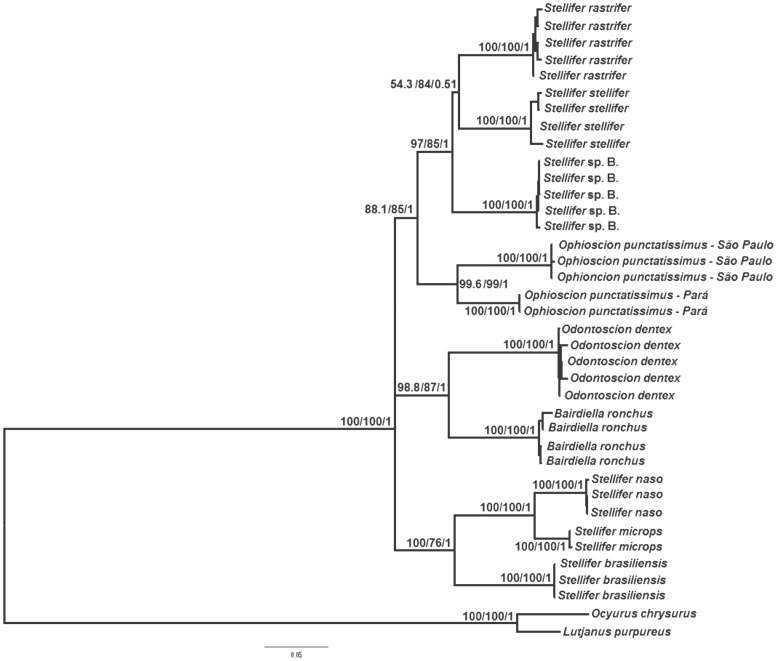
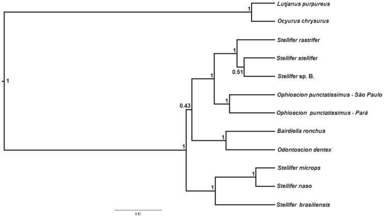
As the maximum parsimony, maximum likelihood, and Bayesian inference trees all presented similar topologies, only the maximum likelihood tree is shown here (Figure 1). The principal difference among the trees was in the position of S. stellifer, which grouped with Stellifer sp. B in the Bayesian species tree (Figure 2), but is the sister group of S. rastrifer in the other trees (Figure 1). In both cases, however, the statistical support is weak. All the results suggest the monophyly of the Stellifer group, with significant bootstrap and posterior probability values (Figures 1 and 2). However, it was not possible to determine which of the group's lineages is basal because all three approaches produced a polytomous arrangement (Figures 1 and 2).
Figure 1. Maximum likelihood tree for the Stellifer group, based on mitochondrial (COI and 16S rRNA) and nuclear DNA sequences (rhodopsin, TMO-4C4, and RAG-1).
The numbers above the branches represent the bootstrap values for maximum likelihood and maximum parsimony, and posterior Bayesian probabilities, respectively.
Figure 2. Species tree of the Stellifer group constructed from sequences of mitochondrial (COI and 16S rRNA) and nuclear DNA (rhodopsin, TMO-4C4, and RAG-1).
The numbers above the branches indicate the posterior probabilities for the respective clade.
The close relationship between Bairdiella and Odontoscion was well supported in all of the analyses (Figures 1 and 2). Our results also suggest that the genus Stellifer is not monophyletic because the species S. rastrifer, S. stellifer, and Stellifer sp. B form a clade closely related to Ophioscion, with significant statistical support (Figures 1 and 2), whereas S. microps, S. naso, and S. brasiliensis form a distinct clade, which is also strongly supported by bootstrap and posterior probability values (Figures 1 and 2).
Regarding the interspecific relationships within genus Stellifer, S. naso is a sister group to S. microps, composing a clade along with S. brasiliensis (Figures 1 and 2). In the second clade containing the other species of Stellifer, the low bootstrap and posterior probability values did not allow a reliable definition of the evolutionary relationships among Stellifer sp. B, S. rastrifer and S. stellifer (Figures 1 and 2).
All the analyses supported the separation of the northern (Pará) and southern (São Paulo) lineages of O. punctatissimus, based on high bootstrap and posterior probability values (Figures 1 and 2).
Discussion
This is the first molecular phylogeny that includes species representative of all four genera of the Stellifer group, as proposed by Chao [5]. The results of all of the analyses suggest the monophyly of the group (Figures 1 and 2), and are consistent with those of morphological analyses [5] and a molecular study of 17 sciaenid genera, including those of the Stellifer group [17]. However, as the Sciaenidae is a large family that includes some 70 genera, further analyses including the Stellifer group and other closely-related sciaenids, will be necessary for a more conclusive evaluation of the group's monophyletic status.
Bairdiella is a sister group to Odontoscion in all the topologies generated in the present study (Figures 1 and 2), which corroborate Chao's [5] arrangement, based on morphological traits. By contrast, the findings of Sasaki [1] indicate that Stellifer/Ophioscion/Bairdiella share a common ancestor, whereas Odontoscion would be more closely related to the eastern Pacific Ellatarchus and Corvulla. These results contrast with those obtained in the present study and the phylogenies determined by Chao [5] and Santos et al. [17]. However, Ellatarchus and Corvulla were not included in either the present study or the previous ones [5], [17], which means that further phylogenetic analyses will be necessary to resolve these contradictions.
The results of the present study confirm that Stellifer is not monophyletic. The Stellifer sp. B/S. rastrifer/S. stellifer clade shares a common ancestry with O. punctatissimus, whereas S. microps, S. naso, and S. brasiliensis form a distinct clade, in both cases supported by significant bootstrap and posterior probability values (Figures 1 and 2). These results refute the morphology-based hypotheses [1], [5] and are consistent with the arrangement proposed by Santos et al. [17], who concluded that Stellifer comprises two distinct lineages, and that Stellifer sp. B/S. stellifer/S. rastrifer would be closer to O. punctatissimus than the second clade. Given these findings, we suggest that either that one of the two Stellifer clades should be assigned to a new genus or that the entire group should be subsumed into a single genus. Either way, additional morphological and molecular studies, including more species from the Stellifer group, will be necessary to reach a more conclusive evaluation of the phylogenetic relationship of this group.
Within Stellifer, our results corroborate the close phylogenetic relationship between S. microps and S. naso proposed by Vinson et al. [16], as well as the conclusions of Santos et al. [17] on the S. naso/S. microps/S. brasiliensis clade. However, our findings contrast with those of the latter study [17] with regard to the relationship between S. rastrifer, S. stellifer, and Stellifer sp. B. In the earlier study, S. stellifer was identified as a sister group of O. punctatissimus, whereas in the present one, this species is closer to its congeners than Ophioscion (Figures 1 and 2).
One surprising result of this study was the formation of two distinct and statistically well-supported clades of O. punctatissimus from northern (Pará) and southern (São Paulo) coasts of Brazil (Figure 1). In fact, genetic divergence in both mitochondrial and nuclear genes (2.2% for rRNA 16S, 7.3% for COI, 0.8% for TMO-4C4, 0.2% for Rhod, and 0.9% for RAG-1) is similar to or greater than that found between valid sciaenid species [16] and those of other fish families [23], [45], [46], which leads us to suggest that speciation occurred in the taxa. Ophioscion punctatissimus is the only species of this genus found in Brazil, which eliminates possible errors of identification of the specimens. The northern and southern populations are separated by more than 5000 km of coastline, and inhabitat areas with distinct geomorphological and oceanographic characteristics [47], [48], all of which may have contributed to a reduction in the gene flow between the two populations, and the differentiation observed in the present study.
A number of studies have nevertheless pointed out other factors, such as life-history traits, the ecological requirements of the species [49]–[53], or historic events, such as glaciations, as the primary determinants of genetic differentiation and speciation in fish [45], [54]–[57]. Population differentiation and speciation have been recorded in western Atlantic sciaenids, such as Macrodon [58], [59], which has two highly divergent lineages distributed in the western South Atlantic that were recently differentiated as M. ancylodon and M. atricauda by Carvalho-Filho et al. [60]. Mitochondrial and nuclear DNA sequences also indicate that the two distinct lineages of Larimus breviceps from the western South Atlantic may also represent distinct species [17], [61]. Given these findings, there is a clear need for more comprehensive data on the populations of O. punctatissimus, including additional molecular markers and specimens from a wider geographical area, in order to determine the exact levels of genetic differentiation and the range of each lineage.
In summary, the morphological similarities of the members of the Stellifer group [5] is reinforced by the molecular data from both the present study and previous analyses [16], [17], which have questioned the taxonomic status of the Stellifer group. If, on the one hand, the group is in fact composed of four genera (Stellifer, Ophioscion, Odontoscion, and Bairdiella), one of the two Stellifer clades should be reclassified as a new genus. However, if the close relationship and the reduced genetic diversity (data not shown) found within the group is confirmed in a more extensive study, including representatives of additional taxa, this, together with the morphological evidence, would support downgrading the whole group to a single genus. Obviously, these contradictory findings reinforce the need for a more systematic taxonomic revision of the Stellifer group as a whole.
Conclusions
This study presents the most comprehensive molecular phylogeny yet produced for the genera of the Stellifer weakfish group. The analyses found close relationships among the taxa of the group, as well as two distinct lineages of Stellifer. In addition, marked genetic differentiation was found between the O. punctatissimus populations from northern and southern Brazil, suggesting that speciation occurred in the taxa. All these findings reinforce the need for more comprehensive analyses using both molecular markers and morphological traits for the definition of the phylogenetic relationships within the group.
Acknowledgments
We are grateful to the Brazilian National Council for Scientific and Technological Development (CNPq) for the Master stipend awarded to AJBB. We would also like to thank Grazielle Gomes, Thiony Simon and Alfredo Carvalho-Filho for helping with the collection of specimens, and João Bráullio de Luna Sales for his assistance with some of the analyses.
Data Availability
The authors confirm that all data underlying the findings are fully available without restriction. All sequence files are available from the GenBank database (accession number(s) KJ907197 to KJ907362).
Funding Statement
This study was part of the MS dissertation of AJBB, which was financed by the Brazilian National Council for Scientific and Technological Development (CNPq). This research was also funded by CNPq (Grants 306233/2009-6 to IS; 478027/2007-9 to SS; and 306233/2009-6 to HS), APP064/2011 to IS, FAPESPA (PRONEX 2007 to HS), PROPESP/UFPA (http://www.propesp.ufpa.br), and FADESP (https://www.fadesp.org.br). The funders had no role in study design, data collection and analysis, decision to publish, or preparation of the manuscript.
References
- 1. Sasaki K (1989) Phylogeny of the family Sciaenidae with notes on its zoogeography (Teleostei, Perciformes). Memories of the Faculty of Fisheries of the Hokkaido University 36: 1–37. [Google Scholar]
- 2.Nelson JS (2006) Fishes of the world. New Jersey: John Wiley and Sons. 601 p. [Google Scholar]
- 3.Menezes NA, Figueiredo JL (1980) Manual de peixes marinhos do sudeste do Brasil, IV teleostei. São Paulo: Museu de Zoologia da Universidade de São Paulo. 96 p. [Google Scholar]
- 4.Cervigón F, Cipriani R, Fischer W, Garibaldi L, Hendrickx G, et al.. (1993) FAO species identification sheets for fishery purposes. Field guide to the commercial marine and brackish-water resources of the northern coast of South America. Rome: FAO. 513 p. + XL Plates.
- 5. Chao LN (1978) A basis for classifying western Atlantic Sciaenidae (Teleostei, Perciformes). NOAA. Technical Report Circular 415: 1–64. [Google Scholar]
- 6.Carvalho-Filho A (1999) Peixes: costa brasileira. São Paulo: Melro. 320 p. [Google Scholar]
- 7.Menezes NA, Buckup PA, Figueredo JL, Moura RL (2003) Catálogo das espécies de peixes marinhos do Brasil. São Paulo: Museu de Zoologia da Universidade de São Paulo. 159 p. [Google Scholar]
- 8. Barletta-Bergan A, Barletta M, Saint-Paul U (2002a) Structure and seasonal dynamics of larval fish in the Caeté river estuary in north Brazil. Estuar Coast Shelf Sci 54: 193–206. [Google Scholar]
- 9. Barletta-Bergan A, Barletta M, Saint-Paul U (2002b) Community structure and temporal variability of ichthyoplankton in north Brazilian mangrove creeks. J Fish Biol 61: 33–51. [Google Scholar]
- 10. Barlleta M, Barlleta-Bergan A, Saint-Paul U, Hubold G (2003) Seasonal changes in density, biomass, and diversity of estuarine fishes in tidal mangrove creeks of the lower Caeté estuary (northern Brazilian coast, east Amazon). Mar Ecol Prog Ser 256: 217–228. [Google Scholar]
- 11. Barletta M, Barletta-Bergan A, Saint-Paul U, Hubold G (2005) The role of salinity in structuring the fish assemblages in a tropical estuary. J Fish Biol 66: 45–72. [Google Scholar]
- 12. Barlleta M, Barlleta-Bergan A (2009) Endogenous activity rhythms of larval fish assemblages in a mangrove-fringed estuary in north Brazil. The Open Fish Science Journal 2: 15–24. [Google Scholar]
- 13. Rodrigues-Filho JL, Verani JR, Peret AC, Sabinson LM, Branco JO (2011) The influence of population structure and reproductive aspects of the genus Stellifer (Oken, 1817) on the abundance of species on the southern Brazilian coast. Braz J Biol 71: 991–1002. [Google Scholar]
- 14. Costa AJG, Costa KG, Pereira LCC, Sampaio MI, Costa RM (2011) Dynamics of hydrological variables and the fish larva community in an Amazonian estuary of northern Brazil. J Coastal Res 64: 1960–1964. [Google Scholar]
- 15. Pombo M, Denadai MR, Turra A (2012) Population biology of Stellifer rastrifer, S. brasiliensis and S. stellifer in Caraguatatuba bay, northern coast of São Paulo, Brazil. Braz J Oceanogr 60: 271–282. [Google Scholar]
- 16. Vinson C, Gomes G, Schneider H, Sampaio I (2004) Sciaenidae fish of the Caeté river estuary, northern Brazil: mitochondrial DNA suggests explosive radiation for the western Atlantic assemblage. Genet Mol Biol 27: 174–180. [Google Scholar]
- 17. Santos S, Gomes MF, Ferreira ARS, Sampaio I, Schneider H (2013) Molecular phylogeny of the western South Atlantic Sciaenidae based on mitochondrial and nuclear data. Mol Phylogenet Evol 66: 423–428. [DOI] [PubMed] [Google Scholar]
- 18. Farias IP, Ortí G, Meyer A (2000) Total evidence: molecules, morphology, and the phylogenetics of cichlid fishes. J Exp Zool B Mol Dev Evol 288: 76–92. [PubMed] [Google Scholar]
- 19. Chen W-J, Bonillo C, Lecointre G (2003) Repeatability of clades as a criterion of reliability: a case study for molecular phylogeny of Acanthomorpha (Teleostei) with larger number of taxa. Mol Phylogenet Evol 26: 262–288. [DOI] [PubMed] [Google Scholar]
- 20. Li C, Ortí G (2007) Molecular phylogeny of Clupeiformes (Actinopterygii) inferred from nuclear and mitochondrial DNA sequences. Mol Phylogenet Evol 44: 386–398. [DOI] [PubMed] [Google Scholar]
- 21. Ivanova NV, Zemlak TS, Hanner RH, Hebert PDN (2007) Universal primer cocktails for fish DNA barcoding. Mol Ecol Notes 7: 544–548. [Google Scholar]
- 22. Lakra WS, Goswami M, Gopalakrishnan A (2009) Molecular identification and phylogenetic relationships of seven Indian Sciaenids (Pisces: Perciformes, Sciaenidae) based on 16S rRNA and cytochrome c oxidase subunit I mitochondrial genes. Mol Biol Rep 36: 831–839. [DOI] [PubMed] [Google Scholar]
- 23. Musilová Z, Schindler I, Staeck W (2009) Description of Andinoacara stalsbergi sp. n. (Teleostei: Cichlidae: Cichlasomatini) from Pacific coastal rivers in Peru, and annotations on the phylogeny of the genus. Vertebr Zool 59: 131–141. [Google Scholar]
- 24. Chen D, Guo X, Nie P (2010) Phylogenetic studies of sinipercid fish (Perciformes: Sinipercidae) based on multiple genes, with first application of an immune-related gene, the virus-induced protein (viperin) gene. Mol Phylogenet Evol 55: 1167–1176. [DOI] [PubMed] [Google Scholar]
- 25. Hubert N, Delrieu-Trottin E, Irisson JO, Meyer C, Planes S (2010) Identifying coral reef fish larvae through DNA barcoding: a test case with the families Acanthuridae and Holocentridae. Mol Phylogenet Evol 55: 1195–1203. [DOI] [PubMed] [Google Scholar]
- 26. Viñas J, Bremer JRA, Pla C (2010) Phylogeography and phylogeny of the epineritic cosmopolitan bonitos of the genus Sarda (Cuvier): inferred patterns of intra- and inter-oceanic connectivity derived from nuclear and mitochondrial DNA data. J Biogeogr 37: 557–570. [Google Scholar]
- 27. Cooke GM, Chao NL, Beheregaray LB (2012) Marine incursions, cryptic species and ecological diversification in Amazonia: the biogeographic history of the croaker genus Plagioscion (Sciaenidae). J Biogeogr 39: 724–738. [Google Scholar]
- 28.Palumbi SR (1996) Nucleic acids II: the polymerase chain reaction. In: Hillis DM, Moritz C, Mable BK, editors. Molecular Systematics. Sunderland, MA: Sinauer Associates, Inc. pp. 205–247.
- 29. Ward RD, Zemlak TS, Innes BH, Last PR, Hebert PD (2005) DNA barcoding Australia's fish species. Philos Trans R Soc Lond B Biol Sci 360: 1847–1857. [DOI] [PMC free article] [PubMed] [Google Scholar]
- 30. Streelman JT, Karl SA (1997) Reconstructing labroid evolution with single-copy nuclear DNA. Proc R Soc Lond B Biol Sci 264: 1011–1020. [DOI] [PMC free article] [PubMed] [Google Scholar]
- 31. Sevilla RG, Diez A, Norén M, Mouchel O, Jérôme M, et al. (2007) Primers and polymerase chain reaction conditions for DNA barcoding teleost fish based on the mitochondrial cytochrome b and nuclear rhodopsin genes. Mol Ecol Notes 7: 730–734. [Google Scholar]
- 32. López JA, Chen WJ, Ortí G (2004) Esociform Phylogeny. Copeia 3: 449–464. [Google Scholar]
- 33. Thompson JD, Higgins DG, Gibson TJ (1994) CLUSTAL W: improving the sensitivity of progressive multiple sequence alignment through sequence weighting, position-specific gap penalties and weight matrix choice. Nucleic Acids Res 22: 4673–4680. [DOI] [PMC free article] [PubMed] [Google Scholar]
- 34. Hall TA (1999) BioEdit: a user-friendly biological sequence alignment editor and analysis program for Windows 95/98/NT. Nucleic Acids Symp Ser 41: 95–98. [Google Scholar]
- 35. Xia X, Xie Z (2001) DAMBE: software package for data analysis in molecular biology and evolution. J Hered 92: 371–373. [DOI] [PubMed] [Google Scholar]
- 36. Posada D (2008) jModelTest: phylogenetic model averaging. Mol Biol Evol 25: 1253–1256. [DOI] [PubMed] [Google Scholar]
- 37.Swofford DL (2002) PAUP*. Phylogenetic analysis using parsimony (*and other methods). Version 4.0. Sunderland, MA: Sinauer Associates, Inc.
- 38. Guidon S, Dufayard JF, Lefort V, Anisimova M, Hordjik W, et al. (2010) New algorithms and methods to estimate maximum-likelihood phylogenies: assessing the performance of PhyML 3.0. Syst Biol 59: 307–321. [DOI] [PubMed] [Google Scholar]
- 39.Felsenstein J (1985) Confidence limits on phylogenies: an approach using the bootstrap. Evolution: 783–791. [DOI] [PubMed]
- 40. Ronquist F, Huelsenbeck JP (2003) MrBayes 3: bayesian phylogenetic inference under mixed models. Bioinformatics 19: 1572–1574. [DOI] [PubMed] [Google Scholar]
- 41. Drummond AJ, Rambaut A (2007) BEAST: bayesian evolutionary analysis by sampling trees. BMC Evol Biol 7: 214. [DOI] [PMC free article] [PubMed] [Google Scholar]
- 42.Rambaut A, Drummond AJ (2007) Tracer v. 1.4. Available: http://beast.bio.ed.ac.uk/Tracer. Accessed 2013 May 20.
- 43.Rambaut A (2008) FigTree v. 1.4.0. Available: http://tree.bio.ed.ac.uk/software/figtree/. Accessed 2013 May 20.
- 44. Tamura K, Peterson D, Peterson N, Stecher G, Nei M, et al. (2011) MEGA5: molecular evolutionary genetics analysis using maximum likelihood, evolutionary distance, and maximum parsimony methods. Mol Biol Evol 28: 2731–2739. [DOI] [PMC free article] [PubMed] [Google Scholar]
- 45. Rocha LA, Lindeman KC, Rocha CR, Lessios HA (2008) Historical biogeography and speciation in the reef fish genus Haemulon (Teleostei: Haemulidae). Mol Phylogenet Evol 48: 918–928. [DOI] [PubMed] [Google Scholar]
- 46. April J, Mayden RL, Hanner RH, Bernatchez L (2011) Genetic calibration of species diversity among North America's freshwater fishes. Proc Natl Acad Sci U S A 108: 10602–10607. [DOI] [PMC free article] [PubMed] [Google Scholar]
- 47.Castro BM, Miranda LB (1998) Physical oceanography of the western Atlantic continental shelf located between 4°N and 34°S coastal segment (4, W). In: Robinson AR, Brink KH, editors. The sea VII. New York: John Wiley & Sons, Inc. pp. 209–251.
- 48. Casey KS, Cornillon P (1999) A comparison of satellite and in situ-based sea surface temperature climatologies. J Climate 12: 1848–1863. [Google Scholar]
- 49. Ramsey PR, Wakeman JM (1987) Population structure of Sciaenops ocellatus and Cynoscion nebulosus (Pisces: Sciaenidae): biochemical variation, genetic subdivision and dispersal. Copeia 3: 682–695. [Google Scholar]
- 50. Planes S, Doherty PJ, Bernardi G (2001) Strong genetic divergence among populations of a marine fish with limited dispersal, Acanthochromis polyacanthus, within the great barrier reef and the Coral sea. Evolution 55: 2263–2273. [DOI] [PubMed] [Google Scholar]
- 51. Stepien CA, Rosenblatt RH, Bargmeyer BA (2001) Phylogeography of the spotted sand bass, Paralabrax maculatofasciatus: divergence of Gulf of California and Pacific coast populations. Evolution 55: 1852–1862. [DOI] [PubMed] [Google Scholar]
- 52. Rocha LA, Robertson DR, Roman J, Bowen BW (2005) Ecological speciation in tropical reef fishes. Proc R Soc Lond B Biol Sci 272: 573–579. [DOI] [PMC free article] [PubMed] [Google Scholar]
- 53. Bradbury IR, Campana SE, Bentzen P (2008) Estimating contemporary early life-history dispersal in an estuarine fish: integrating molecular and otolith elemental approaches. Mol Ecol 17: 1438–1450. [DOI] [PubMed] [Google Scholar]
- 54. Brunner PC, Douglas MR, Osinov A, Wilson CC, Bernatchez L (2001) Holartic phylogeography of arctic charr (Salvelinus alpinus L.) inferred from mitochondrial DNA sequences. Evolution 55: 573–586. [DOI] [PubMed] [Google Scholar]
- 55. Beheregaray LB, Sunnucks P, Briscoe DA (2002) A rapid fish radiation associated with the last sealevel changes in southern Brazil: the silverside Odontesthes perugiae complex. Proc R Soc Lond B Biol Sci 269: 65–73. [DOI] [PMC free article] [PubMed] [Google Scholar]
- 56. Grunwald C, Stabile J, Waldman JR, Gross R, Wirgin I (2002) Population genetics of shortnose sturgeon Acipenser brevirostrum based on mitochondrial DNA control region sequences. Mol Ecol 11: 1885–1898. [DOI] [PubMed] [Google Scholar]
- 57. Leray M, Beldade R, Holbrook SJ, Schmitt RJ, Planes S, et al. (2010) Allopatric divergence and speciation in coral reef fish: the three-spot dascyllus, Dascyllus trimaculatus, species complex. Evolution 64: 1218–1230. [DOI] [PubMed] [Google Scholar]
- 58. Santos S, Schneider H, Sampaio I (2003) Genetic differentiation of Macrodon ancylodon (Sciaenidae, Perciformes) populations in Atlantic coastal waters of South America as revealed by mtDNA analysis. Genet Mol Biol 26: 151–161. [Google Scholar]
- 59. Santos S, Hrbek T, Farias IP, Schneider H, Sampaio I (2006) Population genetic structuring of the king weakfish, Macrodon ancylodon (Sciaenidae), in Atlantic coastal waters of South America: deep genetic divergence without morphological change. Mol Ecol 15: 4361–4373. [DOI] [PubMed] [Google Scholar]
- 60. Carvalho-filho A, Santos S, Sampaio I (2010) Macrodon atricauda (Günther, 1880) (Perciformes: Sciaenidae), a valid species from the southwestern Atlantic, with comments on its conservation. Zootaxa 2519: 48–58. [Google Scholar]
- 61.Alencar SSC (2012) Taxonomia, estrutura populacional e filogeografia de Larimus breviceps (Sciaenidae) do Atlântico Sul ocidental através de dados mitocondriais e nucleares. Master's thesis. Universidade Federal do Pará, Pará, Brasil. 84p.
Associated Data
This section collects any data citations, data availability statements, or supplementary materials included in this article.
Data Availability Statement
The authors confirm that all data underlying the findings are fully available without restriction. All sequence files are available from the GenBank database (accession number(s) KJ907197 to KJ907362).


