Figure 1.

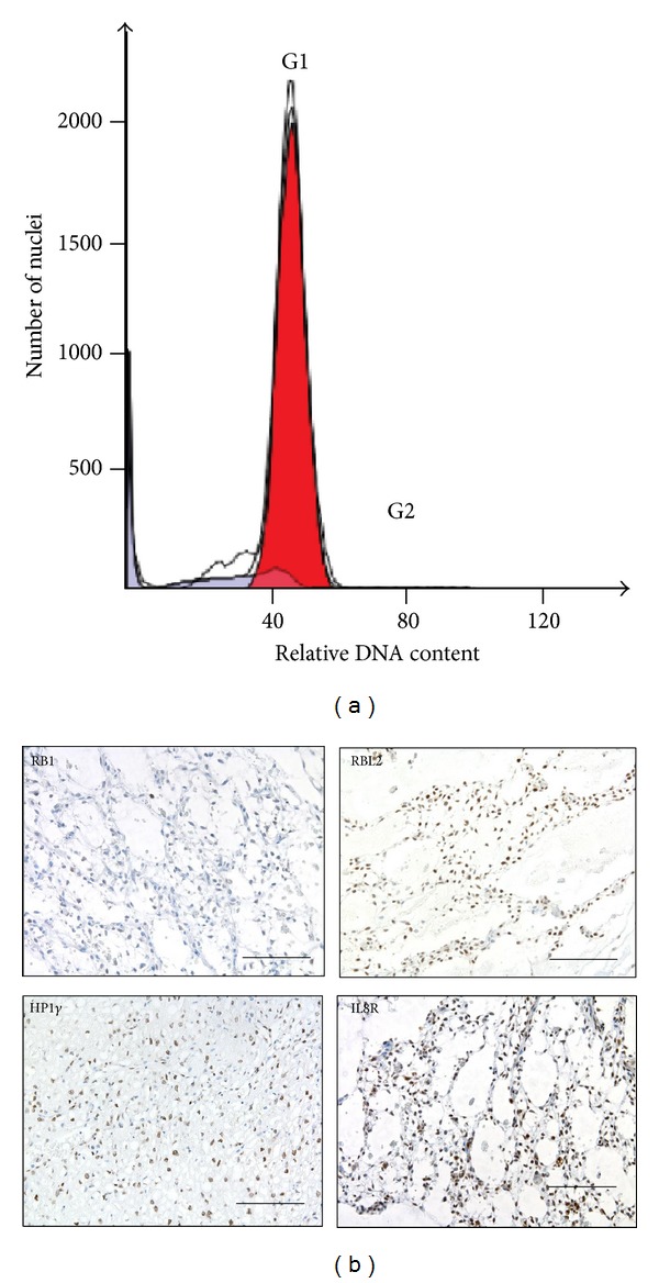
(a) Flow cytometry histogram of cell nuclei extracted from paraffin embedded MLS/RCLS tumor tissue (Cases 8) showing more than 95% of the cells in the G1 phase of the cell cycle (red). (b) Immunohistochemistry analysis of RB1, RBL2, HP1γ, and IL8R in MLS/RCLS tumor tissues. Brown staining shows reactivity with the specific antibodies. Bars are 100 μm.
