Abstract
Context
Mutations of the fragile X mental retardation 1 (FMR1) gene are associated with distinct ovarian aging patterns.
Objective
To confirm in human in vitro fertilization (IVF) that FMR1 affects outcomes, and to determine whether this reflects differences in ovarian aging between FMR1 mutations, egg/embryo quality or an effect on implantation.
Design, Setting, Patients
IVF outcomes were investigated in a private infertility center in reference to patients' FMR1 mutations based on a normal range of CGGn = 26–34 and sub-genotypes high (CGGn>34) and low (CGG<26). The study included 3 distinct sections and study populations: (i) A generalized mixed-effects model of morphology (777 embryos, 168 IVF cycles, 125 infertile women at all ages) investigated whether embryo quality is associated with FMR1; (ii) 1041 embryos in 149 IVF cycles in presumed fertile women assessed whether the FMR1 gene is associated with aneuploidy; (iii) 352 infertile patients (< age 38; in 1st IVF cycles) and 179 donor-recipient cycles, assessed whether the FMR1 gene affects IVF pregnancy chances via oocyte/embryo quality or non-oocyte maternal factors.
Interventions
Standardized IVF protocols.
Main Outcome Measures
Morphologic embryo quality, ploidy and pregnancy rates.
Results
(i) Embryo morphology was reduced in presence of a low FMR1 allele (P = 0.032). In absence of a low allele, the odds ratio (OR) of chance of good (vs. fair/poor) embryos was 1.637. (ii) FMR1 was not associated with aneuploidy, though aneuploidy increased with female age. (iii) Recipient pregnancy rates were neither associated with donor age or donor FMR1. In absence of a low FMR1 allele, OR of clinical pregnancy (vs. chemical or no pregnancy) was 2.244 in middle-aged infertility patients.
Conclusions
A low FMR1 allele (CGG<26) is associated with significantly poorer morphologic embryo quality and pregnancy chance. As women age, low FMR1 alleles affect IVF pregnancy chances by reducing egg/embryo quality by mechanisms other than embryo aneuploidy.
Background
Though currently still considered a gene with primarily adverse neuro-psychiatric associations, the fragile X mental retardation 1 (FMR1) gene, located at Xq27.3 [1], has in recent years also attracted attention because of its apparent involvement in regulating ovarian aging [2]–[5]. A recent publication conclusively demonstrated in the human differences in decline of functional ovarian reserve (FOR) with different FMR1 mutations (genotypes and sub-genotypes) [6].
These mutations are based on a newly described normal range, defined by CGGn = 26–34. This range allows for the definition of mutations based on CGGn on both alleles of the X chromosome, described as genotypes/sub-genotypes [2], [3], [7], [8]. Aside from demonstrating FMR1 mutation-specific declines in FOR [2], [3], [7], [8], different FMR1 mutations also have been associated with varying pregnancy chances in association with in vitro fertilization (IVF) [7], [8], an observation suggesting that, as women age, the FMR1 gene may not only affect FOR, but also chance of conception.
Since FMR1 mutations also appear associated with autoimmune-risk [7], [8], and implantation is an immunologic process [9], how the FMR1 gene affects pregnancy chances (via oocyte/embryo quality or other non-oocyte maternal factors) is, therefore, of interest.
This study investigated this question, utilizing distinct patient populations of young oocyte donors, middle-aged women of presumed normal fertility and infertile middle-aged women in treatment. The latter patient population included a subset of women who used donor eggs, allowing for assessment of possible donor FMR1 effects mediated by oocyte quality alone and allowing comparison to those who were using their own oocytes.
Methods
Study Participants
This study investigated three distinct patient populations: (i) 777 embryos in 168 IVF cycles in 125 infertile women of all ages, assessing whether embryo quality differs in association with FMR1 mutations. (ii) In assessing whether the FMR1 gene is associated with risk of aneuploidy, 1041 embryos in 149 IVF cycles in presumed fertile women undergoing IVF for pre-implantation genetic diagnosis; and (iii) in a third model 179 consecutive oocyte recipient IVF cycles, in determining whether mutations of the FMR1 gene in donors affect IVF pregnancy chances in recipients. This third model in addition utilized as comparison group 352 consecutive infertile patients (mean age 33.4±3.4 years), who used their own eggs in first IVF cycles.
Since low (CGG n<26) FMR1 alleles have previously been associated with significant declines in IVF pregnancy rates [7], [8], two questions were posed by this model: first, whether with low FMR1 alleles a similar decline in pregnancy chances can be confirmed as previously reported, this time in another infertile patient population; and, second, whether a decline in pregnancy chance would also be observed in an egg donation model, where older patients receive oocytes from young oocytes donors with different FMR1 mutations.
All patients and oocyte donors sign informed consents at time of initial consultation, allowing the use of their medical record for research purposes, as long as their identity is protected and the medical record remains confidential. Both conditions were met for this study, qualifying it for expedited review by the center's institutional review board (IRB).
Our center accepts less than five percent of oocyte donor applicants. Donor selection involves an initial screening step by questionnaire, followed by two rounds of face-to-face interviews and a medical testing round. Screening excludes donor candidates with presumed increased reproductive risks, based on their medical, family and genetic histories.
Full mutation (CGGn>200) and premutation range alleles (CGGn = 55–200), long known associated with POI/POF, were absent in investigated populations. Here presented FMR1 data on high alleles (CGGn>34), therefore reflect on women with what currently are considered normal CGG n<45 or “gray-zone” CGG n∼45–54 CGG repeats, and cannot be extrapolated to women with premutation or full mutation range alleles.
Art Protocols
Patients and donors underwent standardized ovarian stimulation protocols as previously described [6], [8]. Briefly, patients under age 40 with normal functional ovarian reserve (FOR) received down regulation with full dose (1.0 mg/0.1 mL) gonadotropin releasing hormone agonist (GnRH-a; Lupron, Abbot Pharmaceuticals, North Chicago, IL) and ovarian stimulation with up to 300 IU of gonadotropins daily, usually half as follicle stimulation hormone (FSH) and half as human menopausal gonadotropins (hMG).
Patients with diminished FOR and/or low serum androgens and those over age 40 received at least six weeks of dehydroepiandrosterone (DHEA) supplementation with 25 mg t.i.d. of pharmaceutical grade, micronized DHEA prior to IVF cycle start, as previously described [10]. Their cycles involved prevention of premature ovulation with microdose GnRH-a (50 µg/0.1 mL, b.i.d.), and ovarian stimulation with 300–450 IU FSH and 150 IU of hMG daily.
Oocyte donors underwent down regulation with full dose GnRH-a (1.0 mg/0.1 mL) and ovarian stimulation with up to 300 IU of hMG daily.
Final oocyte maturation was triggered in all cycles with 5,000–10,000 IU of human chorionic gonadotropin (hCG).
Laboratory Assesments
FMR1 Testing
Assessments of CGGn of the FMR1 gene was performed by commercial assays, as previously described, with FMR1 mutations (genotypes and sub-genotypes) defined as described in prior publications [2], [3], [7], [8]. In brief, by defining a normal CGG n = 26–34 range, CGG counts below and above that range are abnormal. A female with both FMR1 alleles in normal range, therefore, is norm, one with one in and one outside normal range is het and one with both alleles outside norm range is hom. Whether an allele is above (high) or below (low) normal range further sub-divides het and hom genotypes into sub-genotypes (het-norm/high, het-norm/low; hom-high/high, hom-high/low, hom-low/low).
In this study, included oocyte donors and infertility patients had FMR1 testing performed to exclude FXS risks in offspring. Based on the center's IRB instructions, the center during the study period did not consider FMR1 mutations in either selecting egg donors and/or in selecting treatments of infertile patients.
Determination of Embryo Morphology
Cleavage stage embryos were classified as good (4 cells d-2, 8 cells d-3, little or no fragments), poor (arrested embryos or >25% fragmented) or fair (all other embryos).
Determination of Embryo Ploidy
Preimplantation genetic screening (PGS) was performed in a group of 121 fertile women undergoing a total of 149 IVF cycles for non-infertility related reasons, primarily elective gender selection. Embryos were biopsied on day three after fertilization at 6–8 cell stages. Fluorescence in situ hybridization (FISH) was utilized with probes for seven chromosomes (X, Y, 13, 16, 18, 21 and 22). Reported aneuploidy rates are, therefore, incomplete but should not influence FMR1 mutation-associated findings.
Statistical Analysis
This study investigated the relationship between FMR1 mutations (genotypes/sub-genotypes) and embryo morphology, embryo ploidy and clinical pregnancy rates, while accounting for the variability of age. Since presence of low alleles (CGGn<26) in prior studies impacted FOR as well as pregnancy chance with fertility treatment [8], analyses in this study primarily compared patients with low alleles to those without low alleles.
Generalized mixed-effects (GLME) models were applied to examine embryo morphology and ploidy based on FMR1 genotype. Generalized estimating equation (GEE) models were utilized to confirm GLME results. A logistic regression model was used to study the clinical pregnancy rate of first IVF treatments in infertility patients under age 38.
All statistical analyses were adjusted for female age. The analysis of embryo morphology was also adjusted for number of prior treatment cycles. Covariates were considered statistically significant when P values were <0.05 using SAS 9.2. The center's senior statistician (Y.Y.) performed all analyses.
Results
(i) Fmr1 And Morphological Embryo Quality
We here investigated 777 embryos from 125 women in 168 IVF cycles in infertile women of all ages. Table 1 summarizes patient characteristics: Mean age of this patient population was 39.7±5.7 years. This infertile patient group, therefore, was not restricted in age. A little less than half (45.4%) of all embryos were considered of good quality, 43.4% fair and 11.2% of poor quality.
Table 1. Characteristics of infertile patients in section (i).
| Variables |
 or n (%) or n (%)
|
||
| Number of patients | 125 | ||
| Number of cycles | 168 | ||
| Age (years) | 39.7±5.7 | ||
| Race (African; Asian; Caucasian; other) | 9.7%; 15.3%; 74.2%; 0.8% | ||
| FSH (mIU/mL) | 11.2±12.5 | ||
| AMH (ng/mL) | 1.5±1.9 | ||
| Number of embryos | 777 | ||
| Embryo quality (Good/Fair/Poor) | 353(45.4%)/337(43.4%)/87(11.2%) | ||
| FMR1 mutations | Patients n(%) | Cycles n(%) | Embryos n%) |
| Norm | 51(40.8%) | 60(35.7%) | 319(41.1%) |
| het-norm/high | 25(20.0%) | 34(20.2%) | 144(18.5%) |
| hom-high/high | 1(0.8%) | 1(0.6%) | 3(0.4%) |
| het-norm/low | 37(29.6%) | 56(33.3%) | 234(30.1%) |
| hom-low/low | 4(3.2%) | 6(3.6%) | 41(5.3%) |
| hom-low/high | 7(5.6%) | 11(6.6%) | 36(4.6%) |
Women with low sub-genotypes (CGGn<26) were overrepresented and those with high sub-genotype (CGGn>34) and norm genotypes underrepresented in comparison to prior reports [5], [7], [8], [11]. Here investigated infertile women, therefore, are likely more adversely selected than previously reported infertile populations.
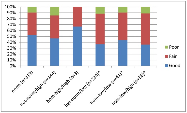
Figure 1 summarizes morphologic embryo quality, based on FMR1 genotypes/sub-genotypes. Comparing availability of good quality embryos to availability of fair and poor quality embryos, morphologic embryo quality in women with at least one low FMR1 allele was statistically different from patients with only norm (CGGn = 26–34) and high (CGGn>34) alleles (P = 0.03). Odds ratio (OR) estimate of having good morphologic quality embryos vs. having fair and/or poor quality embryos between low and norm and/or high genotypes/sub-genotypes was 1.637, indicating that patients with only norm and/or high alleles had a 63.7% higher probability of producing good morphologic quality embryos than patients with at least one low FMR1 allele.
Figure 1. The distribution of morphologic embryo quality for each FMR1 sub-genotype.
*Morphologic embryo quality in sub-genotypes with a low allele present was significantly reduced in comparison to those without a low allele (P = 0.032). OR of chance of good vs. fair/poor embryos in absence of a low allele was 1.637.
(ii) Fmr1 And Embryo Aneuploidy Rates
Since embryo ploidy represents a substantial component of total functional embryo quality [12], embryo ploidy was assessed next. This assessment was made in 1041 embryos from 149 IVF cycles in presumably fertile women undergoing IVF with preimplantation genetic diagnosis (PGD) for non-infertility related reasons, mostly elective gender determination. This patient group represented a mid-range age (33.5±5.5 years), and was, therefore, somewhat younger than in section (i) investigated infertility patients. Table 2 summarizes patient characteristics.
Table 2. Characteristics in section (ii) of women undergoing IVF for non-fertility related indications.
| Variables |
 or n(%) or n(%)
|
||
| Patients | 121 | ||
| Number of cycles | 149 | ||
| Age (years) | 33.5±5.5 | ||
| Race (African; Asian; Caucasian; other) | 11%; 23%; 65%; 1% | ||
| FSH (mIU/mL) | 8.7±3.1 | ||
| AMH (ng/mL) | 3.2±2.6 | ||
| Number of embryos | 1041 | ||
| Ploidy (normal/abnormal) | 571(54.9%)/470(45.2%) | ||
| FMR1 sub-genotypes | Oocyte source n(%) | Cycles n(%) | Embryos n(%) |
| norm | 71(58.7%) | 85(57.1%) | 646(62.1%) |
| het-norm/high | 19(15.7%) | 23(15.4%) | 141(13.5%) |
| hom-high/high | 2(1.7%) | 3(2.0%) | 26(2.5%) |
| het-norm/low | 26(21.5%) | 34(22.9%) | 193(18.5%) |
| hom-low/low | 2(1.7%) | 2(1.3%) | 16(1.5%) |
| hom-low/high | 1(0.8%) | 2(1.3%) | 19(1.8%) |
As the table demonstrates, the FMR1 mutation distribution in this presumed fertile female population differed from in section (i) investigated infertile patients (P<0.001), and is closer to previously reported distribution patterns, demonstrating predominantly more norm genotypes and fewer het as well as hom FMR1 mutations with low as well as high alleles. Most remarkable is, however, the remarkably lower rate of low alleles, suggesting a possible association between low alleles and infertility in older women.
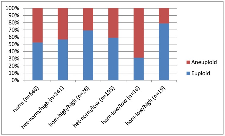
Figure 2 summarizes aneuploidy rates in reference to FMR1 mutations. No statistical differences in aneuploidy rate were noted between women with low and/or norm and high alleles (OR, 0.855; 95% CI 0.578, 1.266; P = 0.434). Interestingly, biallelic low women, however, did demonstrate unusually high aneuploidy rates, suggesting that lack of significant findings in this section of the study could be due to relatively small study subject numbers. Also of interest is the very low aneuploidy number in women with one low and one high allele, suggesting a potential compensatory effect of a high allele on potential negative effects of a low allele. Since both of the latter observation occurred in only small patient subsets, and did not reach statistical significance, they should, however, as of this point only be considered hypothesis generating.
Figure 2. The distribution of embryo ploidy based on patients FMR1 sub-genotype.
FMR1 mutations were not statistically associated with embryo ploidy, though the high aneuploidy rate in biallelic low, hom-low/low women is quite remarkable. Lack of significance may in here-reported findings, therefore, be consequence of small patient numbers. Also of interest is the very low aneuploidy rate in women with one low and one high allele, presenting with only approximately half the aneuploidy rate of even norm women. While these findings, dues to small patient numbers, also need to be viewed with caution, they could suggest a compensatory benefit from a high allele on negative effects of a low allele.
As would be expected, age of women yielding oocytes was statistically related to aneuploidy (OR 1.041; 95%CI 1.011, 1.072; P = 0.007): one-year increase of advancing female age resulted in a 4.1% higher chance of embryo aneuploidy.
These results suggest that the relationship demonstrated in section (i) between morphologic embryo quality and FMR1 mutations is not, likely, based on embryo ploidy.
(iii) Effect Of Fmr1 On Clinical Pregnancy Rates
In infertile women FMR1 mutations (genotypes/sub-genotypes) have previously been demonstrated predictive of IVF pregnancy chances [7], [8]. Since IVF pregnancy chances are known associated with embryo quality [13] and, since results in section (i) demonstrate that norm and/or high FMR1 alleles are associated with significantly more good quality embryos than low alleles, one can conclude that the FMR1 gene affects IVF pregnancy chances via oocyte/embryo quality.
IVF pregnancy chances can, however, also be significantly affected by implantation, considered an immunologically-influenced process [9], [14]. Low FMR1 alleles in infertile women have also been associated with abnormal immune laboratory findings in infertile women, suggestive of immune system activation [7], [8]. Adverse effects of the FMR1 gene on implantation, therefore, cannot be ruled out.
Section (iii) of this study was, therefore, designed to differentiate between egg/embryo quality or implantation effects as cause for lowered IVF pregnancy rates in association with low FMR1 alleles. It represents a study, investigating 352 first autologous IVF cycles in middle-aged infertile women under age 38 (mean 33.4±3.4 years) and 179 donor/recipient IVF cycles, utilizing donated eggs from 162 young oocyte donors (mean donor age 25.0±2.9 years). As expected, the clinical pregnancy rate was higher in donor (55.9%) than autologous IVF cycles (28.4%; P<0.001).
Due to the small sample sizes of hom-low/low, hom-high/high and hom-low/high women, we in this study section combined all hom patients into one group when comparing the distribution of FMR1 mutations between the two sub-study groups in this section.
Distribution of FMR1 genotypes/sub-genotypes significantly differed between the two study groups in this section (P<0.001). Interestingly, the here investigated younger infertile patient group demonstrated a similar distribution of FMR1 mutations to the presumed fertile women in section (ii), while young oocyte donors presented with a distribution in-between these two middle-aged groups and in section (i) investigated much older infertile patients (Table 3). The distribution pattern seen in oocyte donors, therefore, likely is typical for young normal female populations, suggesting in young women an approximately 22% prevalence of low FMR1 alleles but a much higher prevalence in older infertile women. Once again, the increasing prevalence of low alleles, comparing young donors and older infertile women points towards a potential association of low FMR1 alleles with infertility at advanced ages.
Table 3. Donor egg recipients and infertility patients using autologous oocytes included in section (iii).
| Donor/Recipients | Infertile patients | |
| Variables |
 or n(%) or n(%)
|
 or n(%) or n(%)
|
| Donors | 127 | - |
| Recipients and Infertile women | 162 | 352 |
| Cycles | 179 | 352 |
| Age – Oocyte Source (years) | 25.0±2.9 | 33.4±3.4 |
| Race – Oocyte Source (African; Asian; Caucasian; other) | 7.3%; 12.9%; 75.3%; 4.5% | 11.9%; 15.2%; 70.0%; 2.9% |
| AMH – Oocyte Source (ng/mL) | 4.0±2.3 | 1.9±2.0 |
| Clinical pregnancy rate | 100(55.9%) | 100(28.4%) |
| FMR1 sub-genotypes | n (%) | n (%) |
| (of women reaching retrieval) | ||
| norm | 90(50.3%) | 217(61.7%) |
| het-norm/low | 51(28.5%) | 63(17.9%) |
| het-norm/high | 17(9.5%) | 55(15.6%) |
| hom-low/low | 5(2.8%) | 10(2.8%) |
| hom-high/high | 9(5.0%) | 3(0.9%) |
| hom-low/high | 7(3.9%) | 4(1.14%) |
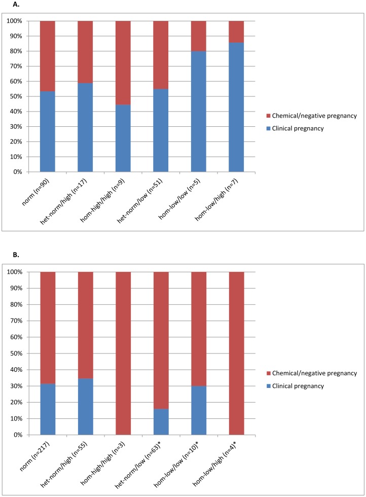
Figures 3A and 3B demonstrate clinical pregnancy rates in association with FMR1 mutations. Once again comparing women with at least one low FMR1 allele to those with only norm and high alleles, Figure 3A demonstrates that in donor-recipient cycles the FMR1 mutation of the donor did not affect recipient pregnancy rates (OR 0.738; 95% CI 0.387, 1,405; P = 0.347). Moreover, donor ages also did not affect pregnancy chances (OR 0.970; 95%CI 0.874, 1.078; P = 0.568).
Figure 3. The distribution of clinical pregnancies based on FMR1 sub-genotype of Clinical pregnancy rates in A oocyte donors and B middle-aged infertility patients.
Oocyte recipient pregnancy rates were not associated with donor FMR1. Using a logistic regression model in middle-aged infertility patients, OR of clinical pregnancy vs. chemical or no pregnancy was 2.244 in absence of a low FMR1 allele vs. presence of low alleles (P = 0.0015), suggesting 1.244-times odds of clinical IVF pregnancy in absence of a low FMR1 allele.
Using a logistic regression model, in young infertility patients under age 38 years, odds of clinical pregnancy, however did differ significantly between patients with single low FMR1 alleles and those with only norm and/or high alleles (OR 2.244; 95%CI 1.168, 4.312; P = 0.015; Figure 3B). Odds of clinical pregnancy vs. biochemical or no pregnancy between both groups were 2.244. The odds ratio estimate, thus, indicates that women with only norm and/or high FMR1 alleles have 1.244-times higher probability of clinical pregnancy than women with low alleles, and confirming our earlier reports [7], [8].
Figure 4 demonstrates that the difference in odds of clinical pregnancy in this relatively young group of infertile women remains remarkably stable with advancing female age between women with low and with norm/high alleles, though it does minimally narrow as women age.
Figure 4. Predicted probabilities of clinical IVF pregnancy in infertile patients based on age and FMR1.
Prior IVF cycle numbers (at other centers) and patient age were statistically not related to morphologic embryo quality (data not shown).
Discussion
By demonstrating that specific FMR1 gene mutations are associated with morphologic embryo quality and with chance of clinical pregnancy in association with IVF, this study establishes the FMR1 gene as the first gene statistically associated with IVF outcomes. This also means that the gene is not only, as previously reported, associated with ovarian aging by affecting FOR [2], [3], [5], [7], [8], but also affects embryo quality. Since embryo quality is largely dependent on egg quality [15], it, as of this point, remains to be determined whether the FMR1 gene impacts the oocyte or, directly, the embryo. The study also demonstrates that this genetic effect persists at all ages (Figure 4).
Interestingly, while embryo ploidy is generally considered to represent a large part of functional embryo competence, this study suggests that the morphologic differences in embryo quality between FMR1 mutations were, likely, not ploidy-related. These observations may at least partially explain why embryo morphology is only relatively poorly associated with embryo ploidy [16], and why the procedure of PGS so far has failed to improve IVF outcomes [17].
Here reported ploidy-related finding should, however, be viewed with some caution since they may not be applicable to all infertility patients undergoing IVF: Section (ii) of the study was performed in presumably fertile women, most undergoing IVF for non-fertility relates issues. Our center routinely supplements women with low age-specific FOR with DHEA [10] in order to raise androgen levels, in such patients reported to be low [18]. DHEA supplementation, in turn, appears to lower aneuploidy rates in patients with low FOR [19]. Treatment with DHEA in some patients in this study group may, therefore, have affected the results of here reported ploidy investigation.
Significant differences in FMR1 mutation distribution between the different study groups investigated in the three sections of this study were an unexpected finding. In previous studies infertile patients demonstrated in slightly more than half of cases norm genotypes, in slightly more than 40% het sub-genotype, het-low slightly exceeding het-high, and in under 10% the three hom sub-genotypes [2], [3], [5], [7], [8].
In this study infertility patients of very advanced age (mean 39.7±5.7 year) in section (i) deviated from this distribution most, demonstrating few norm genotypes (41.1%) and a very high prevalence of low alleles. In contrast, presumed fertile middle-aged patients in section (ii) (mean age of 33.5±5.5) presented with high numbers of norm genotypes (62.1%) and relatively low numbers of monoalleleic low patients. Middle-aged infertility patients in section (iii) of the study also presented with a high number of norm genotypes (61.7%) and a relative low number of monoalleleic low patients.
Interestingly, young and presumed healthy oocyte donors (mean age 25.0±2.9 years) presented with exactly half (50.3%) norm genotypes, 28.5% het-norm/low sub-genotypes and only 9.5% het-norm/high sub-genotypes. Overall, 35.2% of the center's oocyte donors undergoing egg retrieval carried a monoalleleic low FMR1 gene.
Biallelic low carriers even at young ages already demonstrate abnormally low FOR, and low monoalleleic carriers, already at young ages lose FOR more rapidly than other FMR1 mutations [6]. This study now demonstrates that monoalleleic low FMR1 alleles are also associated with poor embryo quality; yet despite approximately one-third of all here investigated oocytes donors carrying a low FMR1 allele, donor/recipient pregnancy rates were not affected by a donor's low allele. The clinical pregnancy rate was, indeed, higher in donors carrying a low FMR1 allele (60.3%) than donors with norm and/or high FMR1 alleles (53.5%), though this difference did not reach statistical significance (P = 0.3767).
On first impression contradictory, these results actually confirm prior publications. We [20] and others [21] previously demonstrated how difficult it is to detect adverse FMR1 effects in young egg donors. Here presented data confirm this fact and again suggest that at such young ages enough ovarian function redundancy may exists to obviate existing defects.
An alternative explanation would be that, as noted earlier, FMR1 effects occur only later in life and, therefore, are not yet apparent in young oocyte donors. Such an explanation, however, appears less likely since FMR1 related differences in IVF pregnancy rates are already apparent at relative young ages, and differences between monoalleleic low and all other FMR1 mutation carriers do not change dramatically with age (Figure 4).
Moreover, at least in rodents, fragile X mental retardation protein (FXMRP) and FMR1 mRNA appear already expressed during all stages of follicle development [22], thus suggesting a possible direct FMR1 effect on oocytes.
While at young ages redundancy of ovarian reserve, likely, among those with low FRM1 alleles accounts for no observed decrease in pregnancy rates in donor cycles, redundancy does not necessarily also protect cumulative pregnancy chances over sequential IVF cycles, utilizing fresh and frozen embryos. Unless egg donors are very young, exclusion of those with low FMR1 alleles and/or low ovarian reserve may, therefore, be appropriate to optimize cumulative pregnancy rates based on here presented and recently reported data [6].
Many IVF programs currently test oocyte donor's FMR1 status to prevent transmission of maternal premutation range (CGGn∼55–200) and/or expansions to full mutations (CGGn>200; fragile X syndrome; FXS) and other associated neuro-psychiatric complications, mostly affecting males [1]. Such testing is, however, usually only performed after a donor has already been selected. Here we suggest that, if further studies confirm here reported data, FMR1 testing should be performed in oocyte donor candidates as a tool of primary selection.
Finally, here presented differences in distribution of FMR1 mutations also further demonstrate the negative impact of low alleles on the FMR1 gene on female fertility. While middle aged infertility patients in section (iii) of this study and presumed fertile women in section (ii) present with quite similar genotype/sub-genotype distribution, older infertile women in section (i), with mean age 39.7±5.7 years, demonstrated approximately 20 percentage points lower norm FMR1 genotype prevalence (41.1%) and a much higher prevalence in het-norm/low sub-genotype (30.1%. equaling the prevalence of het-low and het-high sub-genotypes combined in the younger group of infertile women).
These data, therefore, suggest that, as infertile women age, those who remain in treatment are increasingly adversely selected: those with norm genotypes and best pregnancy chances decline in prevalence, and women with low alleles, and poorer pregnancy chances, increase in prevalence. As noted before, this patient distribution is also likely reflective of our center's highly adversely selected patient population. Older reproductive age women with low FMR1 alleles, who disproportionally fail to conceive, can be expected to “accumulate” in a center like ours, which generally is considered a center of “last resort” for patients who have previously failed elsewhere. A younger patient population, demonstrated in section (ii) reflects women with lower dropout rates and, therefore, fewer low FMR1 alleles and more norm genotypes. This is consistent with our recent finding that young infertile women with low alleles disproportionately dropout from infertility treatment [6].
We in this discussion outlined the principal weaknesses of this study. Likely the most important being the small size of some patient subgroups in the three investigated patient populations. The statistical robustness of here reported findings is, therefore, that more remarkable. Our data, nevertheless, require confirmation. They, however, suggest an increasingly important impact of the FMR1 gene on ovarian aging and female fertility and infertility.
Data Availability
The authors confirm that all data underlying the findings are fully available without restriction. Relevant data are included within the paper.
Funding Statement
This work was supported by Foundation for Reproductive Medicine and Center for Human Reproduction. The funders had no role in study design, data collection and analysis, decision to publish, or preparation of the manuscript.
References
- 1. Bagni C, Tassone F, Neri G, Hagerman R (2012) Fragile X syndrome: causes, diagnosis, mechanisms, and therapeutics. J Clin Invest 122: 4314–22. [DOI] [PMC free article] [PubMed] [Google Scholar]
- 2. Gleicher N, Weghofer A, Barad DH (2010) Ovarian reserve determinations suggest new function of FMR1 (fragile X gene) in regulating ovarian ageing. Reprod Biomed Online 20: 768–75. [DOI] [PubMed] [Google Scholar]
- 3. Gleicher N, Weghofer A, Kim A, Barad DH (2012) The impact in older women of ovarian FMR1 genotypes and sub-genotypes on ovarian reserve. PloS one 7: e33638. [DOI] [PMC free article] [PubMed] [Google Scholar]
- 4. Gleicher N, Kim A, Barad DH, Shohat-Tal A, Lazzaroni E, et al. (2013) FMR1-dependent variability of ovarian aging patterns is already apparent in young oocyte donors. Reprod Biol Endocrinol 11: 80. [DOI] [PMC free article] [PubMed] [Google Scholar]
- 5. Gleicher N, Kim A, Weghofer A, Barad DH (2012) Differences in ovarian aging patterns between races are associated with ovarian genotypes and sub-genotypes of the FMR1 gene. Reprod Biol Endocrinol 10: 77. [DOI] [PMC free article] [PubMed] [Google Scholar]
- 6.Kushnir VA, Himaya E, Barad DH, Weghofer A, Gleicher N (2013) Functional ovarian reserve assessments in young oocyte donors based on FMR1 genotypes and sub-genotypes. Fertil Steril 100(3): , S114. [Google Scholar]
- 7. Gleicher N, Weghofer A, Lee IH, Barad DH (2011) Association of FMR1 genotypes with in vitro fertilization (IVF) outcomes based on ethnicity/race. PloS one 6: e18781. [DOI] [PMC free article] [PubMed] [Google Scholar]
- 8. Gleicher N, Weghofer A, Lee IH, Barad DH (2010) FMR1 genotype with autoimmunity-associated polycystic ovary-like phenotype and decreased pregnancy chance. PloS one 5: e15303. [DOI] [PMC free article] [PubMed] [Google Scholar]
- 9. Sen A, Kushnir VA, Barad DH, Gleicher N (2014) Endocrine autoimmune diseases and female infertility. Nat Rev Endocrinol. 10(1): 37–50. [DOI] [PubMed] [Google Scholar]
- 10. Gleicher N, Barad DH (2011) Dehydroepiandrosterone (DHEA) supplementation in diminished ovarian reserve (DOR). Reprod Biol Endocrinol 9: 67. [DOI] [PMC free article] [PubMed] [Google Scholar]
- 11. Gleicher N, Weghofer A, Barad DH (2010) Effects of race/ethnicity on triple CGG counts in the FMR1 gene in infertile women and egg donors. Reproductive biomedicine online 20: 485–91. [DOI] [PubMed] [Google Scholar]
- 12. Fragouli E, Wells D (2012) Aneuploidy screening for embryo selection. Semin Reprod Med. 30: 289–301. [DOI] [PubMed] [Google Scholar]
- 13. Goto S, Kadowaki T, Tanaka S, Hashimoto H, Kokeguchi S, et al. (2011) Prediction of pregnancy rate by blastocyst morphological score and age, based on 1,488 single frozen-thawed blastocyst transfer cycles. Fertil Steril 95: 948–52. [DOI] [PubMed] [Google Scholar]
- 14. Gleicher N, Weghofer A, Barad DH (2012) Cutting edge assessment of the impact of autoimmunity on female reproductive success. J Autoimmun 38: J74–80. [DOI] [PubMed] [Google Scholar]
- 15. Setti AS, Figueira RC, Braga DP, Colturato SS, Iaconelli A Jr, et al. (2011) Relationship between oocyte abnormal morphology and intracytoplasmic sperm injection outcomes: a meta-analysis. Eur J Obstet Gynecol Reprod Biol 159: 364–70. [DOI] [PubMed] [Google Scholar]
- 16. Alfarawati S, Fragouli E, Colls P, Stevens J, Gutiérrez-Mateo C, et al. (2011) The relationship between blastocyst morphology, chromosomal abnormality, and embryo gender. Fertil Steril 95: 520–4. [DOI] [PubMed] [Google Scholar]
- 17. Gleicher N, Barad DH (2012) A review of, and commentary on, the ongoing second clinical introduction of preimplantation genetic screening (PGS) to routine IVF practice. J Assist Reprod Genet 29: 1159–66. [DOI] [PMC free article] [PubMed] [Google Scholar]
- 18. Gleicher N, Kim A, Weghofer A, Kushnir VA, Shohat-Tal A, et al. (2013) Hypoandrogenism in association with diminished functional ovarian reserve. Hum Reprod 28: 1084–91. [DOI] [PubMed] [Google Scholar]
- 19. Gleicher N, Weghofer A, Barad DH (2010) Dehydroepiandrosterone (DHEA) reduces embryo aneuploidy: direct evidence from preimplantation genetic screening (PGS). Reprod Biol Endocrinol 8: 140. [DOI] [PMC free article] [PubMed] [Google Scholar]
- 20.Gleicher N, Weghofer A, Barad DH (2012) Intermediate and normal sized CGG repeat on the FMR1 gene does not negatively affect donor ovarian response. Hum Reprod. 27: 2241–2; author reply 2–3. [DOI] [PubMed]
- 21. Lledo B, Guerrero J, Ortiz JA, Morales R, Ten J, et al. (2012) Intermediate and normal sized CGG repeat on the FMR1 gene does not negatively affect donor ovarian response. Hum Reprod 27: 609–14. [DOI] [PubMed] [Google Scholar]
- 22. Ferder I, Parborell F, Sundblad V, Chiauzzi V, Gómez K, et al. (2013) Expression of fragile X mental retardation protein and Fmr1 mRNA during folliculogenesis in the rat. Reproduction 145: 335–43. [DOI] [PubMed] [Google Scholar]
Associated Data
This section collects any data citations, data availability statements, or supplementary materials included in this article.
Data Availability Statement
The authors confirm that all data underlying the findings are fully available without restriction. Relevant data are included within the paper.


