ABSTRACT
The hemagglutinin (H) gene of canine distemper virus (CDV) encodes the receptor-binding protein. This protein, together with the fusion (F) protein, is pivotal for infectivity since it contributes to the fusion of the viral envelope with the host cell membrane. Of the two receptors currently known for CDV (nectin-4 and the signaling lymphocyte activation molecule [SLAM]), SLAM is considered the most relevant for host susceptibility. To investigate how evolution might have impacted the host-CDV interaction, we examined the functional properties of a series of missense single nucleotide polymorphisms (SNPs) naturally accumulating within the H-gene sequences during the transition between two distinct but related strains. The two strains, a wild-type strain and a consensus strain, were part of a single continental outbreak in European wildlife and occurred in distinct geographical areas 2 years apart. The deduced amino acid sequence of the two H genes differed at 5 residues. A panel of mutants carrying all the combinations of the SNPs was obtained by site-directed mutagenesis. The selected mutant, wild type, and consensus H proteins were functionally evaluated according to their surface expression, SLAM binding, fusion protein interaction, and cell fusion efficiencies. The results highlight that the most detrimental functional effects are associated with specific sets of SNPs. Strikingly, an efficient compensational system driven by additional SNPs appears to come into play, virtually neutralizing the negative functional effects. This system seems to contribute to the maintenance of the tightly regulated function of the H-gene-encoded attachment protein.
IMPORTANCE To investigate how evolution might have impacted the host-canine distemper virus (CDV) interaction, we examined the functional properties of naturally occurring single nucleotide polymorphisms (SNPs) in the hemagglutinin gene of two related but distinct strains of CDV. The hemagglutinin gene encodes the attachment protein, which is pivotal for infection. Our results show that few SNPs have a relevant detrimental impact and they generally appear in specific combinations (molecular signatures). These drastic negative changes are neutralized by compensatory mutations, which contribute to maintenance of an overall constant bioactivity of the attachment protein. This compensational mechanism might reflect the reaction of the CDV machinery to the changes occurring in the virus following antigenic variations critical for virulence.
INTRODUCTION
Canine distemper virus (CDV) is an enveloped, negative-sense, single-stranded RNA virus in the genus Morbillivirus within the family Paramyxoviridae. CDV is considered one of the most significant pathogens of carnivores (1). Of the structural proteins encoded by its genome, the hemagglutinin (H; attachment protein) has been thoroughly investigated because of its key role as a receptor-binding protein (2–5). Two receptors are currently known to engage with H, i.e., nectin-4 and the signaling lymphocyte activation molecule (SLAM) (6–8). Of these, SLAM has been shown to be critical for host susceptibility, whereas nectin-4 is required for clinical disease (9).
The attachment protein H is multifunctional. H tetramers or higher-molecular-weight oligomer complexes are believed to first bind to trimeric fusion (F) proteins (F-protein binding activity) within the endoplasmic reticulum and then travel toward the cell surface as preformed hetero-oligomeric complexes (cell surface expression). Upon engagement with a specific receptor expressed on target cells (SLAM binding activity), it is thought that the H protein subsequently undergoes conformational changes that eventually lead to the activation of the F protein (10–15). Once triggered, F-protein trimers in turn refold through a series of structural rearrangements that are associated with membrane merging for viral entry into the cell and/or lateral spread (cell-cell fusion activity).
Evolutionary forces are likely to drive the incessant remodeling of the attachment protein of CDV by the naturally occurring mutations (3, 16–18). However, how and to what extent the functional features of the H protein are affected, if any, by this process and how the virus maintains its infectious efficiency through these changes are, to the best of our knowledge, unknown.
In order to partially answer these questions, we selected two H genes from two distinct but related CDV strains that were part of the same outbreak occurring in wild carnivores (red foxes [Vulpes vulpes] and Eurasian badgers [Meles meles]) and that were detected 2 years apart in Germany (2008) and Switzerland (2010), respectively (19, 20). We analyzed all the missense single nucleotide polymorphisms (SNPs) that differentiated the two H genes of the two strains, i.e., a wild type and a consensus strain, respectively. All the single mutations and their combinations were generated by site-directed mutagenesis. All the single mutants along with a selection of double, triple, and quadruple mutants were then analyzed and compared with the wild type and the consensus strain according to their efficiency of cell surface expression, SLAM receptor binding, and cell-to-cell fusion. Finally, a panel of the mutants showing among the most relevant functional changes was selected to evaluate the interaction between the H and the F proteins.
Here we report the results suggesting the existence of an articulated combinatorial system of SNP selection in CDV-related strains which appears to contribute to the maintenance of a tightly regulated functional efficiency.
MATERIALS AND METHODS
Viruses and hemagglutinin genes.
The wild-type H gene (H301F) was amplified from CDV strain W10/301F (GenBank accession number JF810106) that had been isolated from the lung of a naturally CDV-infected red fox during an outbreak that occurred in Switzerland in 2009 and 2010 (19). The sequence of the second H gene was a consensus sequence obtained from four CDV strains detected during a CDV outbreak that occurred in wild carnivores in Germany in 2008 (20). Three out of the four strains that were selected to obtain the consensus sequence were detected in red foxes, whereas the fourth one was from a Eurasian badger (GenBank accession numbers FJ416336, FJ416337, FJ416338, and FJ416339). The consensus sequence was obtained by considering all the missense SNPs that were common to at least three of the four German strains examined. Consequently, a missense SNP that was present in only one of the four German strains (resulting in the amino acid change H61Q) was not included in the final consensus sequence. The SNP leading to the amino acid change M500L, which was present in three of the four German strains, was included. The sequences of the wild-type H gene and the consensus H gene (HCons) were compared, and the missense SNPs were recorded.
Mutations and sequencing.
Following the identification of the five SNPs differentiating the H gene of Swiss strain W10/301F from that of the consensus sequence obtained from the German strains, we designed 5 pairs of primers (available upon request) for site-directed mutagenesis that was carried out according to the instructions provided by Stratagene (La Jolla, CA) in the QuikChange site-directed mutagenesis kit. Whereas H301F was previously cloned into an expression vector (pCI; Promega, Madison, WI) carrying a FLAG tag located at the 3′ end of the gene (pCI-H301F) (19), HCons was obtained through a series of additive site-directed mutagenesis steps carried out on pCI-H301F. The first run of mutagenesis aimed to obtain all the single mutations, i.e., N71S, V159I, M195V, M500L, and R580Q. All the subsequent mutations were obtained by sequential additive site-directed mutagenesis until the full mutant with the N71S, V159I, M195V, M500L, and R580Q mutations (HCons) was obtained. Every H mutant was sequenced to confirm the presence of the desired mutation(s), cloned, and expanded as previously described (19). Besides H301F, intermediate mutants, and HCons, the H gene from wild-type CDV reference strain A75/17 (Hwt), which was already available as a clone in the pCI vector, was used as an additional control in the functional studies. Additionally, the F-protein gene from wild-type strain A75/17 (Fwt) strain previously cloned in the pCI vector was also available (19, 21).
The amino acid sequences of the H proteins of representative strains belonging to each of the main CDV genotypes were also selected in order to assess if they carried the same substitutions present in H301F and HCons, which are both members of the Europe 1 genotype (see Table 1). The genotypes selected (along with their GenBank accession numbers in parentheses) were as follows: Europe 1 (H301F, JF810106; HCons, FJ4163336, FJ4163337, FJ4163338, FJ4163339), European wildlife (DQ228166, Z47759), Arctic-like (X84998, Z47760, AF172411), Africa (FJ461715, FJ461711), Asia 1 (AB329581, AB212963, EF445051, EU379560), Asia 2 (AB040768, AB040767), Asia 3 (EU743935, EU743934), America 1 (Z35493, D00758, ABI97484, AF259552), America 2 (Z47763, Z54166, Z54156, AY498692, AY649446), phocine distemper virus (PDV; AF479274, AJ224707), cetacean (dolphin) morbillivirus (CAA85427), measles virus (MeV; NC_001498), rinderpest virus (AAD25093), and peste des petits ruminants virus (PPRV; ACQ44671).
TABLE 1.
Distribution of H301F-specific amino acid changes in other CDV genotypes and PDV
| CDV genotype or virus | Amino acid change(s) |
||||
|---|---|---|---|---|---|
| N71S | V159I | M195V | M500L | R580Q | |
| Europe 1 (H301F) | N | V | M | M | R |
| Europe 1 (HCons) | S | I | V | L (3)/M (1)a | Q |
| European wildlife | S | I | V | M | R |
| Africa | S | I | I (1)/M (1) | M | R |
| Asia 1 | S | V | V | M | R |
| Asia 2 | G | V | V | M | R |
| Asia 3 | G | V | V | M | R |
| Arctic-like | S | I | V | M | R |
| America 1 | S | I | V | I (3)/R (1) | R |
| America 2 | S | I | V | M | R |
| PDV | N | F | M | Q | R |
| Cetacean M.b | N | A | R | K | M |
| Measles virus | H | A | R | V | V |
| Rinderpest virus | N | A | K | S | T |
| PPRVc | Q | A | K | V | L |
Numbers in parentheses indicate the number of occurrences of the specific amino acid residue in the viral strains considered.
Cetacean M., cetacean (dolphin) morbillivirus.
PPRV, peste des petits ruminants virus.
Cell cultures.
Two established cell lines were used for the in vitro experiments described below. More specifically, these included Vero cells either constitutively expressing or not constitutively expressing the canine SLAM (cSLAM) receptor (22) and 293T cells (21). All cells were grown in Dulbecco modified Eagle medium (Life Technologies Europe, Zug, Switzerland) and incubated at 37°C in a 5% CO2 atmosphere unless otherwise described.
QFA.
The quantitative fusion assay (QFA) was performed as described previously (19, 21). Briefly, 6 × 10⁵ Vero cells that had been plated 24 h prior to the experiment were transfected with a solution composed of 1 μg of either the cloned wild-type gene or one of the cloned selected mutants (including HCons) along with 1.8 μg of pCI-Fwt (from strain A75/17) and 0.2 μg of pCI-Luc carrying the luciferase (Luc) gene driven by a T7 RNA polymerase-dependent promoter (21) and incubated for 24 h. pCI-Hwt and pCI were also included as an additional reference and negative control, respectively. Next, the transfected Vero cells were split and mixed with Vero-cSLAM cells (at a 3:1 ratio) (22) that had previously been infected with vaccinia virus MVA-T7 carrying the T7 RNA polymerase gene at a multiplicity of infection of 0.1. After incubation for 2 h, cells were visually assessed for the presence of syncytia and then lysed to allow the measurement of the luciferase activity (dual-luciferase kit; Promega, Madison, WI). The samples were assayed in three independent experiments in triplicate. The quantitative fusion results, along with the standard deviation, were determined by calculating the mean of the values obtained. All the values were first compared as raw values and then normalized according to their FLAG expression (intrinsic normalization; see below). Additionally, the values obtained for each mutant were further normalized to the values for the wild type pCI-H301F (extrinsic normalization), whose fusion efficiency was arbitrarily considered to be equal to 100%.
The F protein-H interaction is pivotal for fusion, and different F proteins might result in different fusion efficiencies. In this test, we used a single F protein derived from the A75/17 strain (Fwt). This is an F protein that is heterologous to H301F and all the mutant attachment proteins used in this assay, and because of this, it was considered to uniformly impact them, partially compensating for the potential F-protein selection bias.
Cell surface expression.
The cell surface expression of each H mutant and the wild-type strains that were evaluated in the QFA (see above) was assessed according to an established protocol (19, 21). Briefly, 6 × 10⁵ Vero cells that had been inoculated in 6-well plates on the day prior to the experiment were transfected with either 1 μg of the plasmid expressing the wild-type H gene or one of the mutant genes (including HCons). Hwt and pCI were also included as an additional reference and negative control, respectively. The transfected Vero cells were incubated for 24 h and then stained first with an anti-FLAG monoclonal antibody (F3 165; Sigma, Buchs, Switzerland) diluted 1/1,000 in Opti-MEM medium (Life Technologies Europe, Zug, Switzerland) and then with a goat anti-mouse fluorescein isothiocyanate (FITC)-labeled secondary antibody diluted 1/500 (Sigma, Buchs, Switzerland) in Opti-MEM medium (Life Technologies Europe, Zug, Switzerland). The surface expression of the wild-type and mutant H genes was assessed by fluorescence-assisted cell sorting (FACS; FACSCalibur; BD, Basel, Switzerland). The FLAG values obtained for each sample analyzed were normalized to the expression of H301F, which was arbitrarily considered to be equal to 100%. Experiments were repeated three times in triplicate.
SLAM receptor-binding assay.
The interaction between either the H wild-type or mutant proteins with a recombinant soluble cSLAM (scSLAM) protein was assessed through a semiquantitative assay, as described by Zipperle and colleagues (21). Briefly, a soluble form of the N-terminally influenza virus hemagglutinin epitope (Ha)-tagged cSLAM molecule (Ha-scSLAM) was expressed in 293T cells. Ha-scSLAM was then added to Vero cells that had previously been transfected with either an H mutant gene or the wild-type H gene cloned in pCI. Receptor-bound wild-type and mutant H proteins were stained with an anti-H monoclonal antibody (Sigma, Buchs, Switzerland) diluted 1/500 in Opti-MEM medium (Life Technologies Europe, Zug, Switzerland) and subsequently stained with a goat antimouse FITC-labeled (1/500) antibody (Sigma, Buchs, Switzerland) in Opti-MEM medium (Life Technologies Europe, Zug, Switzerland). The semiquantitative assessment of the occurrence of Ha-scSLAM binding to the H protein was then performed by FACS analysis (FACSCalibur; BD, Basel, Switzerland). All the values were normalized according to their FLAG expression (surface expression; intrinsic normalization) and then to wild-type H301F gene expression (extrinsic normalization), which was arbitrarily considered to be equal to 100%. Experiments were repeated three times in triplicate. All the experiments were carried out with a canine-derived SLAM receptor, whereas the H proteins were derived from CDV strains isolated from or detected in red foxes or, with a single exception, one of the German strains used to obtain the consensus sequence that was detected in a badger. Consequently, it was pivotal to demonstrate that the single amino acid change in the V domain (H-binding site) (22) that differentiates the fox SLAM (fSLAM) and cSLAM molecules (K54R) was not influential for the binding to H proteins derived from fox strains. The fSLAM molecule was then generated as described for the soluble cSLAM molecule (21). The fSLAM binding assay with CDV H was performed as described above for soluble cSLAM and CDV H.
No F protein was used in this assay, and consequently, no bias concerning H-F protein interaction shall be considered.
F protein/H coimmunoprecipitation.
F protein/H coimmunoprecipitations were performed as previously described (23, 24). Briefly, at 24 h posttransfection, Vero cells were washed with cold phosphate-buffered saline and treated with 3,3-dithiobis-sulfosuccinimidyl propionate (final concentration, 1 mM; Sigma-Aldrich), followed by addition of Tris, pH 7.5, to a final concentration of 20 mM for quenching. Subsequently, cells were lysed in radioimmunoprecipitation assay buffer (10 mM Tris, pH 7.4, 150 mM NaCl, 1% deoxycholate, 1% Triton X-100, 0.1% SDS) containing protease inhibitors (Complete, EDTA-free; Roche, Rotkreuz, Switzerland). Cell lysates were cleared by centrifugation (20,000 × g, 20 min, 4°C) and then incubated with a mix of three anti-CDV H monoclonal antibodies (monoclonal antibodies 1C42H11 [VMRD], 2267, and 3900) (25). Following overnight incubation with immunoglobulin G-coupled Sepharose beads (GE Healthcare, Glattbrugg, Switzerland), the samples were then subjected to Western blot analysis as described previously (19) using either polyclonal anti-H or anti-F-protein antibodies (26). The bands were quantified using the Aida software package (Raytest GmbH, Straubenhardt, Germany). For each F protein/H combination, the strength of the interaction was calculated using the following formula: (co-IP F protein/total F protein)/(IP H/total H). The interaction avidity obtained with the wild-type H301F protein was considered to be equal to 100%, and the values of all the other samples tested in the assay were normalized accordingly.
Statistical analysis.
The analysis of variance (ANOVA) test was used to analyze the results of the QFA, cell surface expression, and semiquantitative analysis of SLAM binding efficiency. Post hoc pairwise comparison analysis (the Tukey-Kramer test) was performed when the statistical analysis showed a significant difference between the examined recombinant attachment proteins (P < 0.05).
RESULTS
The glutamine (Q) at position 580 of CDV H is rarely selected among morbilliviruses.
A total of 31 mutants, including HCons and all the intermediate combinatorial sets of mutants (data not shown), were successfully generated.
The amino acid substitutions differentiating the attachment protein of H301F and HCons were compared with the residues present in the homologous positions of the protein sequences of representative CDV strains from the widely accepted CDV genotypes, PDV, and other morbilliviruses. None of the amino acid substitutions differentiating H301F from HCons were unique to the Swiss strain (Table 1). More specifically, at position 580, the R residue that was present in H301F was shared by all the CDV strains evaluated as well as PDV. In contrast, the homologous HCons residue (Q) could be found only in four South American strains (GenBank accession numbers ABW77321, AER29007, AER29009, and AER29011) and not in any of the other genotypes considered, including PDV and the other morbilliviruses (Table 1). Similarly, at position 195, the M residue was carried only by H301F, one of the two African strains considered, and PDV. All the other CDV genotypes instead carried the V residue, similar to all the German strains used to obtain HCons. At position 500, the M residue, which was carried by H301F and by one of the four German CDV strains that were used to derive HCons, was shared by all but one of the other CDV genotypes (Table 1). At position 159, the residue carried by H301F (V) was shared by three other CDV genotypes, while that carried by HCons (I) was also present in five other genotypes. At position 71, the N residue present in the H301F protein was observed only in PDV, while the most common residue at this position in CDV genotypes was that carried by HCons (S). Finally, among the CDV genotypes considered, the V and G residues at positions 159 and 71, respectively, were predominantly detected in Asian genotypes (Table 1).
The loss of cell fusion efficiency caused by R580Q in CDV H is compensated for by three physically distant amino acid changes.
To quantitatively monitor the capacity of each H mutant to promote membrane fusion, we performed a standard gene reporter-based quantitative fusion assay (QFA) (21).
The first round of quantitative fusion was carried out on the single mutants in order to assess which of them had the most impact. The H301F wild-type H gene, HCons, and the reference gene Hwt (from CDV strain A75/17) were also included in the assay. For each mutant, the amino acid residue on the right side of the position number is that carried by the consensus German strain from 2008, while the one on the left side is that of strain W10/301F from 2010.
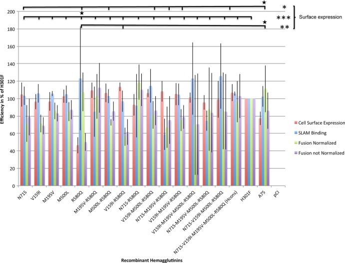
The most striking functional change was observed with CDV H mutant R580Q, which showed a 50% reduction of fusion efficiency compared to that of wild-type H301F, which was arbitrarily set equal to 100%. The other single mutants also showed decreased fusion efficiency, though it was less pronounced, ranging from 69% (V159I) to 87% (M500L) of that of H301F (Fig. 1).
FIG 1.
Combined results of fusion efficiency, cell surface expression, and SLAM binding efficiency of the recombinant H proteins. The combined results of fusion efficiency (both normalized and not normalized to that of wild-type H301F), cell surface expression efficiency, and SLAM binding efficiency are shown. The wild-type, mutant, and control CDV attachment proteins are indicated on the x axis. Efficiency values are expressed as a percentage of the value for wild-type H301F (for which the efficiency was arbitrarily considered to equal 100%) and are shown on the y axis. The values shown here represent the means of triplicate measurements derived from three independent experiments. The error bars show the standard deviations determined for each of the mutant and control attachment proteins. Statistical analysis revealed the presence of significant differences in cell surface expression among the recombinant attachment proteins investigated. Statistical analysis did not reveal statistically significant differences among the mutants when assessed for fusion efficiency (both normalized and not normalized to cell surface expression efficiency), although the P value was relatively low (P = 0.055 for both tests). Similarly, no statistically significant differences were observed (P = 0.1848) among the mutants when their SLAM binding efficiencies were compared. The bars at the top show the statistically significant differences between the recombinant CDV H proteins. Black stars are placed on top of the recombinant that the others mutants are compared to. The asterisks indicate the magnitude of the statistically significant differences (*, P < 0.05; **, P < 0.01; ***, P < 0.001).
However, HCons, which carried all five substitutions, exhibited bioactivity overlapping that of H301F (102%) (Table 2).
TABLE 2.
Amino acid change-associated effects on attachment protein functions
| Amino acid change(s) | Effecta |
||
|---|---|---|---|
| Reducing | Restoring | MS relevance | |
| R580Q | SE | CF | |
| V159I-R580Q | FE | FE | |
| N71S-M195V-R580Q | SLAM B | SLAM B | |
| N71S-M195V-M500L-R580Q | SLAM B | ||
| V159I | SLAM B | ||
| N71S-M195V-M500L | FE | ||
| All but R580Q | SE | ||
SE, surface expression; FE, fusion efficiency; SLAM B, SLAM binding; CF, correct folding; MS, molecular signature.
Based on these findings, we were interested in determining which of the other mutations might have compensated for the monitored defect in cell-to-cell fusion induction of CDV H R580Q. To this aim, a panel of double, triple, and quadruple mutants bearing the R580Q substitution was selected and the mutants were assessed for their ability to induce cell-to-cell fusion; these comprised recombinant mutant H proteins with the following substitutions: M195V-R580Q, M500L-R580Q, V159I-R580Q, N71S-R580Q, N71S-M195V-R580Q, V159I-M195V-R580Q, V159I-M500L-R580Q, V159I-M195V-M500L-R580Q, N71S-M195V-M500L-R580Q, and N71S-V159I-M500L-R580Q. Eight out of these 10 selected CDV H mutants still showed a reduction in cell-to-cell fusion, and this was particularly accentuated for the V159I-R580Q mutant (which had approximately 60% of the fusion efficiency of H301F), although the difference was not statistically significant (Fig. 1). In contrast, two of them (the N71S-R580Q and M195V-R580Q mutants) showed a clear restoration of bioactivity and had fusion values that overlapped or that were slightly higher than the fusion value of H301F. For Hwt, the fusion efficiency was 86% of that of H301F. A complete summary of the statistical results is reported in Fig. 1.
Overall, our results indicate that the R580Q amino acid substitution severely impaired CDV H's bioactivity. However, this negative impact could be efficiently compensated for by the N71S, M500L, and M195V combination of amino acid substitutions.
The CDV H mutant with the R580Q substitution exhibits impaired surface expression that is compensated for by any of the other four coexisting amino acid changes.
It was previously reported that the amount of the H and F proteins expressed at the cell surface also impacted the extent of cell-to-cell fusion (27). We therefore assessed the cell surface expression of the selected panel of H mutants as a measure of a significant step of the whole-cell fusion process (Fig. 1).
Strikingly, among the single mutants, cell surface expression was significantly influenced by CDV H R580Q only, with the efficiency being as low as 46% compared to that for the wild type (H301F) (P < 0.001) (Fig. 1). The other four single mutants did not show any significant variation in cell surface expression.
Of the remaining 10 mutants with multiple substitutions, the substitutions in 7 were associated with a mild increase of cell surface expression (up to 113% for the V159I-R580Q mutant), whereas 3 exhibited a slight reduction, with the reduction being as low as 92% of that for H301F for the N71S-R580Q mutant. While the full mutant (HCons) showed a negligible overexpression (107%), Hwt was characterized by a moderate reduction in intracellular transport competence as low as 77% of that for H301F (Fig. 1).
Taken together, our results suggest that R580Q impacts the overall structure of the attachment protein that translates into folding deficiencies and, consequently, into improper intracellular transport competence. Furthermore, our findings also indicate that folding and, ultimately, transport efficiency could be restored to different extents by any of the additional amino acid changes physically distant from R580Q (Fig. 1; Table 2).
The molecular signature R580Q-V159I of CDV H is detrimental for cell fusion but is fully neutralized by the other three coexisting amino acid changes.
In order to assess if any of the impairment of the fusion support function of the H mutants described above was due only to its deficiency in cell surface expression or was due also to an intrinsic lack of fusion efficiency, the results of the QFA were normalized according to cell surface expression for the respective mutants.
Interestingly, the results shown in Fig. 1 indicate that the fusion support activity of CDV H R580Q was actually almost comparable to that of H301F (107%), consistent with the lack of a direct impact of the R580Q substitution on the intrinsic overall bioactivity of CDV H. Conversely, and of particular interest, the V159I-R580Q mutant exhibited the most defective activity in fusion promotion (56% of that for H301F). Overall, of the five mutants with multiple substitutions and a fusion efficiency lower than 80% of that for H301 (V159I-R580Q, 56%; M500L-R580Q, 72.7%; N71S-M195V-R580Q, 67%; V159I-M195V-R580Q, 75.2%; V159I-M195V-M500L-R580Q, 69.7%), three carried the molecular signature V159I-R580Q, further supporting a direct negative effect of this molecular pattern on the membrane fusion process. Remarkably, the negative functional impact of the molecular signature V159I-R580Q was neutralized to different extents by any of the other three additional amino acid changes and fully neutralized by all of them, when they were present together. Summarized results are shown in Fig. 1.
The loss of SLAM binding efficiency associated with the molecular signature N71S-M195V-R580Q of CDV H is compensated for by an additional amino acid change.
We then investigated the intrinsic ability of the selected H mutants to physically interact with the cSLAM receptor. To test this, we used a previously described semiquantitative receptor-binding assay, where a soluble form of the receptor (scSLAM) is added to the top of H-expressing receptor-negative Vero cells (Fig. 1).
All the single mutants showed an overall H301F-like SLAM binding efficiency. Of the mutants with multiple substitutions (n = 10), two impacted the SLAM binding efficiency of the H proteins in a negative manner, although not statistically significantly (Fig. 1) (59% of the H301F binding efficiency for the N71S-M195V-R580Q mutant and 74% for the N71S-M195V-M500L-R580Q mutant), whereas the remaining eight mutants showed only a mild to negligible influence. Interestingly, residue I194 in MeV H has been shown to play a critical role in SLAM binding activity (28), thereby suggesting that the M195V substitution, when present in association with N71S and R580Q, may be suboptimal for receptor binding. Interestingly, when the V159I substitution was added to the quadruple H mutant carrying the latter receptor-binding-defective molecular signature, the SLAM binding ability was fully restored (Fig. 1; Table 2). Furthermore, the most relevant increment in binding efficiency was observed in association with two mutants carrying, among other substitutions, the V159I residue (the N71S-V159I-M500L-R580Q mutant had 125.8% of the binding efficiency of H301F, and the V159I-M195V-M500L-R580Q mutant had 123.01% of the binding efficiency of H301F). None of the other mutants with multiple substitutions carrying the V159I residue had a SLAM binding efficiency lower than 97% of that for H301F, and none of the other SNPs could similarly compensate for H's receptor-binding activity. Hence, while M195V in combination with N71S and R580Q may directly affect receptor binding, the V159I amino acid substitution might restore receptor binding presumably through a long-range effect. Compared to the binding efficiency of H301F, the full mutant (HCons) and Hwt showed only a minimal increase in binding efficiency (106% and 102%, respectively).
Since the receptor-binding efficiency was assessed with a SLAM molecule of canine origin and because both parental H proteins were derived from CDV strains isolated or detected in foxes, we considered it to be critical to assess the effect of the single amino acid that differs between the fox and canine SLAM sequences (located within the V domain). Both fox and canine SLAM receptors were shown to bind with overlapping efficiency to the two different H proteins, supporting the relevance of our results (data not shown).
The R580Q substitution of the CDV attachment protein impacts the H-F protein interaction but not cell fusion.
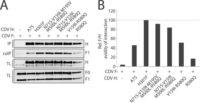
The avidity of the interaction between the H and F proteins was assessed through a coimmunoprecipitation assay for the wild-type H protein and for a selected panel of H mutant proteins shown to have altered functional parameters in the experiments described above (HCons and the R580Q, V159I-R580Q, and N71S-V159I-M500L-R580Q mutants).
Overall, the N71S-V159I-M195V-M500L-R580Q full mutant (HCons) and the N71S-V159I-M500L-R580Q quadruple mutant showed similar avidities for interaction with F protein. However, the V159I-R580Q and R580Q mutants showed reductions in F-protein binding. Intriguingly, although the V159I-R580Q mutant bound F protein with values similar to those for Hwt (about 50% of that for H301F), its fusion efficiency was remarkably lower (Fig. 1 and 2). This finding is consistent with a loss of fusion efficiency (putatively caused by receptor-induced conformational changes of H leading to triggering of F protein), which would be impaired by the presence of the V159I substitution.
FIG 2.

H-F protein coimmunoprecipitation. (A) Western blot. Results for the immunoprecipitated (IP) wild-type and mutant H proteins, along with the coimmunoprecipitated (co-IP) F protein, are shown. As a control, the total H and F proteins present in the lysates were also revealed by immunoblotting (TL). H, H monomers; F0, uncleaved F precursors; F1, cleaved F precursors. (B) Graphic analysis of the Western blotting results. The bars in the histogram express the avidity of the F-H protein interaction. The avidity values were obtained through the following formula: (co-IP F/total F)/(IP H/total H). All the values were normalized to the value for the interaction between H301F and the F protein, which was arbitrarily considered to equal 100%.
As expected for a folding-deficient H mutant, the R580Q mutant showed the lowest avidity for F protein (Fig. 2). Strikingly, the extent of this loss of avidity for F protein did not appear to translate into detrimental fusion promotion, further supporting the finding that the R580Q substitution does not directly affect the intrinsic bioactivity of CDV H (Fig. 1).
Structural mapping of the identified residues reveals remarkable CDV H plasticity though long-range effects.
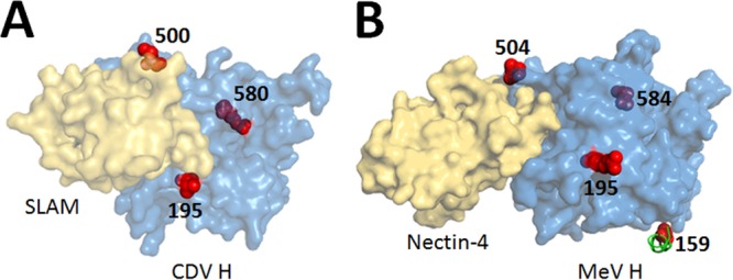
We finally sought to investigate whether the amino acid changes described in our study were mapping to known functional domains encompassed within the H structure. To this aim, we generated a structural homology model based on the recently solved measles virus H/SLAM cocrystal structure (29). In this model, residues 195, 500, and 580 are located in the head domain (Fig. 3). Positions 195 and 500 are mostly solvent exposed and are approximately located at the boundary of the cSLAM binding site, whereas amino acid 580 is buried within the beta-propeller at the C-terminal end of the head at the top of sheet 6 (Fig. 3A). Conversely, residue 71 maps to the section of the stalk domain proximal to the membrane, in a region that was not visible in the H/SLAM cocrystal structure, and, hence, is not visible in our model. Although the residue at position 159 was also absent from the H/SLAM complex (29), it was nevertheless visible in the recently determined atomic structure of MeV H bound to nectin-4 (30). Because the stalk is well conserved among morbilliviruses, we used these atomic coordinates to highlight the putative position 159 in CDV H. In this configuration, residue 159 lies in a short alpha-helix making contact with the beta-propeller in a region that links the head domain distant from the membrane to the stalk domain proximal to the membrane (Fig. 3B).
FIG 3.

Attachment (H) proteins bound to their receptor molecules. (A) Structural homology model of the steric arrangement of one monomeric head domain of CDV H (light blue globular structure) bound to the immune cell receptor (SLAM; light tan globular structure). The amino acid changes identified in this study are highlighted in red (M195V, R580Q, M500L). Note that residues V159I and N71S are not visible in the model. (B) Crystal structure of the measles virus H protein (light blue globular structure) bound to its epithelial cell receptor (nectin-4; light tan globular structure). Note that in this atomic structure, residue V159I is visible, whereas N71S is not. Figures were generated using the PyMOL (v0.99) program.
DISCUSSION
The attachment protein of CDV has been shown to be under major evolutionary pressure, resulting in frequent naturally occurring mutations (3, 16–18). The impact of these mutations on the different functions of this protein is unclear. In an attempt to partially address this question, we investigated the nature of a series of missense SNPs naturally accumulating within the H-gene sequences during the transition between two distinct, yet related, CDV strains. Furthermore, we investigated the effect of these SNPs on the functional properties of the CDV attachment protein. For this purpose, fusion efficiency was dissected into three of its major determinants, i.e., cell surface expression, SLAM binding, and the H-F protein binding necessary for triggering of the F protein.
Despite the theoretically unlimited numbers of amino acid combinations that might have occurred at the five evolutionarily selected positions of the attachment protein of the CDV strains described in this study, our results highlight that the majority of the amino acid substitutions are redundant among the distinct CDV genotypes and to a certain extent also within the other morbilliviruses (Table 1). The only exception to this rule appears to be R580Q, which, with the HCons-specific residue (Q), was observed only in the consensus sequence and in four South American strains but in none of the sequences of the other most well characterized CDV genotypes or of other morbilliviruses (Table 1). This finding is consistent with our experimental results that showed how this substitution severely impaired the attachment protein surface expression, fusion efficiency, and H-F protein interaction efficiency when present as a single amino acid change (Fig. 1 and 2). The nature of the in vitro systems that we used for the assays described in this article did not allow us to conclusively determine if these effects are indeed detrimental to the fitness of CDV. As a matter of fact, for human parainfluenza virus, another paramyxovirus, it has been shown that a loss of fusion efficiency translated into an increase of viral fitness (31). However, here we demonstrated that an overall overlapping functional efficiency is maintained in the attachment protein of related, yet distinct, CDV strains, despite the presence of 5 relevant amino acid changes. This observation suggests that the consensus sequence that emerges among the myriad possible combinations within the quasispecies environment is selected according to functional standards, which remain constant through time, hosts, and geography, similar to what occurs for other viral functions, such as the fidelity of RNA-dependent RNA polymerases, which is fixed within a narrow range (32). It appears, then, that the loss of function secondary to the R580Q substitution would indeed be detrimental to viral fitness.
The selection of a destabilizing change, such as that primed by R580Q, either alone or in combination, which does not appear to contribute positively to viral fitness, would be unlikely to occur if it is not associated with any advantage for the virus. In the case of influenza virus, it was shown that the introduction of an amino acid residue relevant for immune escape had a detrimental effect on viral fitness. A compensatory SNP was shown to occur and partially restored viral fitness (33). Although in our investigation we could not provide a direct line of evidence in this direction, we have shown the existence of a positively selected amino acid change with detrimental functional effects on the attachment protein of CDV that was compensated for by a number of additional amino acid substitutions (Table 2).
Besides the possible antigenic relevance of these changes, we speculate that outcomes from these changes might also be at the base of the molecular plasticity that has allowed CDV to become such a successful viral agent with a continuously growing host range (19, 34–36).
It is not clear in which sequence the selected SNPs would occur in the CDV attachment protein and, more specifically, if the compensatory changes would precede or follow the selection of the destabilizing change. However, it is known that destabilizing molecular changes such as 580Q have been described to be selected only after the occurrence of other compensatory ones, according to the theory of stability-mediated epistasis (37). In this scenario, the negligible functional effect observed for the majority of the amino acid changes investigated might instead represent a critical compensatory platform for the occurrence of destabilizing changes. In this system, specific amino acid combinations would influence the different functional aspects of the attachment protein, with each amino acid change possibly having pleiotropic effects, either functional or structural. The final outcome of such an articulated system may translate into the overall preservation of the biological activity of the attachment protein while allowing the selection of major destabilizing changes likely to be critical for CDV virulence and infectivity. This is the first time, to the best of our knowledge, that a similar finding has been described in CDV.
ACKNOWLEDGMENTS
We thank all the technical personnel of FIWI and of the Division of Experimental Clinical Research of the University of Bern and, in particular, Ljerka Zipperle for her constant help. We also thank Helmut Segner for a critical review of the manuscript and Vanessa Schumacher and Ruth Parham for linguistic editing of the manuscript.
This work was supported in part by the Swiss National Science Foundation (reference no. 310030_153281 to Philippe Plattet).
Footnotes
Published ahead of print 7 May 2014
REFERENCES
- 1.Williams ES, Thorne ET. 1996. Infectious and parasitic diseases of captive carnivores, with special emphasis on the black-footed ferret (Mustela nigripes). Rev. Sci. Tech. 15:91–114 [DOI] [PubMed] [Google Scholar]
- 2.Lednicky JA, Dubach J, Kinsel MJ, Meehan TP, Bocchetta M, Hungerford LL, Sarich NA, Witecki KE, Braid MD, Pedrak C, Houde CM. 2004. Genetically distant American canine distemper virus lineages have recently caused epizootics with somewhat different characteristics in raccoons living around a large suburban zoo in the USA. Virol. J. 1:2. 10.1186/1743-422X-1-2 [DOI] [PMC free article] [PubMed] [Google Scholar]
- 3.Pardo IDR, Johnson GC, Kleiboeker SB. 2005. Phylogenetic characterization of canine distemper viruses detected in naturally infected dogs in North America. J. Clin. Microbiol. 43:5009–5017. 10.1128/JCM.43.10.5009-5017.2005 [DOI] [PMC free article] [PubMed] [Google Scholar]
- 4.von Messling V, Zimmer G, Herrler G, Haas L, Cattaneo R. 2001. The hemagglutinin of canine distemper virus determines tropism and cytopathogenicity. J. Virol. 75:6418–6427. 10.1128/JVI.75.14.6418-6427.2001 [DOI] [PMC free article] [PubMed] [Google Scholar]
- 5.Wyss-Fluehmann G, Zurbriggen A, Vandevelde M, Plattet P. 2010. Canine distemper virus persistence in demyelinating encephalitis by swift intracellular cell-to-cell spread in astrocytes is controlled by the viral attachment protein. Acta Neuropathol. 119:617–630. 10.1007/s00401-010-0644-7 [DOI] [PMC free article] [PubMed] [Google Scholar]
- 6.Noyce RS, Delpeut S, Richardson CD. 2013. Dog nectin-4 is an epithelial cell receptor for canine distemper virus that facilitates virus entry and syncytia formation. Virology 436:210–220. 10.1016/j.virol.2012.11.011 [DOI] [PubMed] [Google Scholar]
- 7.Pratakpiriya W, Seki F, Otsuki N, Sakai K, Fukuhara H, Katamoto H, Hirai T, Maenaka K, Techangamsuwan S, Lan NT, Takeda M, Yamaguchi R. 2012. Nectin4 is an epithelial cell receptor for canine distemper virus and involved in the neurovirulence. J. Virol. 86:10207–10210. 10.1128/JVI.00824-12 [DOI] [PMC free article] [PubMed] [Google Scholar]
- 8.Tatsuo H, Ono N, Yanagi Y. 2001. Morbilliviruses use signaling lymphocyte activation molecules (CD150) as cellular receptors. J. Virol. 75:5842–5850. 10.1128/JVI.75.13.5842-5850.2001 [DOI] [PMC free article] [PubMed] [Google Scholar]
- 9.Sawatsky B, Wong XX, Hinkelmann S, Cattaneo R, von Messling V. 2012. Canine distemper virus epithelial cell infection is required for clinical disease but not for immunosuppression. J. Virol. 86:3658–3666. 10.1128/JVI.06414-11 [DOI] [PMC free article] [PubMed] [Google Scholar]
- 10.Ader N, Brindley MA, Avila M, Origgi FC, Langedijk JP, Örvell C, Vandevelde M, Zurbriggen A, Plemper RK, Plattet P. 2012. Structural rearrangements of the central region of the morbillivirus attachment protein stalk domain trigger F protein refolding for membrane fusion. J. Biol. Chem. 287:16324–16334. 10.1074/jbc.M112.342493 [DOI] [PMC free article] [PubMed] [Google Scholar]
- 11.Bose S, Zokarkar A, Welch BD, Leser GP, Jardetzky TS, Lamb RA. 2012. Fusion activation by a headless parainfluenza virus 5 hemagglutinin-neuraminidase stalk suggests a modular mechanism for triggering. Proc. Natl. Acad. Sci. U. S. A. 109:E2625–E2634. 10.1073/pnas.1213813109 [DOI] [PMC free article] [PubMed] [Google Scholar]
- 12.Brindley MA, Takeda M, Plattet P, Plemper RK. 2012. Triggering the measles virus membrane fusion machinery. Proc. Natl. Acad. Sci. U. S. A. 109:E3018–E3027. 10.1073/pnas.1210925109 [DOI] [PMC free article] [PubMed] [Google Scholar]
- 13.Liu Q, Stone JA, Bradel-Tretheway B, Dabundo J, Benavides Montano JA, Santos-Montanez J, Biering SB, Nicola AV, Iorio RM, Lu X, Aguilar HC. 2013. Unraveling a three-step spatiotemporal mechanism of triggering of receptor-induced Nipah virus fusion and cell entry. PLoS Pathog. 9:e1003770. 10.1371/journal.ppat.1003770 [DOI] [PMC free article] [PubMed] [Google Scholar]
- 14.Navaratnarajah CK, Negi S, Braun W, Cattaneo R. 2012. Membrane fusion triggering: three modules with different structure and function in the upper half of the measles virus attachment protein stalk. J. Biol. Chem. 287:38543–38551. 10.1074/jbc.M112.410563 [DOI] [PMC free article] [PubMed] [Google Scholar]
- 15.Navaratnarajah CK, Oezguen N, Rupp L, Kay L, Leonard VH, Braun W, Cattaneo R. 2011. The heads of the measles virus attachment protein move to transmit the fusion-triggering signal. Nat. Struct. Mol. Biol. 18:128–134. 10.1038/nsmb.1967 [DOI] [PMC free article] [PubMed] [Google Scholar]
- 16.Bolt G, Jensen TD, Gottschalck E, Arctander P, Appel MJ, Buckland R, Blixenkrone-Møller M. 1997. Genetic diversity of the attachment (H) protein gene of current field isolates of canine distemper virus. J. Gen. Virol. 78:367–372 [DOI] [PubMed] [Google Scholar]
- 17.Martella V, Cirone F, Elia G, Lorusso E, Decaro N, Campolo M, Desario C, Lucente MS, Bellacicco AL, Blixenkrone-Møller M, Carmichael LE, Buonavoglia C. 2006. Heterogeneity within the hemagglutinin genes of canine distemper virus (CDV) strains detected in Italy. Vet. Microbiol. 116:301–309. 10.1016/j.vetmic.2006.04.019 [DOI] [PubMed] [Google Scholar]
- 18.Mochizuki M, Hashimoto M, Hagiwara S, Yoshida Y, Ishiguro S. 1999. Genotypes of canine distemper virus determined by analysis of the hemagglutinin genes of recent isolates from dogs in Japan. J. Clin. Microbiol. 37:2936–2942 [DOI] [PMC free article] [PubMed] [Google Scholar]
- 19.Origgi FC, Plattet P, Sattler U, Robert N, Casaubon J, Mavrot F, Pewsner M, Wu N, Giovannini S, Oevermann A, Stoffel MH, Gaschen V, Segner H, Ryser-Degiorgis MP. 2012. Emergence of canine distemper virus strains with modified molecular signature and enhanced neuronal tropism leading to high mortality in wild carnivores. Vet. Pathol. 49:913–929. 10.1177/0300985812436743 [DOI] [PubMed] [Google Scholar]
- 20.Sekulin K, Hafner-Marx A, Kolodziejek J, Janik D, Schmidt P, Nowotny N. 2011. Emergence of canine distemper in Bavarian wildlife associated with a specific amino acid exchange in the haemagglutinin protein. Vet. J. 187:399–401. 10.1016/j.tvjl.2009.12.029 [DOI] [PubMed] [Google Scholar]
- 21.Zipperle L, Langedijk JP, Orvell C, Vandevelde M, Zurbriggen A, Plattet P. 2010. Identification of key residues in virulent canine distemper virus hemagglutinin that control CD150/SLAM-binding activity. J. Virol. 84:9618–9624. 10.1128/JVI.01077-10 [DOI] [PMC free article] [PubMed] [Google Scholar]
- 22.Von Messling V, Springfeld C, Devaux P, Cattaneo R. 2003. A ferret model of canine distemper virus virulence and immunosuppression. J. Virol. 77:12579–12591. 10.1128/JVI.77.23.12579-12591.2003 [DOI] [PMC free article] [PubMed] [Google Scholar]
- 23.Langedijk JP, Janda J, Origgi FC, Örvell C, Vandevelde M, Zurbriggen A, Plattet P. 2011. Canine distemper virus infects canine keratinocytes and immune cells by using overlapping and distinct regions located on one side of the attachment protein. J. Virol. 85:11242–11254. 10.1128/JVI.05340-11 [DOI] [PMC free article] [PubMed] [Google Scholar]
- 24.Plattet P, Langedijk JP, Zipperle L, Vandevelde M, Orvell C, Zurbriggen A. 2009. Conserved leucine residue in the head region of morbillivirus fusion protein regulates the large conformational change during fusion activity. Biochemistry 48:9112–9121. 10.1021/bi9008566 [DOI] [PubMed] [Google Scholar]
- 25.Orvell C, Sheshberadaran H, Norrby E. 1985. Preparation and characterization of monoclonal antibodies directed against four structural components of canine distemper virus. J. Gen. Virol. 66:443–456. 10.1099/0022-1317-66-3-443 [DOI] [PubMed] [Google Scholar]
- 26.Cherpillod P, Beck K, Zurbriggen A, Wittek R. 1999. Sequence analysis and expression of the attachment and fusion proteins of canine distemper virus wild-type strain A75/17. J. Virol. 73:2263–2269 [DOI] [PMC free article] [PubMed] [Google Scholar]
- 27.Whitman SD, Dutch RE. 2007. Surface density of the Hendra G protein modulates Hendra F protein-promoted membrane fusion: role for Hendra G protein trafficking and degradation. Virology 363:419–429. 10.1016/j.virol.2007.01.029 [DOI] [PMC free article] [PubMed] [Google Scholar]
- 28.Navaratnarajah CK, Vongpunsawad S, Oezguen N, Stehle T, Braun W, Hashiguchi T, Maenaka K, Yanagi Y, Cattaneo R. 2008. Dynamic interaction of the measles virus hemagglutinin with its receptor signaling lymphocytic activation molecule (SLAM, CD150). J. Biol. Chem. 283:11763–11771. 10.1074/jbc.M800896200 [DOI] [PMC free article] [PubMed] [Google Scholar]
- 29.Hashiguchi T, Maenaka K, Yanagi Y. 2011. Measles virus hemagglutinin: structural insights into cell entry and measles vaccine. Front. Microbiol. 2:247. 10.3389/fmicb.2011.00247 [DOI] [PMC free article] [PubMed] [Google Scholar]
- 30.Zhang X, Lu G, Qi J, Li Y, He Y, Xu X, Shi J, Zhang CW, Yan J, Gao GF. 2013. Structure of measles virus hemagglutinin bound to its epithelial receptor nectin-4. Nat. Struct. Mol. Biol. 20:67–72. 10.1038/nsmb.2432 [DOI] [PubMed] [Google Scholar]
- 31.Palmer SG, Porotto M, Palermo LM, Cunha LF, Greengard O, Moscona A. 2012. Adaptation of human parainfluenza virus to airway epithelium reveals fusion properties required for growth in host tissue. mBio 3(3):e00137-12. 10.1128/mBio.00137-12 [DOI] [PMC free article] [PubMed] [Google Scholar]
- 32.Domingo E, Sheldon J, Perales C. 2012. Viral quasispecies evolution. Microbiol. Mol. Biol. Rev. 76:159–216. 10.1128/MMBR.05023-11 [DOI] [PMC free article] [PubMed] [Google Scholar]
- 33.Rimmelzwaan GF, Berkhoff EG, Nieuwkoop NJ, Fouchier RA, Osterhaus AD. 2004. Functional compensation of a detrimental amino acid substitution in a cytotoxic/T/lymphocyte epitope of influenza A viruses by comutation. J. Virol. 78:8946–8949. 10.1128/JVI.78.16.8946-8949.2004 [DOI] [PMC free article] [PubMed] [Google Scholar]
- 34.Origgi FC, Sattler U, Pilo P, Waldvogel AS. 2013. Fatal combined infection with canine distemper virus and orthopoxvirus in a group of Asian marmots (Marmota caudata). Vet. Pathol. 50:914–920. 10.1177/0300985813476060 [DOI] [PubMed] [Google Scholar]
- 35.Sakai K, Yoshikawa T, Seki F, Fukushi S, Tahara M, Nagata N, Ami Y, Mizutani T, Kurane I, Yamaguchi R, Hasegawa H, Saijo M, Komase K, Morikawa S, Takeda M. 2013. Canine distemper virus associated with a lethal outbreak in monkeys can readily adapt to use human receptors. J. Virol. 87:7170–7175. 10.1128/JVI.03479-12 [DOI] [PMC free article] [PubMed] [Google Scholar]
- 36.Bieringer M, Han JW, Kendl S, Khosravi M, Plattet P, Schneider-Schaulies J. 2013. Experimental adaptation of wild-type canine distemper virus (CDV) to the human entry receptor CD150. PLoS One 8:e57488. 10.1371/journal.pone.0057488 [DOI] [PMC free article] [PubMed] [Google Scholar]
- 37.Gong LI, Suchard MA, Bloom JD. 2013. Stability-mediated epistasis constrains the evolution of an influenza protein. eLife 2:e00631. 10.7554/eLife.00631 [DOI] [PMC free article] [PubMed] [Google Scholar]


