In this issue of the JCI, J. Alastair Gracie et al. highlight the central importance of IL-18 among several novel cytokines that participate in erosive, inflammatory arthritis (1). The authors base their arguments on experimental results obtained in vitro with human synovial tissue and in vivo with mice immunized with collagen in the presence of incomplete Freund’s adjuvant.
IL-18 and its receptors.
The pioneering work of purifying IL-18 can be attributed to Nakamura and Okamura et al. (see review in refs. 2–4), who in 1989 described an endotoxin-induced serum factor that stimulates IFN-γ production. In 1995, the same group demonstrated that this IFN-γ−inducing factor (IGIF) was present in liver extract, and shortly thereafter, they proceeded to clone its cDNA. Based on its predicted secondary structure, murine IGIF appears to be closely related to human IL-1β and IL-1α, although the primary sequence identity between these proteins is modest. It was originally suggested that the name “IGIF” should be changed to “IL-1γ,” but IGIF does not bind to IL-1RI, and “IL-18” is now the accepted name for this factor. Several excellent review articles on IL-18 have been published recently (2–4).
The first receptor for IL-18 to be identified, now known as “IL-18Rβ,” was characterized earlier as an IL-1 receptor–related protein (5), but IL-1α and -1β bind to this receptor poorly, if at all, and IL-18 also binds it with low affinity (in the range of 10–8 M). However, the recent development of IL-18R–deficient mice confirmed that this receptor is also important for IL-18 signaling (6). A second receptor subunit, initially termed “accessory protein-like receptor” (AcPR) but now called “IL-18Rα,” was also recently cloned and shown to be essential for signaling (7). An IL-18 binding protein (IL-1BP) purified from normal urine is not a variant of a cell surface receptor, but rather is part of a novel family of secreted proteins. This family includes several poxvirus-encoded proteins, which are presumed to act as inhibitors of IL-18 signaling (8). Indeed, administration of IL-18 binding protein prior to LPS injection abrogates the induction of IFN-γ in mice.
Like many other cytokines, IL-18 is pleiotropic, acting in both acquired and innate immunity. In addition to its costimulatory functions on Th1 cytokines, IL-18 helps activate natural killer cells to express Fas ligand, and it acts directly as a proinflammatory cytokine by inducing the CC and CXC chemokines, IL-8, and IL-18 itself, respectively (9). Synergism between IL-18 and IL-12 is mainly due to IL-12 increasing the expression of IL-18 receptors on Th0 cells and B cells (2). Because of the latter mechanism, this synergism is important to the production of IFN-γ in T cells and natural killer cells. While IL-18 itself does not induce naive CD4+ T cells to differentiate, it promotes the action of IL-12, which favors differentiation along the Th1 lineage. IL-18 also induces production of GM-CSF, IL-2, IL-2Rα, TNF-α, prostaglandin E2, and inducible nitric oxide synthase (iNOS) by mononuclear and mesenchymal cells (2, 4).
IL-18 in human disease.
IL-18 mRNA appears to be widely, but not ubiquitously, expressed (4). Based on the results of Gracie et al. in this issue of the JCI, inflamed synovial tissue, particularly in rheumatoid arthritis, can now be added to the list of tissues that express this factor. IL-18 is found primarily in lymphocytic aggregates, but its precise localization remains unclear.
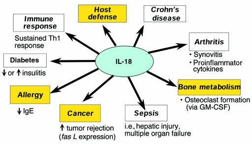
Roles for IL-18 in various pathological conditions are currently of great interest. Figure 1 summarizes several of IL-18’s emerging functions in promoting or suppressing disease. In infectious diseases, IL-18 plays a crucial part in promoting the inflammatory response during bacterial sepsis, a response that can lead to hepatic injury and multiple organ failure (2). On the other hand, like many proinflammatory cytokines, IL-18 appears to act in host defense. For example, in one report, pretreatment with IL-18 prevented or minimized the severity of experimentally induced bacterial infection (10). Therapies that target IL-18 must therefore be approached with caution, because of their possible effects on IFN-γ and hence on the control of intracellular pathogens by macrophages. IL-18 is also instrumental in the suppression of allergy, because it inhibits IgE production (11). With regard to type 1 diabetes, IL-18 favors the development of insulitis in nonobese diabetic mice. Furthermore, the mouse IL-18 gene maps near the Idd2 interval, raising the possibility that recessive defects in the IL-18 gene (along with at least 2 other interacting genes) contribute to the nonobese diabetic phenotype (12). However, in other studies, IL-18 was shown to decrease insulitis (13).
Figure 1.
Potential roles for IL-18 in various pathological conditions. Yellow highlighting indicates a potentially beneficial effect of IL-18.
IL-18 in rheumatoid arthritis.
The observations of Gracie et al. in this issue of the JCI indicate that IL-18 may play an additional pathogenic role in rheumatoid arthritis, a disease that is driven by Th1 responses. IL-18 appears to synergize with IL-12 and IL-15 to activate production by synovial tissue of IFN-γ and of IL-18 itself. Three observations are noteworthy. First, GM-CSF and NO production by macrophages is induced by IL-18 and is not dependent on IL-12 and IL-15. This contrasts with the synergism of these 3 cytokines in regulating TNF-α production. Second, a counter-regulatory loop may exist through NO production, because NO inhibits the IL-1β–converting enzyme (ICE) and thus blocks the processing of proIL-18 into a biologically active cytokine. Thus, by inducing NO, IL-18 decreases its own activity. Third, IL-18 production by synovial tissue is upregulated by TNF-α and IL-1β.
The local activation of monocyte-macrophages leads to the production of IL-1, TNF-α, and matrix metalloproteinases (MMPs) in the inflammatory lesion. This activation is mediated by cytokines and immune complexes, but primarily by cell-cell contact with activated T cells or synovial cells (14–16). Like IL-15, IL-18 may potentiate cell-cell contact in inflamed tissue, such as synovia in rheumatoid arthritis.
A recent study identified active ICE in human articular cartilage (17). ICE was markedly increased in osteoarthritic tissue, and led to the production of both active IL-1β and IL-18. The increased production of IL-18 in synovia from patients with rheumatoid arthritis and in mucosal intestinal tissue in Crohn’s disease has also been reported (18, 19). It has always been surprising that relatively low IFN-γ levels should be detected in rheumatoid arthritis synovitis, despite the presence of Th1 cells. One explanation, proposed by Gracie et al., holds that IL-18 sustains the Th1 phenotype without inducing persistently high levels of IFN-γ production because inhibitory molecules, such as IL-10 and TGF-β, are also highly expressed.
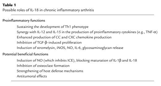
The present report agrees with another recent study (20) that shows that IL-18 induces proinflammatory and catabolic responses in normal human cartilage. Although these findings provide a rationale for blocking IL-18 in treating rheumatoid arthritis, the beneficial effects of this cytokine, shown in Table 1, justify caution. In addition to its above-described roles in host defense and the suppression of allergies, IL-18 induces NO, which can block ICE activity. Therefore, inhibition of IL-18 function could increase ICE activity and promote the maturation of IL-18 and IL-1β, thereby promoting the inflammation and tissue destruction that one is attempting to control. Moreover, IL-18 may inhibit osteoclast formation (21), so blocking IL-18 activity may have long-term effects on bone metabolism, perhaps causing osteoporosis.
Table 1.
Possible roles of IL-18 in chronic inflammatory arthritis
The dilemma regarding the role of this cytokine persists: Does IL-18 protect tissues from infection, or does it mediate deleterious effects during sepsis? Its roles in acute or chronic inflammatory conditions and in long-term physiological tissue remodeling are ambiguous. Careful monitoring of relevant biological markers is necessary, even in phase I and phase II clinical trials, to avoid undesirable long-term consequences of therapies that seek to suppress the activity of this pleiotropic factor.
References
- 1.Gracie JA, et al. A proinflammatory role for IL-18 in rheumatoid arthritis. J Clin Invest. 1999;104:1393–1401. doi: 10.1172/JCI7317. [DOI] [PMC free article] [PubMed] [Google Scholar]
- 2.Okamura H, Tsutsui H, Kashiwamura S-I, Yoshimoto T, Nakanishi K. Interleukin-18: a novel cytokine that augments both innate and acquired immunity. Adv Immunol. 1998;70:281–312. doi: 10.1016/s0065-2776(08)60389-2. [DOI] [PubMed] [Google Scholar]
- 3.Dinarello CA, et al. Overview of interleukin-18: more than an interferon-γ inducing factor. J Leukoc Biol. 1998;63:658–664. [PubMed] [Google Scholar]
- 4.Gillespie MT, Horwood NJ. Interleukin-18: perspectives on the newest interleukin. Cytokine Growth Factor Rev. 1998;9:109–116. doi: 10.1016/s1359-6101(98)00004-5. [DOI] [PubMed] [Google Scholar]
- 5.Parnet P, Garka KE, Bonnert TP, Dower SK, Sims JE. IL-1Rrp is a novel receptor-like molecule similar to the type I interleukin-1 receptor and its homologues T1/ST2 and IL-1R AcP. J Biol Chem. 1996;271:3967–3970. doi: 10.1074/jbc.271.8.3967. [DOI] [PubMed] [Google Scholar]
- 6.Hoshino K, et al. Cutting edge: generation of IL-18 receptor-deficient mice: evidence for IL-1 receptor-related protein as an essential IL-18 binding receptor. J Immunol. 1999;162:5041–5044. [PubMed] [Google Scholar]
- 7.Born TL, Thomassen E, Bird TA, Sims JE. Cloning of a novel receptor subunit, AcPL, required for interleukin-18 signaling. J Biol Chem. 1998;273:29445–29450. doi: 10.1074/jbc.273.45.29445. [DOI] [PubMed] [Google Scholar]
- 8.Novick D, et al. Interleukin-18 binding protein: a novel modulator of the Th1 cytokine response. Immunity. 1999;10:127–136. doi: 10.1016/s1074-7613(00)80013-8. [DOI] [PubMed] [Google Scholar]
- 9.Puren AJ, Fantuzzi G, Gu Y, Su MSS, Dinarello CA. Interleukin-18 (IFN-—inducing factor) induces IL-1β and IL-8 via TNF-α production from non-CD14+ human blood mononuclear cells. J Clin Invest. 1998;101:711–721. doi: 10.1172/JCI1379. [DOI] [PMC free article] [PubMed] [Google Scholar]
- 10.Kawakami K, et al. IL-18 protects mice against pulmonary and disseminated infection with Cryptococcus neoformans by inducing IFN-γ production. J Immunol. 1997;159:5528–5534. [PubMed] [Google Scholar]
- 11.Yoshimoto T, Okamura H, Tagawa Y, Iwakura Y, Nakanishi K. Interleukin 18 together with interleukin 12 inhibits IgE production by induction of interferon-γ production from activated B cells. Proc Natl Acad Sci USA. 1997;94:3948–3953. doi: 10.1073/pnas.94.8.3948. [DOI] [PMC free article] [PubMed] [Google Scholar]
- 12.Rothe H, Jenkins NA, Copeland NG, Kolb H. Active stage of autoimmune diabetes is associated with the expression of a novel cytokine, IGIF, which is located near Idd2. J Clin Invest. 1997;99:469–474. doi: 10.1172/JCI119181. [DOI] [PMC free article] [PubMed] [Google Scholar]
- 13.Rothe H, et al. IL-18 inhibits diabetes development in nonobese diabetic mice by counterregulation of Th1-dependent destructive insulitis. J Immunol. 1999;163:1230–1236. [PubMed] [Google Scholar]
- 14.Vey E, Zhang J-H, Dayer J-M. IFN-γ and 1,25(OH)2D3 induce on THP-1 cells distinct patterns of cell surface antigen expression, cytokine production, and responsiveness to contact with activated T cells. J Immunol. 1992;149:2040–2046. [PubMed] [Google Scholar]
- 15.Burger D, et al. Imbalance between interstitial collagenase (MMP-1) and tissue inhibitor of metalloproteinases-1 (TIMP-1) in synoviocytes and fibroblasts upon direct contact with stimulated T lymphocytes: involvement of membrane-associated cytokines. Arthritis Rheum. 1998;41:1748–1759. doi: 10.1002/1529-0131(199810)41:10<1748::AID-ART7>3.0.CO;2-3. [DOI] [PubMed] [Google Scholar]
- 16.McInnes IB, Leung BP, Sturrock RD, Field M, Liew FY. Interleukin-15 mediates T cell-dependent regulation of tumour necrosis factor-alpha production in rheumatoid arthritis. Nat Med. 1997;3:189–195. doi: 10.1038/nm0297-189. [DOI] [PubMed] [Google Scholar]
- 17.Saha N, et al. Interleukin-1β-converting enzyme/caspase-1 in human osteoarthritic tissues. Localization and role in the maturation of interleukin-1β and interleukin-18. Arthritis Rheum. 1999;42:1577–1587. doi: 10.1002/1529-0131(199908)42:8<1577::AID-ANR3>3.0.CO;2-Z. [DOI] [PubMed] [Google Scholar]
- 18.Monteleone G, et al. Bioactive IL-18 expression is up-regulated in Crohn’s disease. J Immunol. 163:143–147. [PubMed] [Google Scholar]
- 19.Pizarro TT, et al. IL-18, a novel immunoregulatory cytokine, is up-regulated in Crohn’s disease: expression and localization in intestinal mucosal cells. J Immunol. 1999;162:6829–6835. [PubMed] [Google Scholar]
- 20.Olee T, Hashimoto S, Quach J, Lotz M. IL-18 is produced by articular chondrocytes and induces proinflammatory and catabolic responses. J Immunol. 1999;162:1096–1100. [PubMed] [Google Scholar]
- 21.Horwood NJ, et al. Interleukin 18 inhibits osteoclast formation via T cell production of granulocyte macrophage colony-stimulating factor. J Clin Invest. 1998;101:595–603. doi: 10.1172/JCI1333. [DOI] [PMC free article] [PubMed] [Google Scholar]


