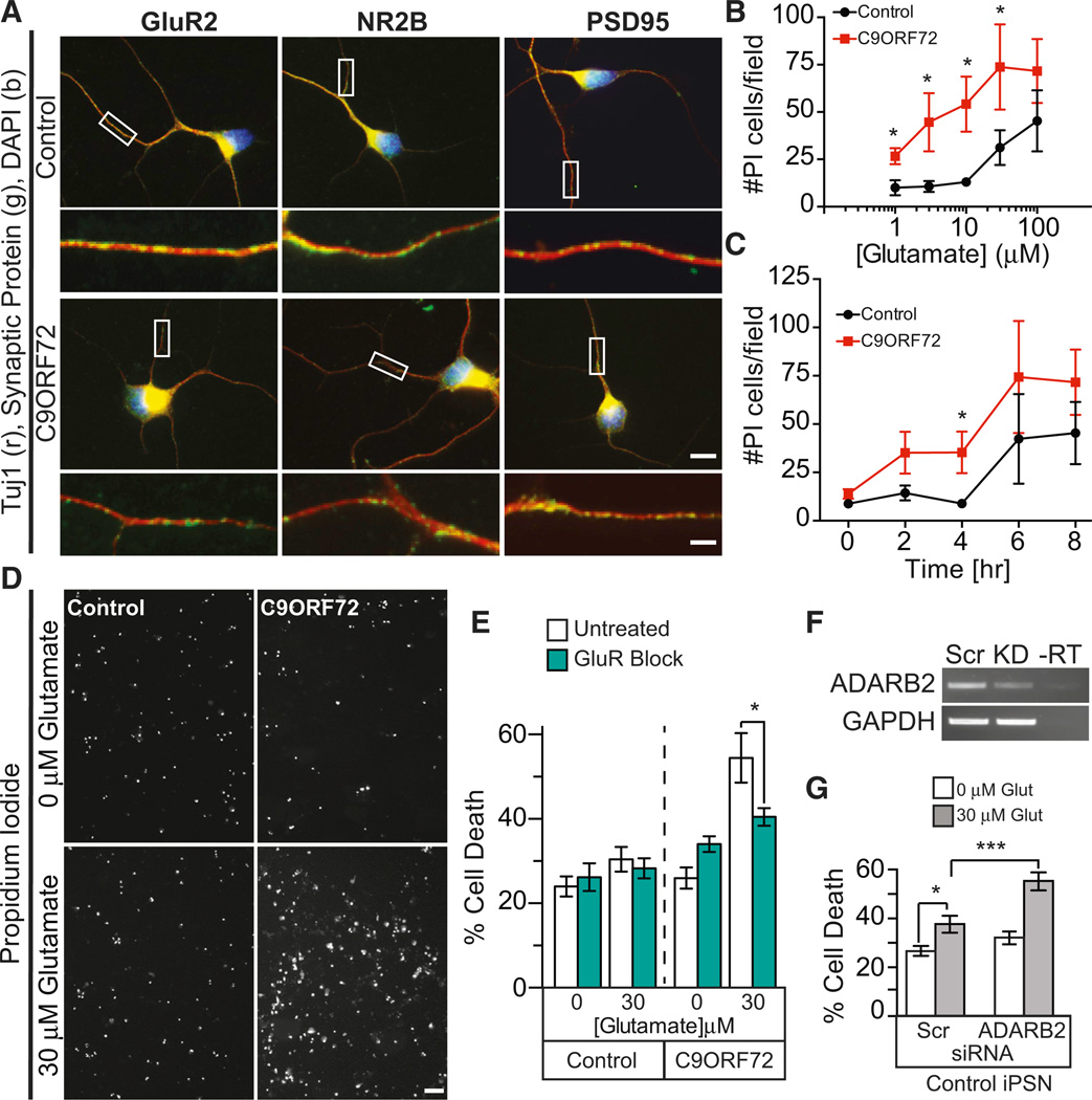
Figure 6. C9ORF72 ALS iPSC Neurons Are Highly Susceptible to Glutamate Toxicity.
(A) Immunofluorescent staining of control and C9ORF72 iPSN cultures show expression of glutamate receptors GluR2, NR2B, and post-synaptic density protein PSD95 at comparable levels as determined by qualitative analysis. Box indicates region of high magnification seen below each image (scale = 10 mm [top] and 2.5 mm [bottom]).
(B) Dose response curve of control and C9ORF72 iPSC neurons revealed that C9ORF72 iPSNs are highly susceptible to glutamate excitotoxicity at 1, 3, 10, and 30 µM concentrations after 8 hr of treatment by popidium iodide staining.
(C) Glutamate-induced excitotoxicity of C9ORF72 iPSNs shows statistically significant cell death after 4 hr of 30 µM glutamate treatment when compared to control iPSNs.
(D) Representative image of propidium iodide staining of control and C9ORF72 ALS iPSN after 4 hr of 0 and 30 µM glutamate treatment; note the increased prodium iodide signal in the C9ORF72 iPSNs as compared to the control iPSNs.
(E) Blocking glutamate receptors prevents glutamate-induced C9ORF72 iPSN cell death (4 hr, 30 µM glutamate).
(F and G) Knockdown of ADARB2 via siRNA treatment resulted in a statistically significant increased susceptibility to glutamate-induced excitotoxicity in control non-C9ORF72 iPSNs at 4 hr, 30 µM glutamate treatment.
Data in (B), (C), (E), and (G) indicate mean ± SEM (*p < 0.05; ***p < 0.001). See also Figure S8.

