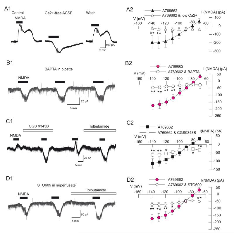
Fig. 6.
Augmentation of NMDA-evoked K-ATP conductance by AMPK is dependent upon Ca2+, calmodulin, and CaMKKβ. All recordings were made with pipettes that contained A769662 (5 μM). (A1) Current traces showing that low Ca2+ aCSF (0.2 mM) blocks NMDA-induced outward current. (A2) Summarized I-V plots for NMDA currents in control and low Ca2+ aCSF. (B1) Current trace shows that NMDA failed to evoke outward current (at −70 mV) when pipettes contained BAPTA (10 mM) in place of EGTA. (B2) Summarized I-V plots show that the presence of BAPTA significantly altered the voltage-dependence of currents evoked by NMDA (n = 6) compared to the A769662 control. (C1) Current trace shows that bath application of the calmodulin inhibitor CGS9343B (40 μM) blocked NMDA-evoked outward current (at –70 mV). (C2) Summarized I-V plots show that CGS9343B (40 μM) significantly altered the voltage-dependence of currents evoked by NMDA (n = 7) compared to A769662 control. (D1) Current trace shows that STO609 (10 μM), an inhibitor of CaMKKβ, blocked NMDA-evoked outward current (at −70 mV). Slices were superfused with STO609 at least 30 min prior to beginning whole-cell recording. (D2) Summarized I-V plots showing that STO609 significantly altered the voltage dependence of NMDA current (n = 7) compared to the A769662 control. STO609 was superfused at least 30 min prior to construction of the I-V plot. I-V plots for A769662 control in B2 and D2 are historical controls and are the same as that shown in Fig. 2A2. The data shown in A2 and C2 are paired data in which data for the A769662 control were obtained from the same neurons treated with CGS9343B or low Ca2+ aCSF. Asterisks: *, P < 0.05; **, P < 0.01.

