Abstract
We investigated the relative biological effectiveness (RBE) of therapeutic proton beams at six proton facilities in Japan with respect to cell lethality of HSG cells. The RBE of treatments could be determined from experimental data. For this purpose, we used a cell survival assay to compare the cell-killing efficiency of proton beams. Among the five linear accelerator (LINAC) X-ray machines at 4 or 6 MeV that were used as reference beams, there was only a small variation (coefficient of variation CV = 3.1% at D10) in biological effectiveness. The averaged value of D10 for the proton beams at the middle position of the spread-out Bragg peak (SOBP) was 4.98. These values showed good agreement, with a CV of 4.3% among the facilities. Thus, the average RBE10 (RBE at the D10 level) at the middle position of the SOBP beam for six facilities in Japan was 1.05 with a CV of 2.8%.
Keywords: proton therapy, relative biological effectiveness, cell survival, spread-out Bragg-peak
INTRODUCTION
Particle radiotherapy is currently an attractive option for oncologists as a new and promising modality for treating malignant tumors. The Heavy-Ion Medical Accelerator in Chiba (HIMAC) facility was constructed in 1993, and clinical trials with carbon beams were initiated there in June 1994, together with academic research activities, including those related to treatment, diagnosis, physics, engineering and biology. Following the HIMAC cancer therapy facility, several ion beam therapy facilities were set up in Japan in the 2000s. We had performed radiobiological studies involving ion beams prior to the initiation of the HIMAC clinical trials [1–3]. We had also performed a relative biological effectiveness (RBE) study at new ion beam facilities, such as the Hyogo Ion Beam Medical Center, the Wakasa Wan Energy Research Center and the Shizuoka Cancer Center, before patient treatment started [4, 5].
New proton treatment facilities are emerging, and previous data on RBE is very useful to them because they can obtain the RBE values obtained at existing Japanese facilities using the same biological systems. However, the standard range for RBE has not been published. Here, we present a unified method for radiobiological testing, coupled with the results of previous RBE studies. This report may not provide new scientific insights, but it may provide important and useful information for new facilities. We have described a variation of RBE and other parameters at a number of therapeutic proton facilities to confirm whether RBE shows good agreement between the facilities.
MATERIALS AND MTEHODS
Cell culture
A human salivary gland tumour (HSG) cell line (JCRB1070: HSGc-C5, Japanese Collection of Research Bioresources Cell Bank) was used in this study. The cell line is a standard reference cell line of RBE used at the NIRS (National Institute of Radiological Sciences)-HIMAC in carbon beam research. The cells were subcultured according to the method described below and were stored in liquid nitrogen at a concentration of 1 × 10⁶ cells/ml in the complete culture medium described below with 10% DMSO. The cells were subcultured in a bottle twice a week until a final concentration of 1 × 10⁴ cells/cm2 was obtained and were then used for experiments within 20 passages after purchase from JCRB to obtain stable results.
Cells were cultured in Eagle's minimum essential medium (E-MEM; SIGMA, M4655) supplemented with 10% fetal bovine serum (FBS) and antibiotics (100 U/ml penicillin and 100 µg/ml streptomycin) under humidified conditions with 5% CO2 and 95% air at 37°C. The FBS was tested before use such that the delay in growth after seeding was <12 h. Exponentially growing cells were trypsinized and seeded in plastic flasks (NUNC 152094 or other flasks without a slanted neck) at 2 × 10⁴ cells/cm2 and incubated for 1 d prior to the irradiation at each facility.
Irradiation
All proton 6-cm spread-out Bragg peak (SOBP) beams were generated from 180–235 MeV beams. High-energy LINAC X-rays at 4 or 6 MeV were used as the reference at each facility, and the energy depended on the specific instrumentation of each facility. The samples were placed at the isocenter of the SOBP beam, and the depth was adjusted to be in the middle position of the SOBP beam. The cells were then irradiated at room temperature. Dosimetry was performed according to the standard protocol by IAEA [6] for both proton and X-ray beams. Briefly, either Type 30001 Farmer Chamber or Type 23343 Markus Chamber (PTW, Freiburg) was placed at sample position. Monitor chambers placed upstream of the sample were calibrated by output from the above-stated chambers. Cell samples were irradiated using the preset values of the monitor chamber. A summary of the proton and X-ray beams for each facility is given in Table 1.
Table 1.
Summary of facilities and D10, D37, SF2 and RBEs
| Facility | A | B | C | D | E | F | Average | CV (%) |
|---|---|---|---|---|---|---|---|---|
| Year of experiment | 2000 | 2001 | 2002 | 2002 | 2002 | 2011 | ||
| Proton energy (MeV) | 190 | 180 | 190 | 190 | 200 | 235 | ||
| Dose rate (Gy/min) | ∼1.5 | ∼2.0 | 1.6–2.7 | 1.0–1.5 | 0.9–1.3 | ∼3.0 | ||
| Reference X-ray (MeV) | 4 | 4 | 6 | 4 | 4 | |||
| Proton D10 (Gy) | 5.1 | 4.97 | 4.91 | 5.32 | 4.92 | 4.68 | 4.98 | 4.3 |
| Proton D37 (Gy) | 2.87 | 2.44 | 2.72 | 3.09 | 2.9 | 2.44 | 2.74 | 9.5 |
| Proton SF2 | 0.655 | 0.557 | 0.687 | 0.707 | 0.628 | 0.458 | 0.615 | 15.1 |
| X-ray D10 (Gy) | 5.25 | 5.29 | 5 | 5.42 | b | 5.1 | 5.21 | 3.1 |
| X-ray D37 (Gy) | 2.88 | 2.97 | 2.88 | 3.13 | b | 2.87 | 2.95 | 3.8 |
| X-ray SF2 | 0.655 | 0.655 | 0.621 | 0.704 | 0.541 | 0.635 | 9.5 | |
| RBE10a | 1.03 ± 0.06 | 1.06 ± 0.05 | 1.02 ± 0.06 | 1.02 ± 0.05 | 1.07 ± 0.06 | 1.09 ± 0.03 | 1.05 | 2.8 |
| RBE37a | 1.00 ± 0.05 | 1.22 ± 0.14 | 1.06 ± 0.05 | 1.01 ± 0.06 | 1.02 ± 0.07 | 1.18 ± 0.10 | 1.08 | 8.6 |
The width of the SOBP was 60 mm for all facilities. aRBE at 10% and 37% survival levels; values are the average and SD for each facility. bThere was no High-energy LINAC X-ray machine available at facility E. Averaged values from facilities A–D were used in the RBE calculation.
Colony formation assay
Irradiated cells were kept in the dark at a low temperature until they were moved to the laboratory. The cells were rinsed twice with PBS(-) and trypsinised (0.2% trypsin in PBS(-)) to harvest them and were then resuspended in the culture medium. After the cell concentration was determined with a particle analyzer (Coulter counter), cells were diluted adequately and plated onto triplicate 6-cm plastic dishes, aiming for 100 colonies per dish for the cell survival assay. After 13 days' incubation in the CO2 incubator, the colonies were rinsed with PBS(-) once, fixed with 10% formalin solution for 10–15 min, and stained with 1% methylene blue. Any colony consisting of >50 cells was counted under a stereomicroscope as a surviving colony.
Survival curves were fitted using the linear–quadratic (LQ) model equation: SF = exp (-αD-βD2), where SF is the surviving fraction and D is the physical dose at the sample position. The parameters α and β were calculated with the least squares method described below. The α and β values were used to recalculate the biological equivalent dose D10 and D37 values, that is, the doses at which the cell survival is 10% and 37%, respectively, and the SF2 values after 2-Gy exposure. RBE10 values were obtained from a comparison of the D10 value for both proton and X-ray beams. The RBE values of proton beams were calculated from the X-ray data in each facility. The α and β values were obtained by the least square method. At least three independent experiments were performed using both proton beams and X-rays at each facility.
RESULTS AND DISCUSSION
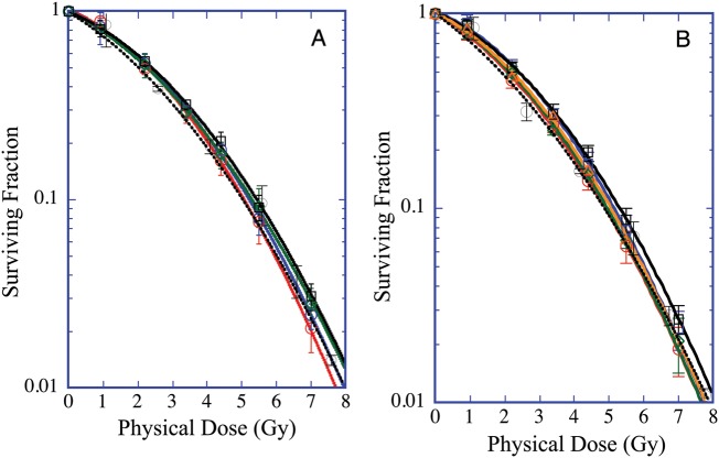
The survival curves relative to the experiments conducted with X-rays and the proton SOBP beam for each facility are shown in Figs 1A and B. The survival curves from each facility are almost identical. In the case of X-rays, the D10 and D37 values on HSG cells varied among facilities between 5.00 and 5.42 Gy and between 2.87 and 3.13Gy, respectively (Table 1), with averaged values of 5.21 Gy (3.1% coefficient of variation, CV) and 2.95 Gy (3.8% CV), respectively. Similarly, in the case of proton SOBP beams, average values of D10 and D37 were 4.98 Gy (4.3% CV) and 2.74 (9.5% CV) Gy, respectively.
Fig. 1.
Survival curves of HSG cells obtained A) from LINAC high-energy X-ray machines at five different therapy facilities located at the corresponding proton facility, and B) at the middle position of 6-cm SOBP beams at six different proton therapy facilities. The red, blue, green, black, brown and broken lines correspond to each facility A to F, respectively. The symbols and bars are the mean and the standard deviation, respectively, obtained from at least three independent experiments.
The RBE10 values that were calculated from the survival curves (Table 1) showed good agreement among the facilities, with a 2.8% CV. The average RBE10 at the middle of the proton SOBP obtained from all the facilities was 1.05 ± 0.03. Similar to the value obtained from another study, the RBE value at the middle of the 6-cm SOBP in the crypt survival assay ranged from 1.01–1.05 in vitro or 1.01–1.08 in vivo [5] or from 1.0–1.11 in vivo [7].
The experimental RBE10 (RBE values at D10) values of the proton SOBP beams from each facility, obtained from the cell survival assay performed in this study, ranged from 1.02–1.09, while a representative clinical RBE value of either 1.0 or 1.1 is used in proton treatment facilities worldwide [8]. Experimental RBE differs from clinical RBE, and the RBE of a given type of radiation will vary with particle type and energy, dose, dose per fraction, degree of oxygenation, cell or tissue type, biological endpoint, etc. [9]. There must be differences between expected clinical RBE and experimental RBE, and the clinical RBE of a carbon beam at HIMAC was determined based on both biological experiments with neutron and carbon beams, and clinical results from neutron therapy conducted at NIRS [10]. The experimental RBEs varied from generic clinical RBE [11, 12]. Thus the RBE10 that we have obtained in this study (1.05) was smaller than the representative clinical RBE value of 1.1. Generic RBE [12] is an averaged value from many radiobiological experiments including different cells and different endpoints. RBE in this paper is a specific value of cell killing at D10 level on HGS cells.
The highest values of D10, D37 and SF2 were found in facility D for both proton and X-ray irradiation. However, these values were in good agreement with those of the other facilities with acceptable variance (except for SF2). Surprisingly, the range of the survival parameter values (averaged values ± SD or CV) at facility F was similar to that of the other facilities and in the range of CVs, even though the experiment was conducted 10 years after those at the other facilities, the cells were older, serum and other chemicals were purchased from different manufacturers, and some of our experimental staff had changed.
CONCLUSION
D10 values of X-rays among the five therapy facilities in Japan showed good agreement, with deviations of 5.21 Gy with 3.1% CV and ranged from 5.0–5.42 Gy. Similarly, proton beams at six facilities also showed D10 values of 4.98 Gy with 4.3% CV and ranged from 4.68–5.32 Gy. The RBE10 values of the six facilities were identical, and the averaged value was 1.05 ± 0.03 with a small deviation (2.8% CV).
ACKNOWLEDGEMENTS
We thank all the crews for operating the proton beams, and are grateful to the various staff for the management of the beam time for our biological experiments at each facility.
REFERENCES
- 1.Kanai T, Furusawa Y, Fukutsu K, et al. Irradiation of mixed beam and design of spread-out Bragg peak for heavy-ion radiotherapy. Radiat Res. 1997;147:78–85. [PubMed] [Google Scholar]
- 2.Fukutsu K, Kanai T, Furusawa Y, et al. Response of mouse intestine after single and fractionated irradiation of accelerated carbon ions with a spread-out Bragg peak. Radiat Res. 1997;148:168–74. [PubMed] [Google Scholar]
- 3.Furusawa Y, Fukutsu K, Aoki M, et al. Inactivation of aerobic and hypoxic cells from three different cell lines by accelerated 3He-, 12C- and 20Ne-ion beams. Radiat Res. 2000;154:485–96. doi: 10.1667/0033-7587(2000)154[0485:ioaahc]2.0.co;2. Update 2012;177:129–31. [DOI] [PubMed] [Google Scholar]
- 4.Ando K, Furusawa Y, Suzuki M, et al. Relative biological effectiveness of the 235 MeV proton beams at the National Cancer Center Hospital East. J Radiat Res. 2001;42:79–89. doi: 10.1269/jrr.42.79. [DOI] [PubMed] [Google Scholar]
- 5.Kagawa K, Murakami M, Hishikawa Y, et al. Preclinical biological assessment of proton and carbon ion beams at Hyogo Ion Beam Medical Center. Int J Radiat Oncol Biol Phys. 2002;54:928–38. doi: 10.1016/s0360-3016(02)02949-8. [DOI] [PubMed] [Google Scholar]
- 6.IAEA. Technical Report Series No. 398. Vienna: International Atomic Energy Agency; 2000. Absorbed Dose Determination in External Beam Radiotherapy, An International Code of Practice for Dosimetry Based on Standards of Absorbed Dose to Water. [Google Scholar]
- 7.Uzawa A, Ando K, Furusawa Y, et al. Biological intercomparison using gut crypt survivals for proton and carbon-ion beams. J Radiat Res. 2007;48S doi: 10.1269/jrr.48.a75. A75–8. [DOI] [PubMed] [Google Scholar]
- 8.Raju MR. Proton radiobiology, radiosurgery and radiotherapy. Int J Radiat Biol. 1995;67:237–59. doi: 10.1080/09553009514550301. [DOI] [PubMed] [Google Scholar]
- 9.IAEA. Technical Reports Series No. 461. Vienna: International Atomic Energy Agency; 2008. Relative Biological Effectiveness in Ion Beam Therapy. [Google Scholar]
- 10.Kanai T, Endo M, Minohara S, et al. Biophysical characteristics of HIMAC clinical irradiation system for heavy-ion radiation therapy. Int J Radiat Oncol Biol Phys. 1999;44:201–10. doi: 10.1016/s0360-3016(98)00544-6. [DOI] [PubMed] [Google Scholar]
- 11.Gerweck L, Kozin S. Relative biological effectiveness of proton beams in clinical therapy. Radiother Oncol. 1999;50:135–42. doi: 10.1016/s0167-8140(98)00092-9. [DOI] [PubMed] [Google Scholar]
- 12.Paganetti H, Niemierko A, Ancukiewicz M, et al. Relative biological effectiveness (RBE) values for proton beam therapy. Int J Radiat Oncol Biol Phys. 2002;53:407–21. doi: 10.1016/s0360-3016(02)02754-2. [DOI] [PubMed] [Google Scholar]


