Abstract
In chronic liver disease leading to fibrosis, hepatic stellate cells (HSC) differentiate into myofibroblasts. Myofibroblastic HSC have taken center stage during liver fibrogenesis, due to their remarkable synthesis of extracellular matrix proteins, their secretion of profibrogenic mediators and their contribution to hypertension, due to elevated contractility. MicroRNAs (miRNAs) are small, noncoding RNA molecules of 19–24 nucleotides in length. By either RNA interference or inhibition of translational initiation and elongation, each miRNA is able to inhibit the gene expression of a wide panel of targeted transcripts. Recently, it was shown that altered miRNA patterns after chronic liver disease highly affect the progression of fibrosis by their potential to target the expression of extracellular matrix proteins and the synthesis of mediators of profibrogenic pathways. Here, we underline the role of miRNAs in the interplay of the profibrogenic cell communication pathways upon myofibroblastic differentiation of hepatic stellate cells in the chronically injured liver.
Keywords: liver fibrosis, myofibroblastic transition, TGF-β, miRNA
1. Introduction
MicroRNAs are small, 19–24 nucleotides-long, non-coding RNA molecules. They are transcribed by RNA polymerase II as primary transcripts (pri-miRNA). Pri-miRNAs are subsequently processed by the RNase III enzyme, Drosha, forming stem-loop structured miRNA precursors (pre-miRNAs) of about 80 nucleotides. After the export of pre-miRNAs into the cytoplasm, the highly conserved RNase III enzyme, Dicer, releases the pre-miRNAs to form the mature miRNAs, which then are incorporated into the RNA-induced silencing complex (RISC). The interaction of the miRNA/RISC complex by complementary annealing of the mostly seven nucleotide-long, so-called miRNA seed sequence with the untranslated region (UTR) of mRNA, leads to the inhibition of translation [1,2,3]. In total, more than 2500 human miRNAs have been discovered [4]. Since one miRNA might target hundreds of different mRNA transcripts [2], it is suggested that miRNAs regulate more than one third of the human genes [5,6].
Due to their high impact on gene regulation, miRNAs are involved in most cellular alteration processes, such as cell proliferation, migration and differentiation. Thus, many previous reports recognized miRNAs as central players in the oncogene and tumor suppressor networks, contributing to the initiation and progression of many human malignancies [7,8]. Furthermore, miRNA-mediated gene regulation is also an important feature in acute and chronic inflammatory diseases [9,10,11,12]. Recent studies on chronic liver diseases of different etiologies revealed a prominent dysregulation of many miRNAs, leading to an altered gene expression profile and progression of liver fibrosis, previously summarized by references [13,14,15,16,17,18]). In the present short review, we summarize recent findings on the role of miRNAs in the pathophysiology of fibrosis after chronic liver injury.
2. Altered miRNA Expression upon Liver Fibrosis
Liver fibrosis has become one of the most serious problems for human health, which can lead to hepatocellular carcinoma. It is characterized by the hepatic accumulation of biomatrix as a pathophysiological response to chronic liver injuries independently of the causative noxa [19,20,21]. In addition to viral infections, such as chronic hepatitis B (HBV) and C (HCV) infections or excessive alcohol abuse, in the recent past, fatty liver disease leading to steatohepatitis is also a frequent agent of chronic liver injury and fibrosis, primarily due to altered food habits in Western countries [22].
Extracellular matrix (ECM) accumulation during liver fibrogenesis is mainly distinguished by a dramatically enhanced deposition of collagen I and collagen III. Especially TGF-β triggers the interstitial ECM accumulation by an induced synthesis, but also by a decreased ECM turn-over after repression of matrix-metalloproteinases [19,20,21]. In addition, the function of TGF-β as a central mediator of fibrosis is strongly manifested by its evoked stimulation of profibrogenic growth factor profiles, including TGF-β itself, the platelet-derived growth factors (PDGF), connective tissue growth factor (CTGF), endothelin (ET) and many others [23].
During liver fibrogenesis caused by different etiologies, an altered miRNA expression was observed [24,25,26,27]. miR-122 is the most abundant miRNA in normal liver, highly enriched in the liver parenchyma, accounting for more than 70% of the total miRNA population in hepatocytes [28,29]. However, the progression of fibrosis after chronic liver disease, such as chronic HCV infection or non-alcoholic liver disease, is accompanied by a predominant decrease of miR-122 [24,26,30]. These reduced miR-122 levels after fibrosis are suggested to be based on hepatocyte injury followed by miR-122 release into the blood stream, on one hand, and by transcriptional repression, due to the loss of liver-specific transcription factors, such as HNF1a and HNF4a, controlling miR-122 gene expression [31], on the other. Of special interest is the downregulation of miR-122 after chronic HCV infection, because miR-122 was shown to be involved in HCV replication. miR-122 triggers HCV replication by interaction with the 5´UTR of HCV RNA genome, resulting in the high stability of the HCV RNA [29,32,33]. These data impose miR-122 as a first target of a novel therapeutic strategy to treat chronic HCV infection [34]. Indeed, a recent clinical trial has demonstrated the successful application of miR-122 antagonists for HCV repression [35].
Furthermore, many other miRNAs are downregulated after liver fibrosis. Hence, a predominant repression of the miR-29 family members, miR-29a and miR-29b, was found in experimental fibrosis after liver intoxication or cholestasis [27]. Additionally, downregulation of miR-29c was also demonstrated in a dietary non-alcoholic steatohepatitis (NASH) mouse model [36] or in human liver fibrosis of chronically HCV-infected patients [37]. Previous findings have shown that the members of the miR-29 family act as tumor suppressor miRNAs, inhibiting the synthesis of the anti-apoptotic proteins, Bcl-2 and Mcl-1, and the DNA methyltransferases, 3A and 3B, involved in the epigenetic methylation machinery of epithelial cell types [38,39]. Most notably, the members of the miR-29 family also function as antifibrotic miRNAs, first described in cardiac fibrosis by their inhibitory role on collagen I and III, elastin and fibrillin-1 expression [40]. Inhibition of ECM synthesis and its downregulation during fibrogenesis was additionally shown in pulmonary fibrosis [41] and systemic sclerosis [42]. Furthermore, the findings of Cushing et al. suggested that miR-29 inhibits expression of a wide variety of fibrosis-associated genes and confirmed that numerous components of ECM are negatively regulated by miR-29, including fibrillin-1, follistatin, nidogen 1 and laminin [41].
Moreover, since the loss of miR-133 in liver fibrosis after TGF-β exposure causes prominent enhancement of collagen 1A1 and collagen 5A3 deposition, Roderburg et al. suggested miR-133 as a main antifibrotic miRNA [43].
On the contrary to these downregulated miRNAs, others are upregulated during fibrogenesis. Thus, miR-34a, miR-199/200 and miR-221/222 are known to be increased during liver fibrosis with different pathogenesis, such as non-alcoholic and alcoholic steatohepatitis (NASH/ASH), HCV infection or experimental fibrosis, including CCl4 intoxication and a fat diet mouse model [36,44,45,46]. The role of the miR-200 family members in liver fibrogenesis is still not completely understood. Although Murakami et al. have shown that miR-200a/b were positively associated with the progression of liver fibrosis in chronic hepatitis C patients [45], Sun et al. have reported that miR-200a is downregulated during hepatic stellate cell activation, as well as after CCl4 intoxication-based experimental fibrosis [47]. However, in agreement with Murakami et al., miR-200c levels were found to be increased in the fibrotic liver after HCV-infection and NASH [48]. These opposing results may be due to diversity in disease progression or etiology.
Though the role of increased miR-34a levels upon liver fibrogenesis is not yet studied in detail, in cancer, miR-34a was shown to repress the deacetylase SIRT1 expression, leading to a marked increase of acetylated p53, followed by elevated apoptosis [49,50]. Interestingly, miR-34a is also involved in ethanol-induced apoptosis and in hepatic remodeling by targeting matrix metalloproteinases, MMP2 and MMP9 [44]. In addition, the increase of miR-199 during fibrogenesis is suggested to contribute to hepatic remodeling by targeting the expression of ECM turnover-involved genes, like collagen Col1A1, the tissue inhibitor of metalloproteinase (TIMP-1) and the matrix metalloproteinase, MMP13.
3. Linkage of miRNA Alteration to Myofibroblastic Transition
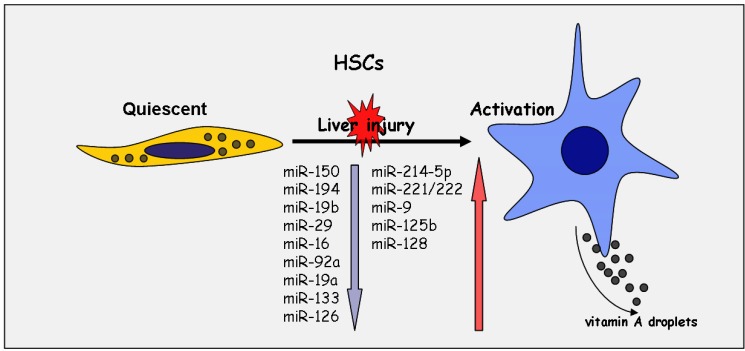
During liver fibrogenesis, myofibroblastic cells are the central fibrotic cell type responsible for ECM accumulation. They derive from different cell types, but most of the myofibroblasts originate from hepatic stellate cells (HSCs) [51,52]. HSCs account for approximately one-third of the non-parenchymal cells and 15% of the total number of resident cells in the healthy liver [53,54]. They store around 80% of the vitamin A of the human body. However, in response to liver injury, HSCs undergo phenotypical and functional changes, including the loss of vitamin A storage, proliferation, cytoskeleton alteration and synthesis of ECM, leading to a myofibroblast phenotype with enhanced matrix deposition and contractility [51,52,55]. This transition process of HSC into a myofibroblastic cell type is accompanied by an altered growth factor profile, resulting in autocrine and paracrine profibrotic stimulation by, e.g., TGF-β, PDGF-BB, endothelin, chemokines and cytokines, and in the enhancement of fibrosis [51,52]. Importantly, previous reports have shown that several miRNA species are involved in the transdifferentiation process of quiescent HSC into a myofibroblastically activated cell type [56,57,58,59,60,61,62] (Figure 1). First, Guo et al. and Ji et al. assumed that miRNAs may be involved in the altered gene expression profile of myofibroblastic HSC [57,59].
Figure 1.
Altered expression pattern of microRNAs during myofibroblastic activation of hepatic stellate cells (HSCs).
Quiescent hepatic stellate cells (HSCs) store 80% of the body vitamin A in fat droplets. In response to chronic liver injury, HSCs undergo myofibroblastic transition. During this process, the phenotype and function of HSCs is considerably changed, including loss of vitamin A storage, proliferation, cytoskeleton alteration and synthesis of ECM, leading to a myofibroblast phenotype with enhanced matrix deposition and contractility. This process is accompanied by an altered miRNA expression pattern. miRNAs, prominently downregulated (blue arrow), and miRNAs shown to be upregulated (red arrow) during myofibroblastic activation are indicated.
Whereas the antifibrotic miRNAs, such as the members of the miR-29 family, miR-19, miR-150 and miR-133, repressing the myofibroblastic features, such as collagen synthesis or smooth muscle actin (SMA) synthesis, decrease after myofibroblastic transdifferentiation [25,43,63,64,65], others, such as miR-221/222, the neuronal miRNAs, miR-9, miR-125b, miR-128 or the miR-214, are increased [46,61,66]. Particularly, miR-221/222 expression is increased in cultivated primary HSC upon induction by the NF-kb activator, which is suggested to be a potential biomarker of stellate cell activation and linked to increased Col1A1 expression during fibrosis [46]. Interestingly, miR-214 upregulated in activated HSC was shown to be controlled by the master transcription factor, Twist-1, [66]. miR-214 has to be considered as a main therapeutic target, because genetically or therapeutically silencing resulted in highly efficient inhibition of renal fibrosis [67]. Recent data of Noetel et al. could demonstrate a prominent upregulation of the neuronal miRNAs, miR-9, miR-125, and miR-128 in HSC upon myofibroblastic activation. These miRNAs were suggested to target several members of the chemokine and the chemokine receptor family [61].
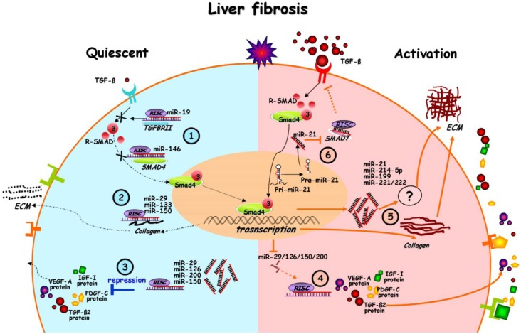
Lakner et al. identified 55 miRNAs that are divergently expressed in quiescent versus activated HSCs by microarray analyses. Hereby, miR-19b was proven to repress TGF-β signaling by targeting TGF-β receptor type II expression, which, in turn, resulted in a decrease of TGF-β signaling followed by reduced expression of collagen subunits (Col1A1 and ColA2) [25]. Furthermore, the miR-29 and miR-21 function is closely linked to the profibrogenic TGF-β pathway [27,65,68,69]. Hence, miRNAs definitely contribute to the profibrogenic changes during fibrogenesis, not only by targeting ECM production, but also by interaction with the TGF-β signaling pathway (Figure 2).
Figure 2.
miRNAs in the interplay of signaling in quiescent and activated HSCs after chronic liver injury.
In quiescent HSCs, antifibrotic miRNAs, like miR-19, miR-146, miR-29, miR-133, miR-150, miR-126 and miR-200, are highly expressed. High levels of these miRNAs, in turn, lead to reduced synthesis of the TGF-β receptor II or smad-4 (1) of ECM, mainly the collagen subunits (2); or synthesis of profibrogenic mediators, e.g., VEGF-A, TGF-β2, IGF-I, PDGF-C (3); After chronic liver injury, HSCs get activated and transduce into a myofibroblastic phenotype. Myofibroblastic transition and autocrine TGF-β stimulation changes the miRNA profile, as shown in Figure 1. In response to TGF-β exposure, miR-29, miR-126, miR-150 and miR-200 are downregulated, resulting in the abolished repression of profibrogenic mediators (4); The reduced miR-29/133/150 levels then contribute to enhanced synthesis and deposition of ECM. Whereas some miRNAs are reduced by TGF-β, others, such as miR-21/214-5p/199/221/222, are stimulated and elevate ECM synthesis via an unknown pathway (5); TGF-β induced miR-21 expression results in a decrease of the TGF-β inhibitory smad-7 protein (6) (Dashed arrow lines indicate the suppression of pathways and gene expression, whereas solid arrow lines indicate stimulation).
4. miRNA in the Interplay of Profibrogenic Pathways
miR-29 expression is strongly regulated by TGF-β, as well as oppositionally by the TGF-β antagonist, hepatocyte growth factor (HGF). Whereas TGF-β inhibits epithelial proliferation, HGF exerts high mitogenic potential on epithelial cells and strong antifibrogenic functions on fibroblasts. Thus, the studies of Kwiecinski et al. revealed that HGF mediates antifibrogenic effects by induction and restoring of miR-29 expression, which, in turn, is followed by a significant decrease of collagen I and collagen IV subtypes [64]. However, miR-29 is not only regulated by the interplay of TGF-β and HGF, but also by other factors involved in inflammation and fibrosis, like PDGF-BB [65] and interferon-α [70].
In addition to its role in ECM repression, miR-29 regulates the growth factor profile of HSC by targeting profibrogenic mediators, such as IGF-I and PDGF-C [71]. Moreover, Sekyia et al. collected evidence that also the PDGF-β receptor is targeted by miR-29 regulation [65]. This is of special interest, because miR-29 itself is downregulated in HSC after PDGF-BB stimulation [65].
Additionally to miR-29, miR-146 is also decreased by TGF-β. TGF-β mediates its profibrogenic functions mainly by the smad pathway. In particular, after ligand binding and TGF-β receptor I phosphorylation, smad-3 activation and its interaction with smad-4 is crucial for HSC activation [58]. After smad-3/-4 transduction into the nucleus, the smad complexes are involved in the transcriptional control of a wide range of genes. While miR-146 is repressed by TGF-β signals, it suppresses TGF-β signals by targeting smad-4 expression. Furthermore, TGF-β signaling is also inhibited by the antifibrotic microRNAs, miR-150 and miR-200a, which, in addition to their function in ECM regulation, target the expression of the signal transducer, smad-3, as well as of TGF-β2 [47,63] (Figure 2).
miR-126 is closely linked to the vascular endothelial growth factor (VEGF) signaling, which is a crucial in angiogenesis, but affects fibrosis by the induction of HSC proliferation and ECM synthesis. During myofibroblastic HSC transition, miR-126 is lost, causing an increased synthesis of ECM proteins, but also of VEGF-A [72,73] (Figure 2).
Whereas the antifibrotic miRNAs are repressed by TGF-β, others, such as miR-214-5p and miR-21, are induced [74] in agreement with their enrichment during liver fibrosis [66,75]. Especially miR-21 was shown to be positively regulated in cancer cells by the TGF-β/smad pathway on both, the transcriptional and the miRNA processing level [68]. miR-21 further triggers the TGF-β/smad pathway by targeting the inhibitory smad protein, smad-7 (Figure 2). Furthermore, in stellate cells, the profibrogenic function of miR-21 is suggested to be also mediated by Pten inhibition, which, in turn, leads to Akt activation [76]. In conclusion, the altered expression of a wide panel of miRNAs affects fibrosis progression by its inhibition of ECM synthesis and its influence on central signaling pathways in HSC.
5. Perspectives
The high impact of miRNAs on the progression of fibrosis by the inhibition of ECM or by interfering with profibrogenic pathways announces the promising potential of miRNAs as biomarkers and targets of novel antifibrotic therapeutic strategies. Although, presently, miRNAs are not yet used as diagnostic biomarkers for fibrosis, in liver cancer, the diagnostic potential of miRNAs is already demonstrated by the low levels of miR-26, indicating the successful response to interferon-a therapy [77]. However, since miRNAs are released into the blood stream, circulating miRNA levels might serve even more efficiently as indicators of fibrosis [78,79]. Furthermore, the important function of many miRNAs acting as anti- or pro-fibrogenic factors emphasizes their role as therapeutic targets. The successful inhibition of miR-122 by antagonizing oligonucleotides [35] has successfully illustrated the proof of principle of this novel therapeutic strategy. However, whereas drug delivery to liver parenchyma is highly efficient, organ and cell-type specific miRNA targeting has to be achieved in liver fibrosis. Thus, in order to avoid cell unspecific side effects of antifibrotic therapies, that target miRNAs involved in liver fibrosis, an HSC- or myofibroblast specific approach of drug delivery is needed.
Acknowledgments
Jia Huang and Xiaojie Yu hold a fellowship from the China Scholarship Council, and Li’ang Zhang is a PhD student supported by a fellowship from the German Academic Exchange Service.
Author Contributions
J.H., X.Y., L.Z., J.W.U.F. and M.O. contribute to the concept of the review. J.H. and M.O. were mainly involved in the preparation of the main text and J.H. performed all illustrations.
Conflicts of Interest
The authors declare no conflict of interest.
References
- 1.Bartel D.P. MicroRNAs: Target recognition and regulatory functions. Cell. 2009;136:215–233. doi: 10.1016/j.cell.2009.01.002. [DOI] [PMC free article] [PubMed] [Google Scholar]
- 2.Lewis B.P., Burge C.B., Bartel D.P. Conserved seed pairing, often flanked by adenosines, indicates that thousands of human genes are microRNA targets. Cell. 2005;120:15–20. doi: 10.1016/j.cell.2004.12.035. [DOI] [PubMed] [Google Scholar]
- 3.Ruby J.G., Jan C.H., Bartel D.P. Intronic microRNA precursors that bypass Drosha processing. Nature. 2007;448:83–86. doi: 10.1038/nature05983. [DOI] [PMC free article] [PubMed] [Google Scholar]
- 4.Kozomara A., Griffiths-Jones S. miRBase: Annotating high confidence microRNAs using deep sequencing data. Nucleic Acids Res. 2014;42:D68–D73. doi: 10.1093/nar/gkt1181. [DOI] [PMC free article] [PubMed] [Google Scholar]
- 5.Friedman R.C., Farh K.K., Burge C.B., Bartel D.P. Most mammalian mRNAs are conserved targets of microRNAs. Genome Res. 2009;19:92–105. doi: 10.1101/gr.082701.108. [DOI] [PMC free article] [PubMed] [Google Scholar]
- 6.Krek A., Grun D., Poy M.N., Wolf R., Rosenberg L., Epstein E.J., MacMenamin P., da Piedade I., Gunsalus K.C., Stoffel M., et al. Combinatorial microRNA target predictions. Nat. Genet. 2005;37:495–500. doi: 10.1038/ng1536. [DOI] [PubMed] [Google Scholar]
- 7.Fabbri M., Ivan M., Cimmino A., Negrini M., Calin G.A. Regulatory mechanisms of microRNAs involvement in cancer. Expert Opin. Biol. Ther. 2007;7:1009–1019. doi: 10.1517/14712598.7.7.1009. [DOI] [PubMed] [Google Scholar]
- 8.Kong Y.W., Ferland-McCollough D., Jackson T.J., Bushell M. microRNAs in cancer management. Lancet Oncol. 2012;13:e249–e258. doi: 10.1016/S1470-2045(12)70073-6. [DOI] [PubMed] [Google Scholar]
- 9.Garbacki N., di Valentin E., Huynh-Thu V.A., Geurts P., Irrthum A., Crahay C., Arnould T., Deroanne C., Piette J., Cataldo D., et al. MicroRNAs profiling in murine models of acute and chronic asthma: A relationship with mRNAs targets. PLoS One. 2011;6:e16509. doi: 10.1371/journal.pone.0016509. [DOI] [PMC free article] [PubMed] [Google Scholar]
- 10.Mallick B., Ghosh Z., Chakrabarti J. MicroRNome analysis unravels the molecular basis of SARS infection in bronchoalveolar stem cells. PLoS One. 2009;4:e7837. doi: 10.1371/journal.pone.0007837. [DOI] [PMC free article] [PubMed] [Google Scholar]
- 11.Recchiuti A., Krishnamoorthy S., Fredman G., Chiang N., Serhan C.N. MicroRNAs in resolution of acute inflammation: Identification of novel resolvin D1-miRNA circuits. FASEB J. 2011;25:544–560. doi: 10.1096/fj.10-169599. [DOI] [PMC free article] [PubMed] [Google Scholar]
- 12.Zhou T., Garcia J.G., Zhang W. Integrating microRNAs into a system biology approach to acute lung injury. Transl. Res. 2011;157:180–190. doi: 10.1016/j.trsl.2011.01.010. [DOI] [PMC free article] [PubMed] [Google Scholar]
- 13.Cheung O., Sanyal A.J. Role of microRNAs in non-alcoholic steatohepatitis. Curr. Pharm. Des. 2010;16:1952–1957. doi: 10.2174/138161210791208866. [DOI] [PubMed] [Google Scholar]
- 14.Haybaeck J., Zeller N., Heikenwalder M. The parallel universe: microRNAs and their role in chronic hepatitis, liver tissue damage and hepatocarcinogenesis. Swiss Med. Wkly. 2011;141:w13287. doi: 10.4414/smw.2011.13287. [DOI] [PubMed] [Google Scholar]
- 15.Noetel A., Kwiecinski M., Elfimova N., Huang J., Odenthal M. microRNA are central players in anti- and profibrotic gene regulation during liver fibrosis. Front. Physiol. 2012;3:49. doi: 10.3389/fphys.2012.00049. [DOI] [PMC free article] [PubMed] [Google Scholar]
- 16.Szabo G., Bala S. MicroRNAs in liver disease. Nat. Rev. Gastroenterol. Hepatol. 2013;10:542–552. doi: 10.1038/nrgastro.2013.87. [DOI] [PMC free article] [PubMed] [Google Scholar]
- 17.Takahashi K., Yan I., Wen H.J., Patel T. microRNAs in liver disease: From diagnostics to therapeutics. Clin. Biochem. 2013;46:946–952. doi: 10.1016/j.clinbiochem.2013.01.025. [DOI] [PMC free article] [PubMed] [Google Scholar]
- 18.Wang X.W., Heegaard N.H., Orum H. MicroRNAs in liver disease. Gastroenterology. 2012;142:1431–1443. doi: 10.1053/j.gastro.2012.04.007. [DOI] [PMC free article] [PubMed] [Google Scholar]
- 19.Brenner D.A. Molecular pathogenesis of liver fibrosis. Trans. Am. Clin. Climatol. Assoc. 2009;120:361–368. [PMC free article] [PubMed] [Google Scholar]
- 20.Lee Y., Friedman S.L. Fibrosis in the liver: Acute protection and chronic disease. Prog. Mol. Biol. Transl. Sci. 2010;97:151–200. doi: 10.1016/B978-0-12-385233-5.00006-4. [DOI] [PubMed] [Google Scholar]
- 21.Zhang D.Y., Friedman S.L. Fibrosis-dependent mechanisms of hepatocarcinogenesis. Hepatology. 2012;56:769–775. doi: 10.1002/hep.25670. [DOI] [PMC free article] [PubMed] [Google Scholar]
- 22.Lade A., Noon L.A., Friedman S.L. Contributions of metabolic dysregulation and inflammation to nonalcoholic steatohepatitis, hepatic fibrosis, and cancer. Curr. Opin. Oncol. 2014;26:100–107. doi: 10.1097/CCO.0000000000000042. [DOI] [PMC free article] [PubMed] [Google Scholar]
- 23.Dooley S., ten Dijke P. TGF-beta in progression of liver disease. Cell Tissue Res. 2012;347:245–256. doi: 10.1007/s00441-011-1246-y. [DOI] [PMC free article] [PubMed] [Google Scholar]
- 24.Cheung O., Puri P., Eicken C., Contos M.J., Mirshahi F., Maher J.W., Kellum J.M., Min H., Luketic V.A., Sanyal A.J. Nonalcoholic steatohepatitis is associated with altered hepatic MicroRNA expression. Hepatology. 2008;48:1810–1820. doi: 10.1002/hep.22569. [DOI] [PMC free article] [PubMed] [Google Scholar]
- 25.Lakner A.M., Steuerwald N.M., Walling T.L., Ghosh S., Li T., McKillop I.H., Russo M.W., Bonkovsky H.L., Schrum L.W. Inhibitory effects of microRNA 19b in hepatic stellate cell-mediated fibrogenesis. Hepatology. 2012;56:300–310. doi: 10.1002/hep.25613. [DOI] [PMC free article] [PubMed] [Google Scholar]
- 26.Morita K., Taketomi A., Shirabe K., Umeda K., Kayashima H., Ninomiya M., Uchiyama H., Soejima Y., Maehara Y. Clinical significance and potential of hepatic microRNA-122 expression in hepatitis C. Liver Int. 2011;31:474–484. doi: 10.1111/j.1478-3231.2010.02433.x. [DOI] [PubMed] [Google Scholar]
- 27.Roderburg C., Urban G.W., Bettermann K., Vucur M., Zimmermann H., Schmidt S., Janssen J., Koppe C., Knolle P., Castoldi M., et al. Micro-RNA profiling reveals a role for miR-29 in human and murine liver fibrosis. Hepatology. 2011;53:209–218. doi: 10.1002/hep.23922. [DOI] [PubMed] [Google Scholar]
- 28.Jopling C. Liver-specific microRNA-122: Biogenesis and function. RNA Biol. 2012;9:137–142. doi: 10.4161/rna.18827. [DOI] [PMC free article] [PubMed] [Google Scholar]
- 29.Jopling C.L., Yi M., Lancaster A.M., Lemon S.M., Sarnow P. Modulation of hepatitis C virus RNA abundance by a liver-specific MicroRNA. Science. 2005;309:1577–1581. doi: 10.1126/science.1113329. [DOI] [PubMed] [Google Scholar]
- 30.Trebicka J., Anadol E., Elfimova N., Strack I., Roggendorf M., Viazov S., Wedemeyer I., Drebber U., Rockstroh J., Sauerbruch T., et al. Hepatic and serum levels of miR-122 after chronic HCV-induced fibrosis. J. Hepatol. 2013;58:234–239. doi: 10.1016/j.jhep.2012.10.015. [DOI] [PubMed] [Google Scholar]
- 31.Coulouarn C., Factor V.M., Andersen J.B., Durkin M.E., Thorgeirsson S.S. Loss of miR-122 expression in liver cancer correlates with suppression of the hepatic phenotype and gain of metastatic properties. Oncogene. 2009;28:3526–3536. doi: 10.1038/onc.2009.211. [DOI] [PMC free article] [PubMed] [Google Scholar]
- 32.Jopling C.L. Targeting microRNA-122 to treat hepatitis C virus infection. Viruses. 2010;2:1382–1393. doi: 10.3390/v2071382. [DOI] [PMC free article] [PubMed] [Google Scholar]
- 33.Roberts A.P., Lewis A.P., Jopling C.L. The role of microRNAs in viral infection. Prog. Mol. Biol. Transl. Sci. 2011;102:101–139. doi: 10.1016/B978-0-12-415795-8.00002-7. [DOI] [PubMed] [Google Scholar]
- 34.Lanford R.E., Hildebrandt-Eriksen E.S., Petri A., Persson R., Lindow M., Munk M.E., Kauppinen S., Orum H. Therapeutic silencing of microRNA-122 in primates with chronic hepatitis C virus infection. Science. 2010;327:198–201. doi: 10.1126/science.1178178. [DOI] [PMC free article] [PubMed] [Google Scholar]
- 35.Janssen H.L., Reesink H.W., Lawitz E.J., Zeuzem S., Rodriguez-Torres M., Patel K., van der Meer A.J., Patick A.K., Chen A., Zhou Y., et al. Treatment of HCV infection by targeting microRNA. N. Engl. J. Med. 2013;368:1685–1694. doi: 10.1056/NEJMoa1209026. [DOI] [PubMed] [Google Scholar]
- 36.Pogribny I.P., Starlard-Davenport A., Tryndyak V.P., Han T., Ross S.A., Rusyn I., Beland F.A. Difference in expression of hepatic microRNAs miR-29c, miR-34a, miR-155 and miR-200b is associated with strain-specific susceptibility to dietary nonalcoholic steatohepatitis in mice. Lab. Investig. 2010;90:1437–1446. doi: 10.1038/labinvest.2010.113. [DOI] [PMC free article] [PubMed] [Google Scholar]
- 37.Bandyopadhyay S., Friedman R.C., Marquez R.T., Keck K., Kong B., Icardi M.S., Brown K.E., Burge C.B., Schmidt W.N., Wang Y., et al. Hepatitis C virus infection and hepatic stellate cell activation downregulate miR-29: miR-29 overexpression reduces hepatitis C viral abundance in culture. J. Infect. Dis. 2011;203:1753–1762. doi: 10.1093/infdis/jir186. [DOI] [PMC free article] [PubMed] [Google Scholar]
- 38.Mott J.L., Kobayashi S., Bronk S.F., Gores G.J. mir-29 regulates Mcl-1 protein expression and apoptosis. Oncogene. 2007;26:6133–6140. doi: 10.1038/sj.onc.1210436. [DOI] [PMC free article] [PubMed] [Google Scholar]
- 39.Xiong Y., Fang J.H., Yun J.P., Yang J., Zhang Y., Jia W.H., Zhuang S.M. Effects of microRNA-29 on apoptosis, tumorigenicity, and prognosis of hepatocellular carcinoma. Hepatology. 2010;51:836–845. doi: 10.1002/hep.23380. [DOI] [PubMed] [Google Scholar]
- 40.Van Rooij E., Sutherland L.B., Thatcher J.E., DiMaio J.M., Naseem R.H., Marshall W.S., Hill J.A., Olson E.N. Dysregulation of microRNAs after myocardial infarction reveals a role of miR-29 in cardiac fibrosis. Proc. Natl. Acad. Sci. USA. 2008;105:13027–13032. doi: 10.1073/pnas.0805038105. [DOI] [PMC free article] [PubMed] [Google Scholar]
- 41.Cushing L., Kuang P.P., Qian J., Shao F., Wu J., Little F., Thannickal V.J., Cardoso W.V., Lu J. miR-29 is a major regulator of genes associated with pulmonary fibrosis. Am. J. Respir. Cell Mol. Biol. 2011;45:287–294. doi: 10.1165/rcmb.2010-0323OC. [DOI] [PMC free article] [PubMed] [Google Scholar]
- 42.Maurer B., Stanczyk J., Jungel A., Akhmetshina A., Trenkmann M., Brock M., Kowal-Bielecka O., Gay R.E., Michel B.A., Distler J.H., et al. MicroRNA-29, a key regulator of collagen expression in systemic sclerosis. Arthritis Rheum. 2010;62:1733–1743. doi: 10.1002/art.27443. [DOI] [PubMed] [Google Scholar]
- 43.Roderburg C., Luedde M., Vargas Cardenas D., Vucur M., Mollnow T., Zimmermann H.W., Koch A., Hellerbrand C., Weiskirchen R., Frey N., et al. miR-133a mediates TGF-beta-dependent derepression of collagen synthesis in hepatic stellate cells during liver fibrosis. J. Hepatol. 2013;58:736–742. doi: 10.1016/j.jhep.2012.11.022. [DOI] [PubMed] [Google Scholar]
- 44.Meng F., Glaser S.S., Francis H., Yang F., Han Y., Stokes A., Staloch D., McCarra J., Liu J., Venter J., et al. Epigenetic regulation of miR-34a expression in alcoholic liver injury. Am. J. Pathol. 2012;181:804–817. doi: 10.1016/j.ajpath.2012.06.010. [DOI] [PMC free article] [PubMed] [Google Scholar]
- 45.Murakami Y., Toyoda H., Tanaka M., Kuroda M., Harada Y., Matsuda F., Tajima A., Kosaka N., Ochiya T., Shimotohno K. The progression of liver fibrosis is related with overexpression of the miR-199 and 200 families. PLoS One. 2011;6:e16081. doi: 10.1371/journal.pone.0016081. [DOI] [PMC free article] [PubMed] [Google Scholar]
- 46.Ogawa T., Enomoto M., Fujii H., Sekiya Y., Yoshizato K., Ikeda K., Kawada N. MicroRNA-221/222 upregulation indicates the activation of stellate cells and the progression of liver fibrosis. Gut. 2012;61:1600–1609. doi: 10.1136/gutjnl-2011-300717. [DOI] [PubMed] [Google Scholar]
- 47.Sun X., He Y., Ma T.T., Huang C., Zhang L., Li J. Participation of miR-200a in TGF-beta1-mediated hepatic stellate cell activation. Mol. Cell. Biochem. 2014;388:11–23. doi: 10.1007/s11010-013-1895-0. [DOI] [PubMed] [Google Scholar]
- 48.Ramachandran S., Ilias Basha H., Sarma N.J., Lin Y., Crippin J.S., Chapman W.C., Mohanakumar T. Hepatitis C virus induced miR200c down modulates FAP-1, a negative regulator of Src signaling and promotes hepatic fibrosis. PLoS One. 2013;8:e70744. doi: 10.1371/journal.pone.0070744. [DOI] [PMC free article] [PubMed] [Google Scholar]
- 49.Vinall R.L., Ripoll A.Z., Wang S., Pan C.X., deVere White R.W. MiR-34a chemosensitizes bladder cancer cells to cisplatin treatment regardless of p53-Rb pathway status. Int. J. Cancer. 2012;130:2526–2538. doi: 10.1002/ijc.26256. [DOI] [PMC free article] [PubMed] [Google Scholar]
- 50.Yamakuchi M., Ferlito M., Lowenstein C.J. miR-34a repression of SIRT1 regulates apoptosis. Proc. Natl. Acad. Sci. USA. 2008;105:13421–13426. doi: 10.1073/pnas.0801613105. [DOI] [PMC free article] [PubMed] [Google Scholar]
- 51.Friedman S.L. Mechanisms of hepatic fibrogenesis. Gastroenterology. 2008;134:1655–1669. doi: 10.1053/j.gastro.2008.03.003. [DOI] [PMC free article] [PubMed] [Google Scholar]
- 52.Lee U.E., Friedman S.L. Mechanisms of hepatic fibrogenesis. Best Pract. Res. Clin. Gastroenterol. 2011;25:195–206. doi: 10.1016/j.bpg.2011.02.005. [DOI] [PMC free article] [PubMed] [Google Scholar]
- 53.Friedman S.L. Hepatic stellate cells: Protean, multifunctional, and enigmatic cells of the liver. Physiol. Rev. 2008;88:125–172. doi: 10.1152/physrev.00013.2007. [DOI] [PMC free article] [PubMed] [Google Scholar]
- 54.Li D., Friedman S.L. Liver fibrogenesis and the role of hepatic stellate cells: New insights and prospects for therapy. J. Gastroenterol. Hepatol. 1999;14:618–633. doi: 10.1046/j.1440-1746.1999.01928.x. [DOI] [PubMed] [Google Scholar]
- 55.Dechene A., Sowa J.P., Gieseler R.K., Jochum C., Bechmann L.P., El Fouly A., Schlattjan M., Saner F., Baba H.A., Paul A., et al. Acute liver failure is associated with elevated liver stiffness and hepatic stellate cell activation. Hepatology. 2010;52:1008–1016. doi: 10.1002/hep.23754. [DOI] [PubMed] [Google Scholar]
- 56.Guo C.J., Pan Q., Cheng T., Jiang B., Chen G.Y., Li D.G. Changes in microRNAs associated with hepatic stellate cell activation status identify signaling pathways. FEBS J. 2009;276:5163–5176. doi: 10.1111/j.1742-4658.2009.07213.x. [DOI] [PubMed] [Google Scholar]
- 57.Guo C.J., Pan Q., Jiang B., Chen G.Y., Li D.G. Effects of upregulated expression of microRNA-16 on biological properties of culture-activated hepatic stellate cells. Apoptosis. 2009;14:1331–1340. doi: 10.1007/s10495-009-0401-3. [DOI] [PubMed] [Google Scholar]
- 58.He Y., Huang C., Sun X., Long X.R., Lv X.W., Li J. MicroRNA-146a modulates TGF-beta1-induced hepatic stellate cell proliferation by targeting SMAD4. Cell Signal. 2012;24:1923–1930. doi: 10.1016/j.cellsig.2012.06.003. [DOI] [PubMed] [Google Scholar]
- 59.Ji J., Zhang J., Huang G., Qian J., Wang X., Mei S. Over-expressed microRNA-27a and -27b influence fat accumulation and cell proliferation during rat hepatic stellate cell activation. FEBS Lett. 2009;583:759–766. doi: 10.1016/j.febslet.2009.01.034. [DOI] [PubMed] [Google Scholar]
- 60.Maubach G., Lim M.C., Chen J., Yang H., Zhuo L. miRNA studies in in vitro and in vivo activated hepatic stellate cells. World J. Gastroenterol. 2011;17:2748–2773. doi: 10.3748/wjg.v17.i22.. [DOI] [PMC free article] [PubMed] [Google Scholar]
- 61.Noetel A., Elfimova N., Altmuller J., Becker C., Becker D., Lahr W., Nurnberg P., Wasmuth H., Teufel A., Buttner R., et al. Next generation sequencing of the Ago2 interacting transcriptome identified chemokine family members as novel targets of neuronal microRNAs in hepatic stellate cells. J. Hepatol. 2013;58:335–341. doi: 10.1016/j.jhep.2012.09.024. [DOI] [PubMed] [Google Scholar]
- 62.Zheng J., Lin Z., Dong P., Lu Z., Gao S., Chen X., Wu C., Yu F. Activation of hepatic stellate cells is suppressed by microRNA-150. Int. J. Mol. Med. 2013;32:17–24. doi: 10.3892/ijmm.2013.1356. [DOI] [PubMed] [Google Scholar]
- 63.Honda N., Jinnin M., Kira-Etoh T., Makino K., Kajihara I., Makino T., Fukushima S., Inoue Y., Okamoto Y., Hasegawa M., et al. miR-150 down-regulation contributes to the constitutive type I collagen overexpression in scleroderma dermal fibroblasts via the induction of integrin beta3. Am. J. Pathol. 2013;182:206–216. doi: 10.1016/j.ajpath.2012.09.023. [DOI] [PubMed] [Google Scholar]
- 64.Kwiecinski M., Noetel A., Elfimova N., Trebicka J., Schievenbusch S., Strack I., Molnar L., von Brandenstein M., Tox U., Nischt R., et al. Hepatocyte growth factor (HGF) inhibits collagen I and IV synthesis in hepatic stellate cells by miRNA-29 induction. PLoS One. 2011;6:e24568. doi: 10.1371/journal.pone.0024568. [DOI] [PMC free article] [PubMed] [Google Scholar]
- 65.Sekiya Y., Ogawa T., Yoshizato K., Ikeda K., Kawada N. Suppression of hepatic stellate cell activation by microRNA-29b. Biochem. Biophys. Res. Commun. 2011;412:74–79. doi: 10.1016/j.bbrc.2011.07.041. [DOI] [PubMed] [Google Scholar]
- 66.Iizuka M., Ogawa T., Enomoto M., Motoyama H., Yoshizato K., Ikeda K., Kawada N. Induction of microRNA-214–5p in human and rodent liver fibrosis. Fibrogenesis Tissue Repair. 2012;5:12. doi: 10.1186/1755-1536-5-12. [DOI] [PMC free article] [PubMed] [Google Scholar]
- 67.Denby L., Ramdas V., Lu R., Conway B.R., Grant J.S., Dickinson B., Aurora A.B., McClure J.D., Kipgen D., Delles C., et al. MicroRNA-214 antagonism protects against renal fibrosis. J. Am. Soc. Nephrol. 2014;25:65–80. doi: 10.1681/ASN.2013010072. [DOI] [PMC free article] [PubMed] [Google Scholar]
- 68.Davis B.N., Hilyard A.C., Lagna G., Hata A. SMAD proteins control DROSHA-mediated microRNA maturation. Nature. 2008;454:56–61. doi: 10.1038/nature07086. [DOI] [PMC free article] [PubMed] [Google Scholar]
- 69.Qin W., Chung A.C., Huang X.R., Meng X.M., Hui D.S., Yu C.M., Sung J.J., Lan H.Y. TGF-beta/Smad3 signaling promotes renal fibrosis by inhibiting miR-29. J. Am. Soc. Nephrol. 2011;22:1462–1474. doi: 10.1681/ASN.2010121308. [DOI] [PMC free article] [PubMed] [Google Scholar]
- 70.Ogawa T., Iizuka M., Sekiya Y., Yoshizato K., Ikeda K., Kawada N. Suppression of type I collagen production by microRNA-29b in cultured human stellate cells. Biochem. Biophys. Res. Commun. 2010;391:316–321. doi: 10.1016/j.bbrc.2009.11.056. [DOI] [PubMed] [Google Scholar]
- 71.Kwiecinski M., Elfimova N., Noetel A., Tox U., Steffen H.M., Hacker U., Nischt R., Dienes H.P., Odenthal M. Expression of platelet-derived growth factor-C and insulin-like growth factor I in hepatic stellate cells is inhibited by miR-29. Lab. Investig. 2012;92:978–987. doi: 10.1038/labinvest.2012.70. [DOI] [PubMed] [Google Scholar]
- 72.Corpechot C., Barbu V., Wendum D., Kinnman N., Rey C., Poupon R., Housset C., Rosmorduc O. Hypoxia-induced VEGF and collagen I expressions are associated with angiogenesis and fibrogenesis in experimental cirrhosis. Hepatology. 2002;35:1010–1021. doi: 10.1053/jhep.2002.32524. [DOI] [PubMed] [Google Scholar]
- 73.Yoshiji H., Kuriyama S., Yoshii J., Ikenaka Y., Noguchi R., Hicklin D.J., Wu Y., Yanase K., Namisaki T., Yamazaki M., et al. Vascular endothelial growth factor and receptor interaction is a prerequisite for murine hepatic fibrogenesis. Gut. 2003;52:1347–1354. doi: 10.1136/gut.52.9.1347. [DOI] [PMC free article] [PubMed] [Google Scholar]
- 74.Denby L., Ramdas V., McBride M.W., Wang J., Robinson H., McClure J., Crawford W., Lu R., Hillyard D.Z., Khanin R., et al. miR-21 and miR-214 are consistently modulated during renal injury in rodent models. Am. J. Pathol. 2011;179:661–672. doi: 10.1016/j.ajpath.2011.04.021. [DOI] [PMC free article] [PubMed] [Google Scholar]
- 75.Marquez R.T., Bandyopadhyay S., Wendlandt E.B., Keck K., Hoffer B.A., Icardi M.S., Christensen R.N., Schmidt W.N., McCaffrey A.P. Correlation between microRNA expression levels and clinical parameters associated with chronic hepatitis C viral infection in humans. Lab. Investig. 2010;90:1727–1736. doi: 10.1038/labinvest.2010.126. [DOI] [PubMed] [Google Scholar]
- 76.Wei J., Feng L., Li Z., Xu G., Fan X. MicroRNA-21 activates hepatic stellate cells via PTEN/Akt signaling. Biomed. Pharmacother. 2013;67:387–392. doi: 10.1016/j.biopha.2013.03.014. [DOI] [PubMed] [Google Scholar]
- 77.Ji J., Yu L., Yu Z., Forgues M., Uenishi T., Kubo S., Wakasa K., Zhou J., Fan J., Tang Z.Y., et al. Development of a miR-26 companion diagnostic test for adjuvant interferon-alpha therapy in hepatocellular carcinoma. Int. J. Biol. Sci. 2013;9:303–312. doi: 10.7150/ijbs.6214. [DOI] [PMC free article] [PubMed] [Google Scholar]
- 78.Ferracin M., Veronese A., Negrini M. Micromarkers: miRNAs in cancer diagnosis and prognosis. Expert Rev. Mol. Diagn. 2010;10:297–308. doi: 10.1586/erm.10.11. [DOI] [PubMed] [Google Scholar]
- 79.Kosaka N., Iguchi H., Ochiya T. Circulating microRNA in body fluid: A new potential biomarker for cancer diagnosis and prognosis. Cancer Sci. 2010;101:2087–2092. doi: 10.1111/j.1349-7006.2010.01650.x. [DOI] [PMC free article] [PubMed] [Google Scholar]


