Abstract
Rapidly developing renal milk of calcium, diagnosed by computed tomography (CT), X-ray and ultrasound, should be considered as a rare differential diagnosis in patients with apparent ureteric obstruction to prevent unnecessary interventions.
Keywords: milk of calcium, imaging, renal
Introduction
Milk of calcium is a viscous colloidal suspension of calcium carbonate, phosphate or oxalate, or a mixture of these compounds.1 It can be found in the gastrointestinal tract (gall-bladder and duplicate cysts), urinary tract (pyelogenic cysts, caliceal diverticula, ureteroceles and in the collecting systems of long standing hydronephrosis), bronchogenic cysts and adrenal cysts.2
The aetiology of milk of calcium is unclear; however, obstruction and infection are usually significant contributing factors.3 Obstruction and stagnation of urine may cause super-saturation of calcium salts which results in the development of calcium microliths. It has been postulated that there is a disruption in the stone forming and inhibiting factors causing a dynamic equilibrium, stopping the accumulation of the calcium microliths. It is unexplained why the microliths do not increase in size and form a calculus.3
Case report
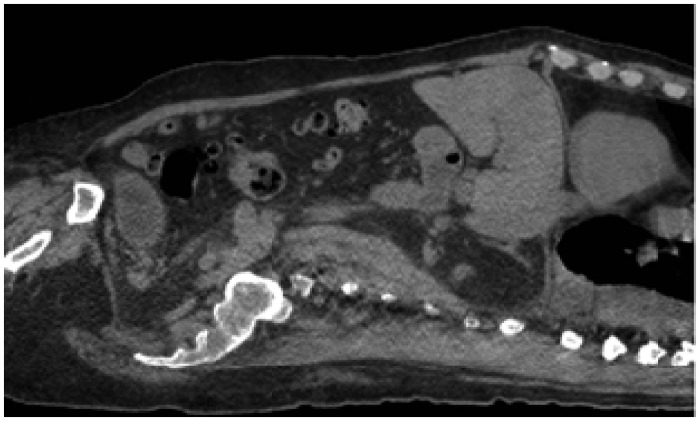
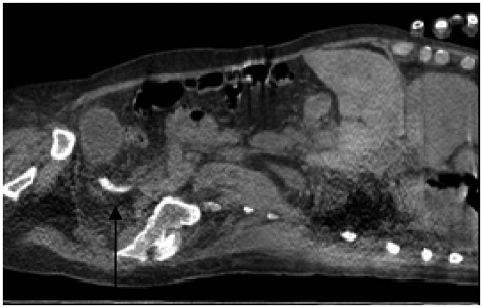
A 77-year-old man recently diagnosed with chronic myelomonocytic leukaemia presented to hospital with left iliac fossa pain and worsening renal function. His medical history included diabetes complicated by underlying chronic kidney disease and heart failure. A non-contrast computed tomography (CT) of the neck, chest, abdomen and pelvis showed an enlarged spleen at 19 cm and multiple cysts were noted in both kidneys. No renal tract calcification was seen (Figure 1). Five weeks later, further decline in renal function associated with left flank pain prompted an urgent abdominal ultrasound scan to be performed, which demonstrated bilateral hydronephrosis without calculi. A second non-contrast CT of his urinary tract was performed which showed bilateral calcification described as milk of calcium in the renal calyces and lower ureter (Figure 2).
Figure 1.
CT of the chest abdomen and pelvis showing no renal tract calcification.
Figure 2.
CT of the urinary tract (non-contrast) showing milk of calcium in the lower ureter five weeks later indicated by arrow.
Discussion
There have been limited case reports and descriptions of renal milk of calcium using different imaging modalities. In most presentations, the diagnosis is provided by an abdominal X-ray, showing a half moon contour with a sharp superior horizontal border.4,5 This represents a fluid–fluid space, with the colloidal suspension or the precipitation of the calcium salts deposited in a dependent manner and acquiring the shape of the collecting system.4,5 Other radiological features on plain abdominal X-rays that may suggest renal milk of calcium are the changing shape of an intra-renal opacity between the supine and erect positions. In addition, faint to moderately opaque densities with fuzzy and indistinct borders in the collecting system are also suggestive of renal milk of calcium on an abdominal X-ray.4,5
In this case, as in similar cases where CT scans have been performed for suspected renal obstruction, renal milk of calcium was identified as a dense layering of the viscous calcium compound in a dependent distribution in the renal collecting system.6 Uniquely in this case, a CT scan performed just five weeks earlier had not demonstrated this phenomenon, indicating the rapidity in which the compounds can form and precipitate.
However, ultrasonography has been found to detect renal milk of calcium more readily than radiography or CT, especially in diverticula and renal cysts.7 Similar to CT scans the echogenic material is seen in the dependent portion of the cyst with reverberation echoes without shadowing.7 But, shadowing can be seen when larger amounts of renal milk of calcium are present.7
Milk of calcium has been previously described in patients with cancers of the urinary tract, urinary diverticuli2 or long-term immobility,1 but no apparent medical treatment methods have been described except symptomatic management. This should therefore be a rare differential diagnosis to be considered in a patient with apparent ureteric obstruction and significant calcification on imaging, to avoid unnecessary interventions such as shock wave lithotripsy and for differentiation from other lesions such as calculi or angiomyolipomas.6,7 In extreme cases of large (giant) renal milk of calcium, urosotomies and nephrectomies have been performed with some success.5 In view of this, our patient was treated with fluid rehydration on a background of acute on chronic renal failure. However, this did not improve his renal function and with his additional co-morbidities he died on this second admission.
Declarations
Competing interests
None declared
Funding
None declared
Ethical approval
Written informed consent for publication was obtained from the patient's next of kin.
Guarantor
MKA
Contributorship
AM was the main contributor, wrote the article and was the junior doctor looking after the patient. MKA was the consultant responsible for the case and helped supervise the case report, SP was the reporting radiologist and selected the radiological figures. GB participated in the writing of the case report.
Acknowledgements
None
Provenance
Not commissioned; peer-reviewed by Shahid Muhammad.
References
- 1. Vaidyanathan S, Hughes PL, Soni BM. Bilateral renal milk of calcium masquerading as nephrolithiasis in patients with spinal cord injury. Adv Ther 2007; 24: 533–544 [DOI] [PubMed] [Google Scholar]
- 2. Bude RO, Korobkin MT. Milk of calcium renal cyst – CT findings. Urology 1992; 40: 149–151 [DOI] [PubMed] [Google Scholar]
- 3. Khan SAA, Khan FR, Fletcher MS, Richenberg JL. Milk of calcium (MoC) cysts masquerading as renal calculi – a trap for the unwary. Cent European J Urol 2012; 65: 170–173 [DOI] [PMC free article] [PubMed] [Google Scholar]
- 4. Murray RL. Milk of calcium in the kidney, diagnostic features on vertical beam roentgenograms. Am J Roentgenol 1971; 113: 455–459 [DOI] [PubMed] [Google Scholar]
- 5. Ulusan S, Koc Z. Milk of calcium collection in the differential diagnosis of giant renal calculus. Br J Radiol 2008; 81: e35–e36 [DOI] [PubMed] [Google Scholar]
- 6. Sandhu G, Bansal A, Chan G, et al. Milk of calcium in the kidney. Nephrol Rev 2011; 3: 42–43 [Google Scholar]
- 7. Yeh HC, Mitty HA, Halton K, Shapiro R, Rabinowitz JG. Milk of calcium in renal cysts: new sonographic features. J Ultrasound Med 1992; 11: 195–203 [DOI] [PubMed] [Google Scholar]


