Fig. 5.
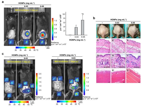
In vivo control of fluc transgene expression in plasmonic hydrogels. Mice were implanted with hydrogels containing the indicated doses of HGNPs and harboring C3H/10T1/2-fLuc cells. One day after, mice were administered rapamycin and NIR-irradiated at 17 mW mm−2 at a single location for 15 min (a) or at three separate locations for 10 min each (c, left panel). Six days later, mice were re-administered rapamycin and subjected to a second irradiation treatment (c, right panel). Bioluminescence imaging was performed 24 h after NIR irradiation. Dashed circles in (c) indicate laser incidence areas. Insets show thermograms of implantation areas at the end of NIR irradiation. Graph shows average luminescence radiance levels detected at the implantation site in (a). *: p<0.05 compared to hydrogels lacking HGNPs; #: p<0.05 compared to hydrogels containing 0.03 mg mL−1 HGNPs; n = 6. (b) Photographs of implantation area (A-C) and histology cross-sections of hydrogels sandwiched by skin (D-I) and gluteal muscle (J-L) 24 h after NIR irradiation for 15 min. Dashed boxes (G-I) show 4x magnified images of epidermis and dermis. Scale bars = 1 cm for photographs and 50 μm for histology captions. p.i.: post-implantation.

