Abstract
• Premise of the study: Demand for fresh-market sweet basil continues to increase, but in 2009 a new pathogen emerged, threatening commercial field/greenhouse production and leading to high crop losses. This study describes a simple and effective staining method for rapid microscopic detection of basil downy mildew (Peronospora belbahrii) from leaves of basil (Ocimum basilicum).
• Methods and Results: Fresh leaf sections infected with P. belbahrii were placed on a microscope slide, cleared with Visikol™, and stained with iodine solution followed by one drop of 70% sulfuric acid. Cell walls of the pathogen were stained with a distinct coloration, providing a high-contrast image between the pathogen and plant.
• Conclusions: This new staining method can be used successfully to identify downy mildew in basil, which then can significantly reduce its spread if identified early, coupled with mitigation strategies. This technique can facilitate the control of the disease, without expensive and specialized equipment.
Keywords: basil, downy mildew, light microscopy, Peronospora belbahrii, staining, visualization
Sweet basil (Ocimum basilicum L., Lamiaceae) is the most important annual culinary herb cultivated in the United States and a source of essential oils and oleoresins, which are used in the manufacturing of foods, flavors, perfumes, and aromatherapy products (Simon et al., 1990; Juliani et al., 2008). Popular commercial basils are not limited to traditional Italian sweet basil (O. basilicum) types, but include a number of different species and varieties. The demand for fresh-market basils has led to more intensive field and greenhouse production systems; however, high losses due to pathogens and pests have been reported (Garibaldi et al., 1997). Basil downy mildew (Peronospora belbahrii Thines) has been previously reported as a destructive disease in the United States (Roberts et al., 2009; Wick and Brazee, 2009) and in several foreign countries, and has recently become a serious concern worldwide (Hansford, 1933; Garibaldi et al., 2004, 2005; McLeod et al., 2006; Khateri et al., 2007; Ronco et al., 2009). A number of commercial varieties belonging to O. ×citriodorum Vis. and O. americanum L. have been identified as highly tolerant to downy mildew (Wyenandt et al., 2010).
Downy mildew (Oomycota) is caused by an obligate biotrophic pathogen and is responsible for diseases of many economically important plant species (Spencer, 1981; Savory et al., 2011). High disease severity can cause complete crop loss in both the greenhouse and field (Roberts et al., 2009; Wick and Brazee, 2009). Downy mildew has been reported to also infect several species of the Lamiaceae family including sage, coleus, and basil (Choi et al., 2009; Thines et al., 2009). Rapid sporulation and dissemination of the pathogen can be observed during periods of high humidity, mild temperatures, poor air circulation, and duration of leaf wetness (Garibaldi et al., 2005, 2007). The pathogen mainly affects the aerial plant organs with initial symptoms of infection identified as chlorosis confined to interveinal regions along the adaxial leaf surface. Thus, early symptoms of downy mildew infection resemble nitrogen deficiency, which can result in misdiagnosis and allow the pathogen to persist under the guise of an abiotic stress. Within a few days under favorable conditions, sporangiophores—visualized as a gray to black fuzzy growth on the abaxial leaf surface epidermis—emerge from stomata and produce asexual spores for secondary infection and disease epidemic in the absence of chemical control (Belbahri et al., 2005; Wyenandt et al., 2010).
The inconspicuous nature of early disease symptoms presents a serious obstacle for growers to prevent disease outbreaks. Although DNA-based assays such as PCR are suitable for the detection of obligate parasites in the laboratory setting (Belbahri et al., 2005; Farahani-Kofoet et al., 2012), these techniques are impractical for field identification purposes and require specialized expertise not available everywhere. Observation of pathogen structures under the microscope remains an effective tool for plant pathogen identification. A simple microscopic method for easy detection of downy mildew can be critical for proper disease management and epidemic prevention. Fluorescence differential staining methods that help the visualization of the pathogen and host plant tissues have been reported (Williamson et al., 1995; Hoch et al., 2005; Diez-Navajas et al., 2007). However, these methods require the use of fluorescence microscopes, as well as a certain level of expertise and multistep procedures.
A simple, reliable staining method that provides high contrast for rapid and early detection of downy mildew can significantly reduce its spread, thus facilitating control of the disease. This technique was originally developed to allow plant breeders in our breeding program to quickly and inexpensively identify the presence of the pathogen not only in the greenhouse but also in the field trials throughout the course of basil downy mildew resistance trials. This procedure will benefit growers, breeders, disease managers, pathologists, and plant diagnostic clinicians because the pathogen can be detected under low magnification (as low as 10×), which will allow a prompt disease response. Early detection and response will allow for reduced spread of the pathogen. This study describes a simple and effective differential staining method for rapid microscopic detection and observation of basil downy mildew (P. belbahrii) using leaves of sweet basil with minimal sample preparation.
METHODS AND RESULTS
Plant material and pathogen inoculum
Diseased plant material was obtained from a 2011 field trial at the Rutgers Agriculture and Research Extension Center (RAREC) in Bridgeton, New Jersey, and maintained in the Rutgers research greenhouse. Diseased leaves were washed with distilled water, and two drops of a suspension containing approximately 5 × 10⁴ spores/mL were used as inoculum. To maintain stock inoculum, host sweet basil (O. basilicum cv. DiGenova Stokes Seed) seedlings were grown out in 7-d successions and repeatedly inoculated with spores harvested from infected plants. Following inoculation, plants were subjected to a 48-h period of 100% leaf wetness and relative humidity with temperatures between 22–24°C and 12-h/12-h light/dark cycle. Dense sporulation was observed 7 d after inoculation. The identity of downy mildew pathogen (P. belbahrii) was confirmed using a real-time PCR assay as previously reported (Belbahri et al., 2005).
Clearing and staining method
Fresh leaf sections (approximately 1 cm2) infected with P. belbahrii from the field trials and from plants artificially inoculated from the greenhouse were collected with a scalpel and placed on a microscope slide with the abaxial side facing up. Two drops of Visikol™ (Phytosys LLC, New Brunswick, New Jersey, USA) clearing solution were applied so as to completely submerge the sample, and a cover slip (0.17 mm thickness) was then applied. To clear the plant material, the microscope slide was placed on a hot plate for approximately 30–60 s until just before boiling, when air bubbles move out to the edges of the slide (Villani et al., 2013). After cooling, the cover slip was removed and two drops of stain were applied. The following four treatments were examined:
1. methylene blue (0.1% in water)
2. aniline blue (0.1% in water)
3. iodine solution (0.5 g I2 plus 1.5 g KI)
4. iodine solution for 1 min followed by one drop of acid (glacial acetic acid, concentrated phosphoric, and 70% sulfuric acid were tested)
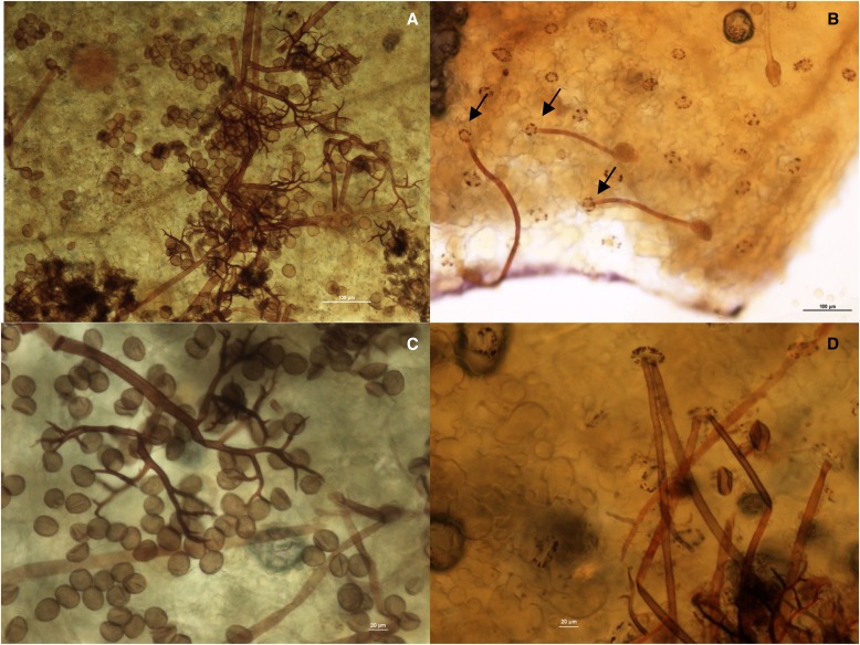
A cover slip was added on the stained samples. Each sample was replicated at least three times. All the microscopic image analyses were taken on a Nikon Eclipse 80i microscope, with NIS Elements D 3.00 SP7 Laboratory Imaging software (Nikon Instruments, Melville, New York, USA). After clearing the leaf with Visikol, the translucent pathogen asexual hyphae and light, elliptical sporangia were clearly observed. Methylene blue or aniline blue stains were nonselective between pathogen and plant tissues. The addition of iodine solution alone was not effective in staining the cell walls of P. belbahrii, but it did stain the starch granules in the guard cells from the basil epidermis. Only after adding two drops of sulfuric acid to the sample cleared with Visikol and stained with iodine solution did the pathogen became a dark brown color in contrast to the leaf tissue. This distinct staining of the pathogen on the surface of the leaf generated an image with high contrast between the pathogen and basil epidermis within 5 min (Fig. 1A). Iodine/potassium iodide stain is commonly used in microscopy to detect the presence of carbohydrates in different organisms or organs (Jackson and Snowdon, 1990). It has been reported that the chemical composition of the cell wall of Peronospora spp. is one of the characteristics that differentiate this organism from true fungi. The major component of Peronospora spp. cell walls is cellulose, β-(1, 3) glucans, and β-(1, 6) glucans (Bartnicki-Garcia, 1968). Other acids (acetic glacial and phosphoric acid) evaluated did not result in high-contrast coloration of the pathogen relative to the host (data not shown).
Fig. 1.
Basil leaf infected with Peronospora belbahrii. (A) Distinct staining of characteristic branched sporangiophores and sporangia after one week of inoculation. (B) Direct germination of sporangia and penetration through stomata (arrows) 48 h after inoculation. (C) High resolution of differential staining of sporangiophores bearing elliptical sporangia. (D) Sporangiophores emerging from stomata.
This method was originally developed as part of an ongoing effort to develop downy mildew–resistant basil plants. Using this simple staining method (protocol is summarized in Appendix S1 (38.3KB, pdf) ), downy mildew was clearly and easily detected. Direct germination of sporangia was observed 48 h after inoculation. A single germ tube was observed per sporangium and could be followed to a stoma by which entry was gained into the host (Fig. 1B). Several days after inoculation, hyphae emerged from the stomata of infected leaves. Extensive hyphal growth was observed with the characteristically well-differentiated sporangiophores showing the determinate growth and dichotomous branches ending in slender, curved, right-angled branchlets (Fig. 1C). In many cases, two to three hyphae could be seen exiting a single stoma (Fig. 1D). In addition to hyphae, light brown oval sporangia were observed at the distal ends of the sporangiophore. The stage of infection can be distinguished easily by the presence or absence of branching in hyphae.
CONCLUSIONS
This technique represents the first practical means by which sweet basil growers and researchers can diagnose and control the spread of basil downy mildew (P. belbahrii). The characteristic branching of P. belbahrii hyphae can be clearly identified using less than 10× magnification after using this staining method. A dissecting microscope or even a magnifying glass would provide the necessary equipment for identification purposes. The distinct contrasting image obtained using this rapid staining method allows for quick identification of the pathogen at the earliest stages of infection with a minimal sample preparation. Compared to molecular techniques such as PCR, this protocol describes an easier, less expensive, and more rapid technique that does not require sophisticated equipment to identify presence of the pathogen. This diagnostic capability allows for appropriate disease control measures to be taken as a function of the pathogen life cycle, which itself can be studied using the technique described in this paper. Early detection of the disease allows for control of its spread by allowing for the identification and quarantine or disposal of infected plants or the application of chemical control agents to reduce and control the disease in the greenhouse or field. Moreover, this simple staining technique would be an effective tool for rapid identification of downy mildew–resistant or susceptible basil plants in breeding programs.
Supplementary Material
LITERATURE CITED
- Bartnicki-Garcia S. 1968. Cell wall chemistry, morphogenesis, and taxonomy of fungi. Annual Review of Microbiology 22: 87–108 [DOI] [PubMed] [Google Scholar]
- Belbahri L., Calmin G., Pawlowski J., Lefort F. 2005. Phylogenetic analysis and real time PCR detection of a presumably undescribed Peronospora species on sweet basil and sage. Mycological Research 109: 1276–1287 [DOI] [PubMed] [Google Scholar]
- Choi Y. J., Shin H. D., Thines M. 2009. Two novel Peronospora species are associated with recent reports of downy mildew on sages. Mycological Research 113: 1340–1350 [DOI] [PubMed] [Google Scholar]
- Diez-Navajas A. M., Greif C., Poutaraud A., Merdinoglu D. 2007. Two simplified fluorescent staining techniques to observe infection structures of the oomycete Plasmopara viticola in grapevine leaf tissues. Micron (Oxford, England) 38: 680–683 [DOI] [PubMed] [Google Scholar]
- Farahani-Kofoet R. D., Römer P., Grosch R. 2012. Systemic spread of downy mildew in basil plants and detection of the pathogen in seed and plant samples. Mycological Progress 10.1007/s11557-012-0816-z. [Google Scholar]
- Garibaldi A., Gullino M. L., Minuto G. 1997. Diseases of basil and their management. Plant Disease 81: 124–132 [DOI] [PubMed] [Google Scholar]
- Garibaldi A., Minuto A., Minuto G., Gullino M. L. 2004. First report of downy mildew on basil (Ocimum basilicum) in Italy. Plant Disease 88: 312. [DOI] [PubMed] [Google Scholar]
- Garibaldi A., Minuto A., Gullino M. L. 2005. First report of downy mildew caused by Peronospora sp. on basil (Ocimum basilicum) in France. Plant Disease 89: 683. [DOI] [PubMed] [Google Scholar]
- Garibaldi A., Bertetti D., Gullino M. L. 2007. Effect of leaf wetness duration and temperature on infection of downy mildew (Peronospora sp.) of basil. Journal of Plant Diseases and Protection 114: 6–8 [Google Scholar]
- Hansford C. G. 1933. Annual report of the mycologist. Review of Applied Mycology 12: 421–422 [Google Scholar]
- Hoch H. C., Galvani C. D., Szarowski D. H., Turner J. N. 2005. Two new fluorescent dyes applicable for visualization of fungal cell walls. Mycologia 97: 580–588 [DOI] [PubMed] [Google Scholar]
- Jackson B. P., Snowdon D. W. 1990. Atlas of microscopy of medicinal plants, culinary herbs and spices. Belhaven Press, London, United Kingdom. [Google Scholar]
- Juliani R., Koroch A., Simon J. E. 2008. Basil: A source of rosmarinic acid. Dietary Supplements ACS Symposium Series, vol. 987, 129–142. American Chemical Society, Washington, D.C., USA. [Google Scholar]
- Khateri H., Calmin G., Moarrefzadeh N., Belbahri L., Lefort F. 2007. First report of downy mildew caused by Peronospora sp. on basil in northern Iran. Journal of Plant Pathology 89: S70 [Google Scholar]
- McLeod A., Coertze S., Mostert L. 2006. First report of a Peronospora species on sweet basil in South Africa. Plant Disease 90: 1115. [DOI] [PubMed] [Google Scholar]
- Roberts P. D., Raid R. N., Harmon P. F., Jordan S. A., Palmateer A. J. 2009. First report of downy mildew caused by a Peronospora sp. on basil in Florida and the United States. Plant Disease 93: 199. [DOI] [PubMed] [Google Scholar]
- Ronco L., Rollán C., Choi Y. J., Shin H. D. 2009. Downy mildew of sweet basil (Ocimum basilicum) caused by Peronospora sp. in Argentina. Plant Pathology 58: 395 [Google Scholar]
- Savory E. A., Granke L. L., Quesada-Ocampo L. M., Varbanova M., Hausbeck M. K., Day B. 2011. The cucurbit downy mildew pathogen Psuedoperonospora cubensis. Molecular Plant Pathology 12: 217–226 [DOI] [PMC free article] [PubMed] [Google Scholar]
- Simon J. E., Quinn J., Murray R. G. 1990. Basil: A source of essential oils. In J. Janick and J. E. Simon (eds.), Advances in new crops, 484–489. Timber Press, Portland, Oregon, USA. [Google Scholar]
- Spencer D. M. 1981. The downy mildews. Academic Press, London, United Kingdom. [Google Scholar]
- Thines M., Telle S., Ploch S., Runge F. 2009. Identity of the downy mildew pathogens of basil, coleus, and sage with implications for quarantine measures. Mycological Research 113: 532–540 [DOI] [PubMed] [Google Scholar]
- Villani T. S., Koroch A. K., Simon J. E. 2013. An improved clearing and mounting solution to replace chloral hydrate in microscopic applications. Applications in Plant Sciences 1 1300016.10.3732/apps.1300016. [DOI] [PMC free article] [PubMed] [Google Scholar]
- Wick R. L., Brazee N. J. 2009. First report of downy mildew caused by a Peronospora species on sweet basil (Ocimum basilicum) in Massachusetts. Plant Disease 93: 318. [DOI] [PubMed] [Google Scholar]
- Williamson B., Breese W. A., Shattock R. C. 1995. A histological study of downy mildew (Peronospora rubi) infection of leaves, flowers and developing fruits of Tummelberry and other Rubus spp. Mycological Research 99: 1311–1316 [Google Scholar]
- Wyenandt C. A., Simon J. E., McGrath M. T., Ward D. L. 2010. Susceptibility of basil cultivars and breeding lines to downy mildew (Peronospora belbahrii). HortScience 45: 1416–1419 [Google Scholar]
Associated Data
This section collects any data citations, data availability statements, or supplementary materials included in this article.


