Abstract
Eugenol is a plant-derived phenolic compound which has recognised therapeutical potential as an antifungal agent. However little is known of either its fungicidal activity or the mechanisms employed by fungi to tolerate eugenol toxicity. A better exploitation of eugenol as a therapeutic agent will therefore depend on addressing this knowledge gap. Eugenol initiates increases in cytosolic Ca2+ in Saccharomyces cerevisiae which is partly dependent on the plasma membrane calcium channel, Cch1p. However, it is unclear whether a toxic cytosolic Ca2+elevation mediates the fungicidal activity of eugenol. In the present study, no significant difference in yeast survival was observed following transient eugenol treatment in the presence or absence of extracellular Ca2+. Furthermore, using yeast expressing apoaequorin to report cytosolic Ca2+ and a range of eugenol derivatives, antifungal activity did not appear to be coupled to Ca2+ influx or cytosolic Ca2+ elevation. Taken together, these results suggest that eugenol toxicity is not dependent on a toxic influx of Ca2+. In contrast, careful control of extracellular Ca2+ (using EGTA or BAPTA) revealed that tolerance of yeast to eugenol depended on Ca2+ influx via Cch1p. These findings expose significant differences between the antifungal activity of eugenol and that of azoles, amiodarone and carvacrol. This study highlights the potential to use eugenol in combination with other antifungal agents that exhibit differing modes of action as antifungal agents to combat drug resistant infections.
Introduction
There is increasing interest in the use of plant-derived antimicrobial compounds as both natural preservatives and in the treatment of fungal infections. A driver for this interest is that antifungal therapies are limited to a few classes of compounds which are undermined by the emergence of resistance and tolerance coupled with innate toxicity of these compounds to the host organism [1].
Eugenol is the major constituent of essential oils from clove, cinnamon and bay leaves and is a member of a wider group of diverse amphipathic phenolic compounds which display antifungal activity [2]. Despite the therapeutic potential of these compounds, relatively little is known about their mode of action and the mechanisms employed by fungi to resist their toxicity. Several modes of action have been proposed including disruption of ion homeostasis [3], nonspecific lesion of the plasma membrane (resulting in leakage of cell contents; [4]), disruption of amino acid metabolism [5] and generation of oxidative stress [6]. However, the mechanism of killing remains to be fully elucidated and consequently we know little of the defence mechanisms employed by fungi to resist the toxic effects of eugenol (and related amphipathic phenolic compounds). Such knowledge will be essential in facilitating the application of these promising therapeutic compounds.
Previous studies have shown that amiodarone and carvacrol, two amphipathic phenolic compounds related to eugenol, induce increases in cytosolic Ca2+ in Saccharomyces cerevisiae and that their toxicity correlates with the amplitude and duration of cytosolic Ca2+ elevation; consequently, it has been proposed that the Ca2+ elevation represents a toxic shock responsible, at least in part, for the antifungal activity of these compounds [7], [8], [3]. Recently however, Roberts et al. [9] have shown that although eugenol also induces a cytosolic Ca2+ elevation in yeast the cch1Δ mutant (which is deficient in a subunit of the yeast high affinity plasma membrane Ca2+ channel) exhibits reduced Ca2+ influx in response to eugenol but is hypersensitive to eugenol. This raises the exciting possibility that the mechanisms by which yeast respond to eugenol and the related compounds amiodarone and carvacrol differ and that a Cch1p-mediated Ca2+ influx forms part of a cell signalling response to enable yeast to survive eugenol stress.
The present study investigates the role of Ca2+ in eugenol toxicity in detail. We show that the eugenol-induced cytosolic Ca2+ elevation in yeast is unlikely to represent a toxic burst of Ca2+. Importantly, Ca2+ influx appears to be limited to a signalling role that is crucial for protecting yeast against eugenol stress.
Materials and Methods
Strains, media and reagents
Single and double mid1Δ and cch1Δ mutants were derived from the parental S. cerevisiae strain JK9-3da (Mata, leu2-3, 112Δ, his4Δ, trp1Δ, ura 3-52Δ, rme1, HMLa) by replacing the MID1 and CCH1 genes by a KanMX cassette [10]. Yeast strains were transformed with pEVP11/AEQ (a plasmid bearing apoaequorin gene under constitutively expression and a LEU2 marker, generously provided by Dr Patrick Masson, University of Wisconsin-Madison, Wisconsin, US) as previously described [11]. Unless otherwise stated, yeast strains were cultured at 30°C in standard synthetic complete media minus the addition of leucine (SCM-leu; Foremedium, UK). SCM-leu-Ca is synthetic minimal glucose media [12] modified to contain no added calcium by replacing the calcium pantothenate with sodium pantothenate and omitting the addition of CaCl2 [10] and supplemented with complete supplement mixture-leu (Foremedium, UK).
vcx1Δ and pmc1Δ mutants were derived from the parental strain S. cerevisiae strain BY4742 (Matα, his3Δ1, leu2Δ0, lys2Δ0, ura3Δ) by gene replacement with a KanMX cassette (EUROSCARF) and cultured in YPD contained 1% yeast extract and 2% peptone. All growth media contained 2% (w/v) glucose (and 2% (w/v) agar for solid media), and where indicated, supplemented with BAPTA (1,2-bis(o-aminophenoxy)ethane-N,N,N′,N′-tetraacetic acid), EGTA (ethylene glycol tetraacetic acid) or CaCl2 using stock solutions of BAPTA (10 mM BAPTA, 10 mM HEPES, 2% glucose, pH 7.5 with Tris base), EGTA (0.5 M EGTA, 10 mM HEPES, 2% glucose, pH 7.0 with NaOH) or 2 M CaCl2. pH was approximately 6.5 for YPD-based growth media and between 5.0 and 5.5 for SCM-based growth media. Eugenol, isoeugenol, estragole, o-eugenol, acetyl eugenol and methyl eugenol (Sigma Aldrich) were in liquid form and made to 100x stocks in ethanol and stored at 4°C.
Luminometry
Cells expressing apoaequorin were grown overnight in SCM-leu in a shaking (150 rpm) incubator to an optical density at 600 nm (OD600) of 8 (approximately 1×10⁸ cells/ml). OD600 was determined after 8x dilution of culture in water. To obtain cells in mid-log growth phase, 0.5 ml of overnight cultures were subcultured into 10 ml of fresh SCM-leu to give an OD600 of 0.8 and incubated shaking at 150 rpm for up to 3 hours until an OD600 between 1.6 and 2.4 was reached. Cells were pelleted (at room temperature using 200 g for 5 minutes) and resuspended in fresh SCM-leu to an OD600 of 2.4. 4 µl of 0.5 mM coelentrazine (Prolume, USA) in absolute methanol was added to 1 ml of cells and incubated in the dark for 2 hours at 30 C shaking at 150 rpm. Coelentrazine loaded cells were pelleted in a microcentrifuge (5000 rpm for 20 seconds) and resuspended in fresh SCM-leu to an OD600 of 8. Luminescence from 20 µl samples of mid-log growth phase cells was recorded as previously reported [9]. Eugenol (and eugenol derivatives) were added to samples at indicated concentrations (containing 1% ethanol) in either 200 µl of Ca buffer (10 mM CaCl2, 10 mM HEPES, 2% glucose, pH 7.5 with Tris base), BAPTA buffer (10 mM BAPTA, 10 mM HEPES, 2% glucose, pH 7.5 with Tris base) or SCM-leu. Luminescence (expressed in arbitrary units (AU) per 0.2 seconds) was measured for up to 10 minutes after which cells were lysed with 1.6 M CaCl2 in 20% (v/v) ethanol to determine total (summed) luminescence. Total luminescence was in much greater excess over luminescence induced by eugenol (or eugenol derivatives) indicating that the availability of aequorin-coelentrazine complex was sufficient for the reporting induced Ca2+ cyt elevations.
Toxicity assays
Transient exposure to eugenol – dot drop assays
1 ml of overnight cultures of JK9-3da (and derived mutants) was added to 9 ml of fresh SCM-leu to an OD600 of 1.6 and incubated for approximately 4 hours shaking at 150 rpm until an OD600 between 3 and 4 was reached and cells were in mid-logarithmic growth phase. Culture was pelleted (200 g for 5 minutes) and resuspended in 5 ml of 2% glucose, repelleted and resuspended in 1 ml of 2% glucose. 0.5 ml cell samples were pelleted in a microcentrifuge (5000 rpm for 20 seconds) and resuspended in 0.5 ml of Ca buffer or BAPTA buffer and adjusted to an OD600 of 20. 100 µl of cells were pelleted and resuspended in 0.5 ml of corresponding buffer containing eugenol and incubated at room temperature for 10 minutes (unless otherwise stated). Following incubation, cells were pelleted in a microcentrifuge and resuspended in 1 ml of SCM-leu, repelleted and resuspended in 100 µl of SCM-leu. 10-fold serial dilution was performed in sterile water and 5 µl of each dilution was placed onto SCM-leu containing 2% agar. Images of plates were taken after two days growth.
Persistent exposure to eugenol - dot drop assays
JK9-3da (and derived mutants) were cultured overnight in 10 ml SCM-leu to an OD600 of 8, pelleted (200 g for 5 minutes) and resuspended in 10 ml of sterile water and resuspended to 0.5×10⁸ cells/ml. Following 10-fold serial dilution of each yeast strain using sterile water, 5 µl drops were spotted onto YPD (1% yeast extract, 2% peptone, 2% glucose, 2% agar) plates containing varying concentrations of eugenol. Images of plates were taken after two days growth at 30 C.
Determination of IC50 values
Sensitivities of yeast growth to eugenol (and derivatives) were assayed by a dilution method in 24 well plates as described by Edlind et al. [13]. Briefly, overnight cultures in SCM-leu were pelleted and washed in 2% glucose, re-pelleted and diluted in either SCM-leu or SCM-leu-Ca supplemented with either Ca2+ or BAPTA to 10−4 cells/ml and 1 ml aliquoted to wells except the initial well in which 2 ml was aliquoted. Eugenol, eugenol derivatives or BAPTA were added to the first well and a two-fold dilution series achieved by mixing, removing 1 ml and adding this to the second well; this was repeated for subsequent wells except the final well which served as a eugenol (or BAPTA)-free control. Plates were sealed and incubated shaking at 150 rpm for 48 hours after which OD600 was determined (for OD values greater than 1, a 10x dilution of the sample was performed). For BY4742 strains, sensitivities to eugenol were determined as above except culture media was YPD. Student' t tests were performed on the IC50 values to determine p values and whether mean IC50 values were significantly differently.
Results and Discussion
Eugenol toxicity is not coupled to Ca2+ influx
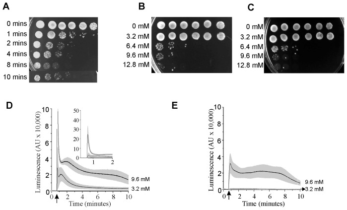
Yeast cells were transiently exposed to toxic levels of eugenol for varying times before transferring on to agar-containing growth media to monitor cell viability. Figure 1A shows that transient exposure to 6.4 mM eugenol was fungicidal and that this toxic effect was apparent within one minute of exposure to eugenol and showed increasing toxicity up to 10 minutes. Therefore, to investigate the role of Ca2+ in eugenol toxicity, 10 minute transient exposure of wild type (Jk9-3da) yeast to varying concentrations of eugenol were performed in the presence (10 mM CaCl2; Figure 1B) and absence (10 mM BAPTA; Figure 1 C) of extracellular Ca2+ before immediately transferring to growth media to monitor cell viability. In both cases transient exposure of up to 3.2 mM eugenol was tolerated by yeast, however, at concentrations greater than 3.2 mM fungicidal effects were apparent. Interestingly, removal of extracellular Ca2+ had no apparent effect on eugenol toxicity towards yeast suggesting that an influx of Ca2+ across the plasma membrane is unlikely to play a role in eugenol toxicity. To gain further sights into the role of Ca2+ in mediating eugenol toxicity in S. cerevisiae, cytosolic Ca2+ was also monitored in cells following exposure to eugenol (in identical conditions to that used to assess cell viability shown in Figures 1B and C) using the genetically encoded reporter, aequorin, reconstituted with its cofactor, coelentrazine. In the presence of extracellular Ca2+, eugenol-induced cytosolic Ca2+ elevations in wild type yeast were characterised by a large transient increase immediately following addition of eugenol followed by a prolonged Ca2+ elevation for up to 10 minutes (Figure 1 D). Increasing the concentration of eugenol increased the magnitude of the Ca2+ elevations (Figure 1 D) whilst removal of extracellular Ca2+ abolished the large transient elevation in cytosolic Ca2+ (and reduced total eugenol-induced cytosolic Ca2+ elevations; Figure 1E). However, there was no correlation between eugenol toxicity and either the amplitude and duration of the Ca2+ elevation or Ca2+ influx across the plasma membrane. Toxicity resulting from transient exposure of eugenol and Ca2+ elevations were also investigated in the yeast Ca2+ channel mutants cch1Δ, mid1Δ and cch1Δ mid1Δ (Figure S1). Interestingly, the mid1Δ mutant consistently exhibited greater tolerance to transient exposure of high concentrations compared to the wild type and cch1Δ mutant yeast although as in wild type yeast there was no correlation between eugenol toxicity and cytosolic Ca2+ elevation; the Ca2+ elevation in the mid1Δ mutant was equivalent to that observed in wild type yeast. It is also notable that the cch1Δ mutants exhibited equivalent tolerance to transient exposure of eugenol compared to wild type yeast despite the cytosolic Ca2+ elevation being consistently reduced in the cch1Δ yeast (Figure S1). Taken together, these results show that eugenol toxicity is unlikely to be mediated by a pancellular “toxic” elevation in cytosolic Ca2+.
Figure 1. Eugenol toxicity is not dependent on Ca2+ influx.
A) Time dependence of eugenol toxicity. Viability of JK9-3da cells after exposure to 6.4 mM eugenol suspended in Ca buffer for 1, 2, 4, 8 and 10 minutes. Yeast cultures are spotted on to SCM-leu media containing 2% agar; left most spots are growth after 2 days following inoculation with 5 µl of culture. Serial 10-fold dilution of the left most inoculum is shown to the right. B) as A except cells were exposed to varying concentrations of eugenol (as indicated) for 10 minutes in Ca buffer. C) as B except cells were exposed to eugenol in BAPTA buffer. D) Eugenol-induced cytosolic Ca2+ elevation in the presence of extracellular Ca2+ in mid log growth phase yeast cells. Ca2+-dependent aequorin luminescence from Jk9-3da cells in response to 3.2 and 9.6 mM eugenol in Ca buffer. Eugenol was added at 40 seconds (indicated by arrow). Traces represent mean (± SEM) from at least 5 independent experiments. SEM values are illustrated using grey shading. Luminescence was recorded every 0.2 seconds and is expressed in arbitrary units (AU). Inset is data from the main figure on an expanded y axis. E) As D except eugenol was in BAPTA buffer.
At higher concentrations (e.g. 9.6 mM) the eugenol-induced cytosolic Ca2+ elevation consists of two distinct components: a rapid transient increase in cytosolic Ca2+ due to Ca2+ influx across the plasma membrane (Figure 1D) followed by a prolonged elevation of cytosolic Ca2+, which is independent of extracellular Ca2+ (i.e. present in BAPTA-containing buffer; Figure 1E) and thus must result from a release of Ca2+ from intracellular stores. The transient Ca2+ influx across the plasma membrane is independent of the presence of Cch1p and Mid1p (Figure S1) revealing that this Ca2+ influx across the plasma membrane is via an unspecified pathway; this could reflect non-specific disruption of the plasma membrane and general cell leakage [14], [4] or hitherto unidentified Ca2+ specific entry pathways [15], [16], [17].
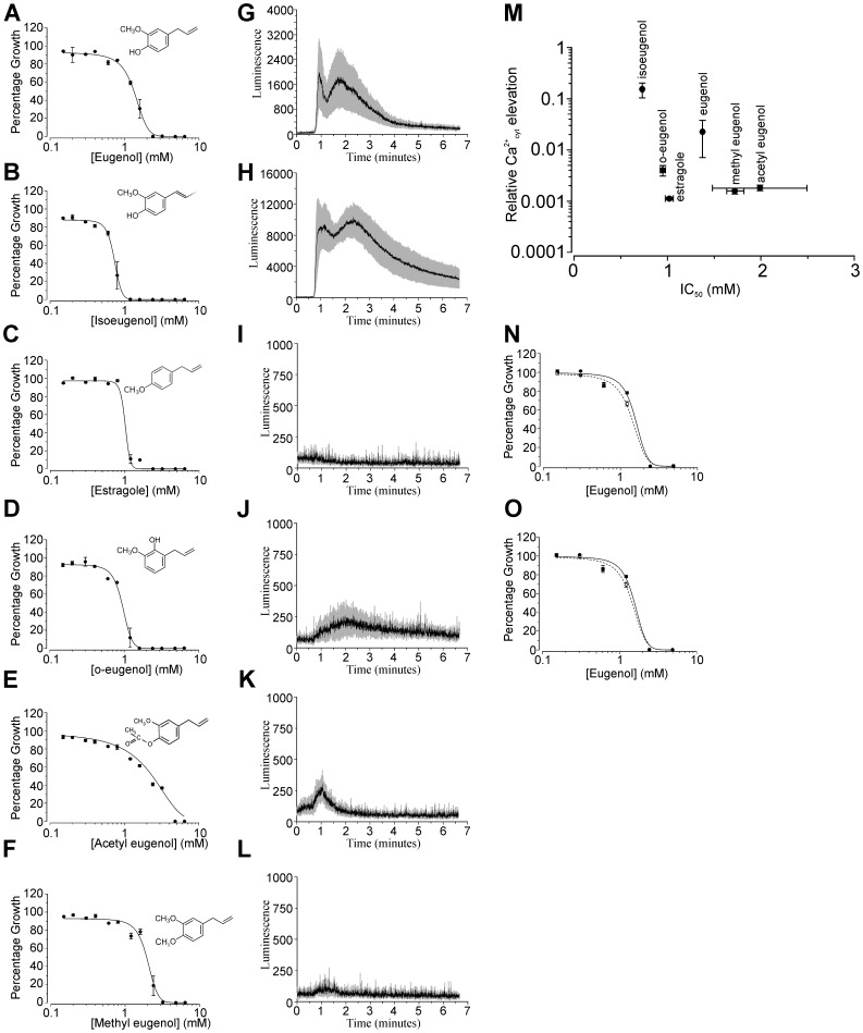
To investigate the relationship between toxicity and cytosolic Ca2+ elevation further, we compared the toxicity of a range of eugenol derivatives with their ability to induce cytosolic Ca2+ elevations (Figure 2). Figure 2 M plots the concentration of eugenol (and derivatives) which results in 50% inhibition of yeast growth (IC50) against the cytosolic Ca2+ elevation induced following addition of 3.2 mM eugenol (or derivative). Notably, both estragole (hydroxyl group replaced with methoxy group) and o-eugenol (hydroxyl group moved to the carbon situated between the methoxy and allyl groups) exhibited significantly greater toxicity towards yeast (1.017±0.0435 mM and 0.949±0.0285 mM respectively) than eugenol (p<0.02 and <0.01 respectively) but with reduced elevation of cytosolic Ca2+. Isoeugenol however (which differs from eugenol in the position of the double bond in the allylic side chain) was significantly more toxic to yeast than eugenol (IC50 was 0.73±0.0096 mM compared to 1.375±0.0349 mM for eugenol; p<0.01) and induced greater cytosolic Ca2+ elevation compared to that for eugenol. Taken together, these results show no correlation between Ca2+ elevation and toxicity and do not support a mode of action based on a toxic elevation in cytosolic Ca2+. Consistent with this, estragole and methyl eugenol both failed to evoke measurable Ca2+ elevation using a concentration (i.e. 3.2 mM) which is 2 to 3 times greater than the determined IC50. It is noteworthy that the IC50 values determined for the eugenol derivatives are unlikely to simply reflect differences in hydrophobicity because acetyl eugenol, isoeugenol and eugenol (which represent the full range of IC50 values observed in the present study) have virtually identical hydrophobicity (log P) values of approximately 2.5 [14].
Figure 2. Eugenol toxicity is not dependent on Ca2+ influx.
(A - F) Jk9-3da growth in SCM-leu containing varying concentrations of eugenol (A) or eugenol derivative (B–F). Absorbance was recorded after 48 hours incubation and is shown as growth (absorbance) relative to growth exhibited in eugenol or eugenol derivative-free control media (SCM-leu containing 1% ethanol). Data are fitted with the dose-response function min + (max-min)/1+ ((x/IC50)−p)) where p is the slope, IC50 is the eugenol concentration inhibiting 50% growth and min and max represent minimum and maximum relative absorbance values respectively. Mean values (± SEM) from 4 experiments are shown. (G–L) Ca2+-dependent aequorin luminescence from Jk9-3da cell in response to addition of 3.2 mM eugenol (G), isoeugenol (H), estragole (I), o-eugenol (J), acetyl eugenol (K) and methyl eugenol (L) added at 40 seconds in SCM-leu. Traces represent mean (± SEM) from at least 4 independent experiments. SEM values are illustrated using grey shading. (M) plot of IC50 values (from data shown in parts A–F) against Ca2+ elevations (determined from data shown in parts G–L). Relative Ca2+ elevations were calculated as the sum of luminescence resulting from addition of eugenol or eugenol derivative divided by total luminescence determined after lysis with 1.6 M CaCl2, 20% ethanol (see Materials and Methods). N–O) Growth of BY4742 (parental strain; solid symbols and line) and pmc1Δ (N) and vcx1Δ (O) yeast mutants (open symbols and dashed line) in YPD containing varying concentrations of eugenol. Growth was recorded as detailed in parts A–F. IC50 values are 1.56±0.189 mM (BY4742), 1.45±0.112 mM (PMC1Δ) and 1.41±0.083 mM (VCX1Δ).
In contrast to the present study, the amplitude and duration of cytosolic Ca2+ elevation in response to amiodarone and carvacrol correlates with drug toxicity [7], [8]. The fungicidal activity of amiodarone has been shown to be tightly coupled to Ca2+ influx across the plasma membrane; reducing extracellular Ca2+ with EGTA blocks cytosolic Ca2+ elevation and rescuses growth inhibition by amiodarone [7]. Furthermore, amiodarone toxicity is also dependent on hyperpolarisation of the yeast plasma membrane and consequently increases the driving force for Ca2+ influx [18]. Consistent with a cytotoxic influx of Ca2+ mediating amiodarone and carvacrol toxicity, vma2Δ mutants which lack vacuolar H+ pumping activity (and as a consequence have impaired Ca2+ sequestration into the vacuole via Ca2+/H+ antiport) exhibit prolonged cytosolic Ca2+ elevations and have increased sensitivity to amiodarone and carvacrol [19], [8]. We adopted a similar approach in order to investigate the role of the vacuole in sequestering Ca2+ from the cytosol and to negate eugenol toxicity using the pmc1Δ (vacuolar Ca2+ ATPase; Figure 2N) and vcx1Δ (vacuolar Ca2+/H+ antiporter; Figure 2O) mutants. Both pmc1Δ and vcx1Δ exhibited similar sensitivity to eugenol as the parental wild type strain (BY4742) indicating that a reduced capacity to remove Ca2+ from the cytosol across the vacuolar membrane did not affect eugenol toxicity. Taken together, these data are consistent with eugenol toxicity being independent of a general cytotoxic Ca2+ elevation.
Tolerance to eugenol is dependent on extracellular Ca2+ influx via Cch1p
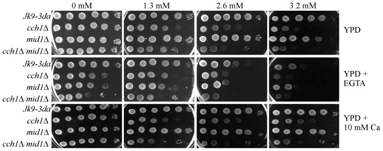
Previous studies showed that Cch1p (but not Mid1p) was a crucial factor in determining the tolerance of S. cerevisiae to eugenol; the hyper-sensitivity of cch1Δ mutants suggested that a Cch1p-mediated Ca2+ influx may be necessary to protect yeast against the toxic effects of eugenol [9]. To investigate this possibility further, dot drop growth assays on YPD media supplemented with eugenol and either 10 mM CaCl2 or 10 mM EGTA (to increase or reduce extracellular Ca2+ respectively) were conducted. Reducing extracellular Ca2+ reduced the tolerance of the wild type (Jk9-3da) and mid1Δ strains to levels similar to that observed for the cch1Δ mutants (Figure 3). Furthermore, the sensitivity of the cch1Δ strains to eugenol was unaffected by the reduction of extracellular Ca2+ using EGTA. These data suggest that Cch1p-mediated Ca2+ influx is necessary for eugenol tolerance in yeast rather than a Ca2+ influx per se. Consistent with this, the eugenol sensitivity of the cch1Δ mutants could not be rescued by supplementing the growth media with additional extracellular Ca2+. Furthermore, the inability to enhance the tolerance of yeast growth to eugenol by increasing extracellular Ca2+ content of the growth media also indicates that there is sufficient Ca2+ in YPD (approximate Ca2+ content of 100–200 µM; [20]) to support Cch1p-mediated Ca2+ influx. This is consistent with previous reports which show that Cch1p mediates high affinity Ca2+ uptake in yeast (reviewed by [21]). Figure 3 is also consistent with the previous observation that Cch1p operates independently of Mid1p in response to eugenol stress [9]. Although it is generally accepted that Mid1p and Cch1p function together as a high affinity Ca2+ influx mechanism in S. cerevisiae (e.g. [10]) Cch1p activity independent of Mid1p has also been reported for Li stress at high temperature [22]. Finally, the observation that the cch1Δ yeast mutants are able to survive 10 minute exposure to eugenol at 3.2 mM (Figure S1, A & G) but not persistent exposure at 3.2 mM eugenol (Figure 3) indicates that the Cch1p-dependent tolerance mechanism operates to enhance the survival of yeast to persistent (long term) exposure to eugenol.
Figure 3. Extracellular Ca2+ is necessary for eugenol tolerance.
Yeast cultures were spotted onto YPD, YPD supplemented with 102 agar plates containing either 0 (1% ethanol), 1.3, 2.6 or 3.2 mM eugenol. Left-most spots on each plate are growth after 2 days at 30 C after inoculation with 5 µl culture at approximately 0.5×10⁸ cells/ml. Serial 10-fold serial dilution of the left-most inoculum is shown to the right.
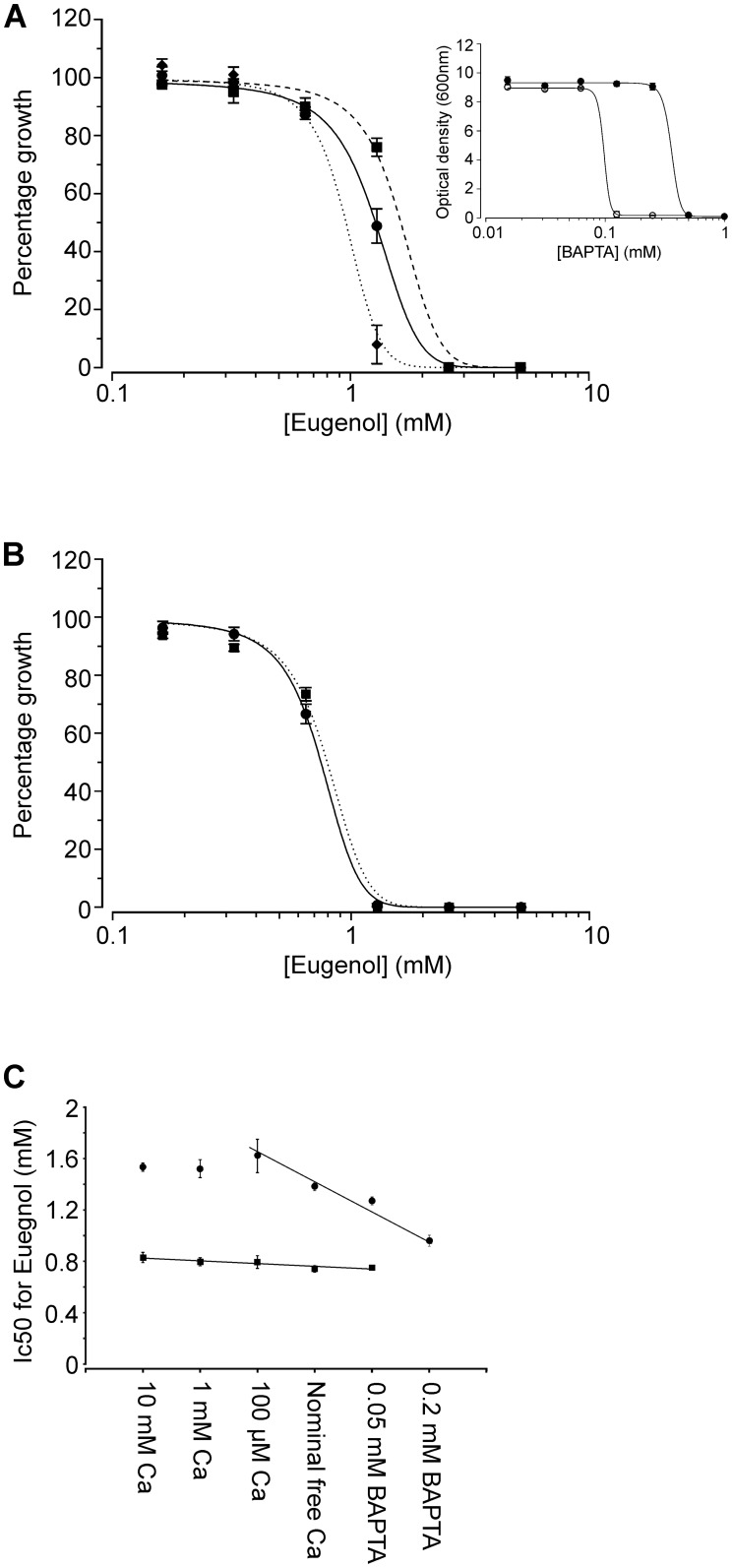
In order to define the dependence of eugenol tolerance on extracellular Ca2+ more precisely, growth experiments were conducted using a Ca2+-free synthetic complete medim (SCM-leu-Ca); this permitted a more robust yet subtle control of extracellular Ca2+ levels using the Ca2+ chelator, BAPTA (which exhibits higher specificity for Ca2+ and is less pH sensitive than EGTA). Figure 4A shows that the IC50 of eugenol (the concentration of eugenol resulting in 50% inhibition of growth when compared to growth in the absence of eugenol) for the wild type strain was strongly dependent on extracellular Ca2+. In the presence of 100 µM extracellular Ca2+, the IC50 of eugenol for wild type yeast was 1.62±0.133 mM and this decreased to 1.27±0.031 and 0.96±0.043 mM in the presence of 0.05 and 0.2 mM BAPTA, respectively. Importantly, the growth of wild type yeast in the absence of eugenol was unaffected by the presence of 0.05 and 0.2 mM BAPTA (Figure 4A, inset).
Figure 4. Dependence of eugenol tolerance on Cch1p-mediated Ca2+ influx.
A) Dilution assays of eugenol activity versus Jk9-3da growth in SCM-leu-Ca supplemented with 100 µM CaCl2 (▪), 50 µM BAPTA (•) and 200 µM BAPTA (♦). Absorbance was recorded after 48 hours incubation and is shown as growth (absorbance) relative to growth exhibited by yeast in eugenol-free control media. Data are fitted with the dose-response function min + (max-min)/1+ ((x/IC50)−p)) where p is the slope, IC50 is the eugenol concentration inhibiting 50% growth and min and max represent minimum and maximum relative absorbance values respectively. Mean values (± SEM) from 4 experiments are shown. Inset: Dilution assays showing growth of jk9-3da (solid symbols) and cch1Δ (open symbols) cells in SCM-leu-Ca supplemented with BAPTA. Growth is shown as optical density (600 nm) after 48 hours and data are fitted with the dose-response function used in ‘A’. IC50 for BAPTA is 359±35.9 µM and 97.7±12.4 µM for jk9-3da and cch1Δ cells respectively. Mean values (± SD) from 3 experiments are shown. B) as ‘A’ except growth of cch1Δ strain in SCM-leu-Ca supplemented with 100 µM CaCl2 (▪), 50 µM BAPTA (•). C) IC50 for eugenol plotted as a function of CaCl2 and BAPTA added to SCM-leu-Ca growth media for Jk9-3da (•) and cch1Δ (▪) strains. IC50 values where obtain from fits of data as shown in parts ‘A’ and ‘B’. Data are fitted with linear regression fits for Jk9-3da (r2 = 0.8775) and cch1Δ (r2 = 0.8694).
In contrast to wild type yeast, the addition or removal of extracellular Ca2+ did not affect the sensitivity of the cch1Δ strain to eugenol (Figure 4B and C). The sensitivity of the cch1Δ mutant to eugenol (IC50 = 0.796±0.045 mM in the presence of 100 µM Ca2+) was similar to that for wild type yeast in the presence of 0.2 mM BAPTA (0.96±0.043 mM) indicating that the Ca2+-dependent tolerance to eugenol exhibited by wild type yeast is dependent on Ca2+ influx via Cch1p. Furthermore, the cch1Δ mutant was more sensitive to reductions in extracellular Ca2+ than wild type yeast as it was unable to grow in media containing more than 0.0625 mM BAPTA (Figure 4A inset). These data support the yeast growth patterns shown in Figure 3 and illustrate that in low Ca2+ conditions, Cch1p is required for Ca2+ homeostasis. In addition, they are also consistent with previous observations that Ca2+ uptake in cch1Δ mutants is approx. 5-fold less than wild type yeast under non-stress conditions [10] and with the widely accepted dogma that Cch1p forms part of the high affinity Ca2+ influx system (HACS; [23], [24]) which is essential to maintain growth in low Ca2+ environments.
Taken together, the data show that Ca2+ influx via Cch1p is necessary for yeast tolerance to eugenol. In addition, the observation that increasing extracellular Ca2+ to levels greater than 100 µM does not enhance the tolerance of wild type yeast to eugenol (Figure 2C) is in agreement with the dot drop experiments on YPD media (Figure 3) and is consistent with Cch1p-mediated Ca2+ transport being saturated at µM levels of Ca2+. Interestingly, similar studies conducted on S. cerevisiae have shown that the activity of antifungal azoles (e.g. miconazole) is also enhanced by extracellular Ca2+ sequestration (using 1 mM EGTA); however, in contrast to that observed with eugenol, the addition of 3 mM Ca2+ to the growth media reduced azole activity by 3-fold [13]. Furthermore, azole activity against yeast is enhanced in the presence of FK506 [13] suggesting a role for extracellular Ca2+ influx and calcineurin activation in the azole tolerance mechanisms employed by yeast. Interestingly, eugenol tolerance is independent of calcineurin activation [9] which serves to highlight differences between the Ca2+-dependent tolerance mechanisms employed by yeast in response to the antifungal properties of eugenol and the commercially available azoles.
In conclusion, our data show that a toxic elevation in cytosolic Ca2+ elevation is unlikely to be responsible for eugenol toxicity in yeast and that the role of Ca2+ in eugenol toxicity appears confined to a Cch1p-dependent Ca2+ influx which is necessary to enhance eugenol tolerance in yeast. Although the downstream targets of the resulting Ca2+ signal remain unknown, our data suggest that the signalling pathways employed by yeast to tolerate eugenol toxicity will be distinct to those employed in azole and amiodarone tolerance. This is supported further by the discovery that eugenol and amiodarone employ different modes of action with respect to antifungal activity. These differences highlight the potential to use eugenol in combination therapies which aim to augment the efficacy of commercially available azoles and other promising antifungal drugs.
Supporting Information
Viability of cch1Δ (A and G), mid1Δ (B and H) and cch1Δmid1Δ (C and I) cells after 10 minutes exposure to varying concentrations of eugenol suspended in Ca buffer (A, B, C) and BAPTA buffer (G, H, I). Yeast cultures are spotted on to SCM-leu media containing 2% agar; left most spots are growth after 2 days following inoculation with 5 µl of culture. Serial 10-fold dilution of the left most inoculum is shown to the right. Ca2+-dependent aequorin luminescence from cch1Δ (D and J), mid1Δ (E and K) and cch1Δmid1Δ (F and L) cells in response to 3.2 and 9.6 mM eugenol in Ca buffer (D, E, F) and BAPTA buffer (J, K, L). Eugenol was added at 40 seconds (indicated by arrow). Traces represent mean (± SEM) from at least 5 independent experiments. SEM vales are illustrated using grey shading. Luminescence was recorded every 0.2 seconds and is expressed in arbitrary units (AU). Inset is data from the main figure on an expanded y axis.
(TIF)
Data Availability
The authors confirm that all data underlying the findings are fully available without restriction. All relevant data are within the paper and its Supporting Information files.
Funding Statement
The authors have no funding or support to report.
References
- 1. Monk BC, Goffeau A (2008) Outwitting multidrug resistance to antifungals. Science 321: 367–369. [DOI] [PubMed] [Google Scholar]
- 2. Bakkali F, Averbeck S, Averbeck D, Waomar M (2008) Biological effects of essential oils - A review. Food and Chemical Toxicology 46: 446–475. [DOI] [PubMed] [Google Scholar]
- 3.Zhang Y, Muend S, Rao R (2012) Dysregulation of ion homeostasis by antifungal agents. Frontiers in Microbiology 3.. [DOI] [PMC free article] [PubMed] [Google Scholar]
- 4. Zore GB, Thakre AD, Jadhav S, Karuppayil SM (2011) Terpenoids inhibit Candida albicans growth by affecting membrane integrity and arrest of cell cycle. Phytomedicine 18: 1181–1190. [DOI] [PubMed] [Google Scholar]
- 5.Darvishi E, Omidi M, Bushehri AAS, Golshani A, Smith ML (2013) The Antifungal Eugenol Perturbs Dual Aromatic and Branched-Chain Amino Acid Permeases in the Cytoplasmic Membrane of Yeast. Plos One 8.. [DOI] [PMC free article] [PubMed] [Google Scholar]
- 6. Khan A, Ahmad A, Akhtar F, Yousuf S, Xess I, et al. (2011) Induction of oxidative stress as a possible mechanism of the antifungal action of three phenylpropanoids. Fems Yeast Research 11: 114–122. [DOI] [PubMed] [Google Scholar]
- 7. Muend S, Rao R (2008) Fungicidal activity of amiodarone is tightly coupled to calcium influx. Fems Yeast Research 8: 425–431. [DOI] [PMC free article] [PubMed] [Google Scholar]
- 8. Rao A, Zhang YQ, Muend S, Rao R (2010) Mechanism of Antifungal Activity of Terpenoid Phenols Resembles Calcium Stress and Inhibition of the TOR Pathway. Antimicrobial Agents and Chemotherapy 54: 5062–5069. [DOI] [PMC free article] [PubMed] [Google Scholar]
- 9.Roberts SK, McAinsh M, Widdicks L (2012) Cch1p Mediates Ca2+ Influx to Protect Saccharomyces cerevisiae against Eugenol Toxicity. Plos One 7.. [DOI] [PMC free article] [PubMed] [Google Scholar]
- 10. Fischer M, Schnell N, Chattaway J, Davies P, Dixon G, et al. (1997) The Saccharomyces cerevisiae CCH1 gene is involved in calcium influx and mating. Febs Letters 419: 259–262. [DOI] [PubMed] [Google Scholar]
- 11. Gietz D, Woods RA (1998) Transformation of yeast by the lithium acetate single-stranded carrier DNA/PEG method. Yeast Gene Analysis 26: 53–66. [Google Scholar]
- 12. Sherman F (2002) Getting started with yeast. Meth. Enzymol 350: 3–41. [DOI] [PubMed] [Google Scholar]
- 13. Edlind T, Smith L, Henry K, Katiyar S, Nickels J (2002) Antifungal activity in Saccharomyces cerevisiae is modulated by calcium signalling. Molecular Microbiology 46: 257–268. [DOI] [PubMed] [Google Scholar]
- 14. Carrasco H, Raimondi M, Svetaz L, Di Liberto M, Rodriguez MV, et al. (2012) Antifungal Activity of Eugenol Analogues. Influence of Different Substituents and Studies on Mechanism of Action. Molecules 17: 1002–1024. [DOI] [PMC free article] [PubMed] [Google Scholar]
- 15. Loukin S, Zhou XL, Kung C, Saimi Y (2008) A genome-wide survey suggests an osmoprotective role for vacuolar Ca2+ release in cell wall-compromised yeast. Faseb Journal 22: 2405–2415. [DOI] [PubMed] [Google Scholar]
- 16. Popa CV, Dumitru I, Ruta LL, Danet AF, Farcasanu IC (2010) Exogenous oxidative stress induces Ca2+release in the yeast Saccharomyces cerevisiae. Febs Journal 277: 4027–4038. [DOI] [PubMed] [Google Scholar]
- 17. Groppi S, Belotti F, Brandao RL, Martegani E, Tisi R (2011) Glucose-induced calcium influx in budding yeast involves a novel calcium transport system and can activate calcineurin. Cell Calcium 49: 376–386. [DOI] [PubMed] [Google Scholar]
- 18. Maresova L, Muend S, Zhang YQ, Sychrova H, Rao R (2009) Membrane Hyperpolarization Drives Cation Influx and Fungicidal Activity of Amiodarone. J Biol Chem 284: 2795–2802. [DOI] [PMC free article] [PubMed] [Google Scholar]
- 19. Sen Gupta S, Ton VK, Beaudry V, Rulli S, Cunningham K, et al. (2003) Antifungal activity of amiodarone is mediated by disruption of calcium homeostasis. J Biol Chem 278: 28831–28839. [DOI] [PubMed] [Google Scholar]
- 20. Blankenship JR, Heitman J (2005) Calcineurin is required for Candida albicans to survive calcium stress in serum. Infection and Immunity 73: 5767–5774. [DOI] [PMC free article] [PubMed] [Google Scholar]
- 21. Cunningham KW (2011) Acidic calcium stores of Saccharomyces cerevisiae. Cell Calcium 50: 129–138. [DOI] [PMC free article] [PubMed] [Google Scholar]
- 22. Liu M, Du P, Heinrich G, Cox GM, Gelli A (2006) Cch1 mediates calcium entry in Cryptococcus neoformans and is essential in low-calcium environments. Eukaryotic Cell 5: 1788–1796. [DOI] [PMC free article] [PubMed] [Google Scholar]
- 23. Muller EM, Locke EG, Cunningham KW (2001) Differential regulation of two Ca2+ influx systems by pheromone signaling in Saccharomyces cerevisiae. Genetics 159: 1527–1538. [DOI] [PMC free article] [PubMed] [Google Scholar]
- 24. Bonilla M, Nastase KK, Cunningham KW (2002) Essential role of calcineurin in response to endoplasmic reticulum stress. Embo Journal 21: 2343–2353. [DOI] [PMC free article] [PubMed] [Google Scholar]
Associated Data
This section collects any data citations, data availability statements, or supplementary materials included in this article.
Supplementary Materials
Viability of cch1Δ (A and G), mid1Δ (B and H) and cch1Δmid1Δ (C and I) cells after 10 minutes exposure to varying concentrations of eugenol suspended in Ca buffer (A, B, C) and BAPTA buffer (G, H, I). Yeast cultures are spotted on to SCM-leu media containing 2% agar; left most spots are growth after 2 days following inoculation with 5 µl of culture. Serial 10-fold dilution of the left most inoculum is shown to the right. Ca2+-dependent aequorin luminescence from cch1Δ (D and J), mid1Δ (E and K) and cch1Δmid1Δ (F and L) cells in response to 3.2 and 9.6 mM eugenol in Ca buffer (D, E, F) and BAPTA buffer (J, K, L). Eugenol was added at 40 seconds (indicated by arrow). Traces represent mean (± SEM) from at least 5 independent experiments. SEM vales are illustrated using grey shading. Luminescence was recorded every 0.2 seconds and is expressed in arbitrary units (AU). Inset is data from the main figure on an expanded y axis.
(TIF)
Data Availability Statement
The authors confirm that all data underlying the findings are fully available without restriction. All relevant data are within the paper and its Supporting Information files.


