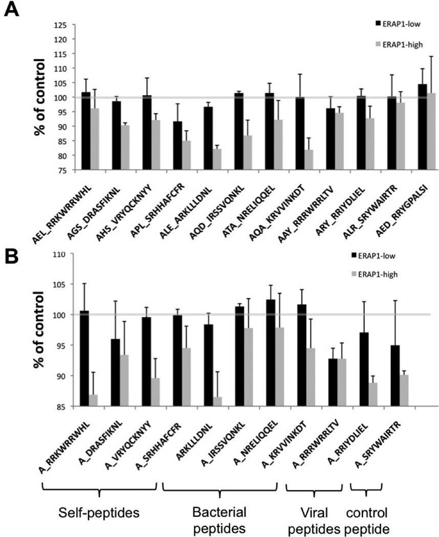
Figure 4. ERAP1_High reduces HLA-B27 antigen presentation in intact cells as compared to ERAP1_Low when peptide precursors or mature B27 peptides are delivered.
HeLa-Kb-B27/47 cells were transiently transfected with plasmids expressing HLA-B27-specific peptides (with ER-signal sequence) with GFP and either empty vector (control) or the indicated ERAP1 allele with RFP. At 48 hours post transfection surface HLA-B27 surface expression on double-transfected (GFP+RFP+) was analyzed by flow cytometry. Peptides were transfected as N3-extended precursors (A) or as “mature” B27 specific epitopes (B), extended with Alanine on N-terminus for all peptides except ARKLLLDNL, which by coincidence already had N-terminal alanine. Mean fluorescent intensity (MFI), normalized to control (empty RFP vector, grey line, representing the level of endogenous antigen presentation) is shown. Several (5–10) independent transfection experiments were performed for every peptide / ERAP1 combination. Combined graph is shown. Bars represent mean ± SEM.

