
Fig.1. Expression of Wee1B during the oocyte-to-embryo transition.
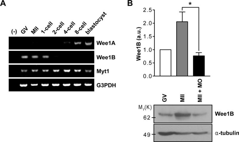
(A) RT-PCR of Wee1 kinases in oocyte and preimplantation embryos at different stages of development. G3PDH was used as an internal control. (B) GV and MII oocytes along with Wee1B MO-injected MII oocytes were immunoblotted for Wee1B and α-tubulin. Each lane contains 100 oocytes. Band intensities for Wee1B were quantified and normalized to α-tubulin level. a.u., arbitrary units. *p=0.0283.