Sir,
Crystal storing histiocytosis (CSH) is a rare manifestation of B-cell lymphoproliferative disorders characterized by intracytoplasmic accumulation of crystallized immunoglobulins within the macrophages.[1] CSH can be generalized involving multiple sites or can manifest as a localized mass lesion, most commonly in the head and neck region. It equally involves both males and females; with a wide age distribution (17-81 years).[2] We report an interesting case of localized CSH associated with marginal zone B-cell lymphoma involving a retroperitoneal lymph node at the left renal hilum.
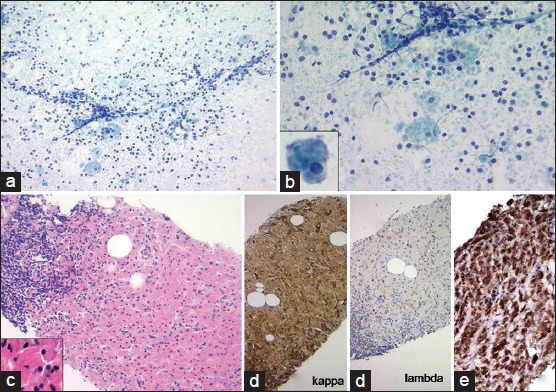
The patient was a 77-year-old male who presented with a slow growing mass at the renal hilum over a period of 3 years, with an increase in size from 4.1 cm to 7.0 cm. The complete blood counts, urine analysis, and comprehensive metabolic profile on a 6-monthly regular follow-up during the 3 year period had always been normal. The patient initially declined undergoing an invasive procedure. However, due to the progressive increase in mass size, he consented, and a fine-needle aspiration (FNA) and core biopsy of the mass was performed. The FNA smears demonstrated a monomorphic population of mononuclear cells, lymphoglandular bodies, and scattered histiocytes. The histiocytes demonstrated low nuclear to cytoplasmic ratio, small round regular eccentric nucleus, and abundant refractile, dense amorphous, needle to rhomboid shaped crystals within the cytoplasm. No mast cells or plasma cells were noted in the background [Figure 1a and b]. This cytologic appearance of the histiocytes was suggestive of CSH, which prompted further workup for a lymphoproliferative process. The histological sections of the core biopsy demonstrated monomorphic lymphoid aggregates admixed with sheets of oval to polygonal histiocytes filled with eosinophilic refractile crystals within the cytoplasm pushing the nucleus to the side [Figure 1c]. Immunohistochemical stain performed on the tissue core biopsy for kappa and lambda light chains demonstrated monoclonal staining pattern for kappa light chains in the lymphocytes as well as in the cytoplasmic crystals within the histiocytes [Figure 1d]. The histiocytes were highlighted with positive staining for CD68 [Figure 1e]. Cyclin D1 immunostain was negative. Flow cytometry performed on the aspirated material demonstrated a clonal B-cell lymphoid population with the following immunophenotype: CD45+, CD19+, CD20+, CD3−, CD5−, CD10−, and kappa light chain restricted. No paraprotein was detected in the serum or urine electrophoresis. No other lymphadenopathy or splenomegaly was observed on imaging. A diagnosis of localized CSH associated with a nodal marginal zone B-cell lymphoma was rendered. The patient declined further therapy and was lost to follow-up.
Figure 1.

(a and b) Fine-needle aspiration of the mass demonstrate monomorphic lymphocyte population admixed with scattered histiocytes in a clear background. Inset: Histiocyte with refractile cytoplasmic crystals (Diff Quik, ×100 and ×200). (c) Core biopsy showing monomorphic lymphoid aggregate admixed with sheets of histiocytes (H and E, ×100). (d) Immunohistochemistry stains on the core biopsy showing kappa light chain restriction in the lymphocytes as well as in cytoplasmic crystals within histiocytes (kappa and lambda light chain, ×100). (e) CD68 stain performed on the core biopsy highlights CD68+ histiocytes (CD68, ×100)
On review of the literature, approximately 80 cases have been reported to date with the majority of them (90%) associated with lymphoproliferative or plasma cell disorder including multiple myeloma (32%), lymphoplasmacytic lymphoma (24%), monoclonal gammopathy of undetermined significance (21%), B-cell lymphoma (15%) or plasma cell dyscrasia/neoplasm, not otherwise specified (6%).[2] CSH is usually associated with kappa light chain restriction as seen in the present case due to high intrinsic stability or a loss of proteolytic site on these light chains. The associated heavy chain is variable. Ultrastructurally, the crystals are dense, membrane bound and elongated, rectangular and/or rhomboid in configuration.[2] The exact mechanism for crystal formation is not well-known and may involve factors ranging from overproduction to impaired secretion or excretion of the immunoglobulin chains.
The common sites involved in generalized CSH include bone marrow (97%), liver (47%), lymph nodes (44%), spleen (44%), and kidney (38%), whereas, head and neck region (parotid and thyroid gland, cornea, or otolaryngeal mucosa) is the most commonly involved in localized CSH.[2,3,4,5] Generalized CSH is usually associated with poor prognosis, regardless of the type of underlying lymphoproliferative process, however, in localized CSH the prognosis is variable depending upon the extent of tissue involved and the underlying condition of the patient. In general, the prognosis and overall survival in patients with localized CSH associated with marginal zone lymphoma is comparable to age and sex matched normal patient control, as described in a recent review by Zhang and Myers.[4] We herewith, describe a rare presentation of a lymphoproliferative process due to the presence of background CSH initially diagnosed on a FNA. A similar report of mucosa-associated lymphoid tissue (MALT) lymphoma involving the parotid gland has been described in an 81-year-old female on FNA, demonstrating immunoglobulin crystals within histiocytes admixed with monocytoid B-cells and plasma cells.[3] MALT lymphoma in these mucosal locations is analogous to marginal zone B-cell lymphoma involving the lymph node described in the present case.
Crystals in CSH are usually described most often of immunoglobulin origin, which are associated with an underlying lymphoproliferative or plasma cell disorder. However, non-immunoglobulin associated CSH also exist, with the differential diagnosis include adult rhabdomyoma, granular cell tumor, Langerhans cell histiocytosis (LCH), Gaucher's disease, malakoplakia, drug-induced CSH (clofazimine), and hereditary cystinosis.[6,7,8,9,10] The crystals in adult rhabdomyoma represent tropomyosin and are thus positive for various muscle makers such as desmin, muscle specific actin, and/or myoglobin; granular cell tumor shows positive staining for S-100; nuclei of histiocytes in LCH are folded or have grooves resembling coffee bean, stain positive for CD1a and S-100, and are usually associated with eosinophils in the background. In Gaucher's disease, the cytoplasm of the histiocyte has striated appearance (crumpled tissue paper) due to the deposition of beta-glucocerebroside and stain positive for PAS. Malakoplakia is most often found in the urinary tract, less commonly in the head and neck region, composed of sheets of CD68-positive epithelioid histiocytes (von Hansemann histiocytes) and scattered pathognomonic Michaelis-Gutmann bodies which contain Gram-negative bacteria with laminated dystrophic calcium and iron deposits. Clofazimine is a drug used to treat leprosy and can present as crystals within the histiocytes, usually limited within the bowel wall and mesenteric lymph nodes, and show prominent red birefringence under polarized light on frozen sections.
Thus, in summary, CSH is a well-recognized entity with an accumulation of intra-cytoplasmic crystalline immunoglobulin light chain deposits within non-neoplastic histiocytes and its presence should alert for a workup of a lymphoproliferative process. In addition, an extensive search including imaging, bone marrow biopsy, and serum/urine electrophoresis should be followed to evaluate the extent of involvement of the lymphoproliferative process associated with CSH. The present case adds CSH associated with rare marginal zone B-cell lymphoma to that list.
COMPETING INTERESTS STATEMENT BY ALL AUTHORS
The authors declare that they have no competing interests.
AUTHORSHIP STATEMENT BY ALL AUTHORS
Each author acknowledges that this final version was read and approved. All authors qualify for authorship as defined by ICMJE http://www.icmje.org/#author. Each author participated sufficiently in the work and takes public responsibility for appropriate portions of the content of this article.
ETHICS STATEMENT BY ALL AUTHORS
As this letter describes a case report without identifiers, our institution does not require approval from Review Ethics Board (REB) or its counterpart.
EDITORIAL/PEER-REVIEW STATEMENT
To ensure the integrity and highest quality of CytoJournal publications, the review process of this manuscript was conducted under a double blind model (authors are blinded for reviewers and vice versa) through automatic online system.
Contributor Information
Karan Saluja, Email: karansaluja1975@gmail.com.
Beenu Thakral, Email: beenuthakral@gmail.com.
Mohamed Eldibany, Email: mmeldibany@yahoo.com.
Robert A. Goldschmidt, Email: rgoldschmidt@northshore.org.
REFERENCES
- 1.Lebeau A, Zeindl-Eberhart E, Müller EC, Müller-Höcker J, Jungblut PR, Emmerich B, et al. Generalized crystal-storing histiocytosis associated with monoclonal gammopathy: Molecular analysis of a disorder with rapid clinical course and review of the literature. Blood. 2002;100:1817–27. [PubMed] [Google Scholar]
- 2.Dogan S, Barnes L, Cruz-Vetrano WP. Crystal-storing histiocytosis: Report of a case, review of the literature (80 cases) and a proposed classification. Head Neck Pathol. 2012;6:111–20. doi: 10.1007/s12105-011-0326-3. [DOI] [PMC free article] [PubMed] [Google Scholar]
- 3.Llobet M, Castro P, Barceló C, Trull JM, Campo E, Bernadó L. Massive crystal-storing histiocytosis associated with low-grade malignant B-cell lymphoma of MALT-type of the parotid gland. Diagn Cytopathol. 1997;17:148–52. doi: 10.1002/(sici)1097-0339(199708)17:2<148::aid-dc12>3.0.co;2-g. [DOI] [PubMed] [Google Scholar]
- 4.Zhang C, Myers JL. Crystal-storing histiocytosis complicating primary pulmonary marginal zone lymphoma of mucosa-associated lymphoid tissue. Arch Pathol Lab Med. 2013;137:1199–204. doi: 10.5858/arpa.2013-0252-CR. [DOI] [PubMed] [Google Scholar]
- 5.Zioni F, Giovanardi P, Bozzoli M, Artusi T, Bonacorsi G, Sighinolfi P. Massive bone marrow crystal-storing histiocytosis in a patient with IgA-lambda multiple myeloma and extensive extramedullary disease. A case report. Tumori. 2004;90:348–51. doi: 10.1177/030089160409000318. [DOI] [PubMed] [Google Scholar]
- 6.Sukpanichnant S, Hargrove NS, Kachintorn U, Manatsathit S, Chanchairujira T, Siritanaratkul N, et al. Clofazimine-induced crystal-storing histiocytosis producing chronic abdominal pain in a leprosy patient. Am J Surg Pathol. 2000;24:129–35. doi: 10.1097/00000478-200001000-00016. [DOI] [PubMed] [Google Scholar]
- 7.Gebrail F, Knapp M, Perotta G, Cualing H. Crystalline histiocytosis in hereditary cysinosis. Arch Pathol Lab Med. 2002;126:1135. doi: 10.5858/2002-126-1135-CHIHC. [DOI] [PubMed] [Google Scholar]
- 8.Amir G, Ron N. Pulmonary pathology in Gaucher's disease. Hum Pathol. 1999;30:666–70. doi: 10.1016/s0046-8177(99)90092-8. [DOI] [PubMed] [Google Scholar]
- 9.Nicholson AG, Florio R, Hansell DM, Bois RM, Wells AU, Hughes P, et al. Pulmonary involvement by Niemann-Pick disease. A report of six cases. Histopathology. 2006;48:596–603. doi: 10.1111/j.1365-2559.2006.02355.x. [DOI] [PubMed] [Google Scholar]
- 10.Friedman MT, Molho L, Valderrama E, Kahn LB. Crystal-storing histiocytosis associated with a lymphoplasmacytic neoplasm mimicking adult rhabdomyoma: A case report and review of the literature. Arch Pathol Lab Med. 1996;120:1133–6. [PubMed] [Google Scholar]


