Abstract
Background
Human studies on exercise, cognition, and apolipoprotein E (APOE) genotype show that ε4 carriers may benefit from regular physical activity.
Methods
We examined voluntary wheel-running, memory, and hippocampal plasticity in APOE ε3 and APOE ε4 transgenic mice at 10–12 months of age.
Results
Sedentary ε4 mice exhibited deficits in cognition on the radial-arm water maze (RAWM), a task dependent on the hippocampus. Six weeks of wheel-running in ε4 mice resulted in improvements on the RAWM to the level of ε3 mice. Hippocampal brain-derived neurotrophic factor (BDNF) levels were similar in ε3 and ε4 mice, and after exercise BDNF was similarly increased in both ε3 and ε4 mice. In sedentary ε4 mice, tyrosine kinase B (Trk B) receptors were reduced by 50%. Exercise restored Trk B in ε4 mice to the level of ε3 mice, and in ε4 mice, exercise dramatically increased synaptophysin, a marker of synaptic function.
Conclusions
Our results support the hypothesis that exercise can improve cognitive function, particularly in ε4 carriers.
Keywords: Exercise, APOE, Behavior, BDNF, Plasticity
1. Introduction
The ε4 allele of the apolipoprotein E (APOE) gene is a known risk factor for Alzheimer’s disease (AD) that is present in 20% to 25% of the population, and in 40% to 50% of late-onset AD cases [1]. However, ε4 is neither necessary nor sufficient for the development of AD. Accumulating evidence in both human studies [2, 3] and animal models [4, 5] suggests that lifestyle factors such as regular exercise may protect against cognitive decline and dementia.
Although physical activity is generally associated with a reduced risk of dementia, several human studies showed the interactive effects of exercise and APOE genotype on cognitive decline. The majority reported that the protective effects of exercise are more robust in carriers of the ε4 allele [6–9], although at least one study reported a protective effect in noncarriers of the ε4 allele, but not in carriers [10]. The specific effects of exercise on brain function have not been examined in an animal model of APOE ε4, which allows greater control of confounding variables and the examination of underlying mechanisms associated with enhanced cognitive performance. Previously published research showed that nerve-growth factor and synaptophysin, indicators of synaptic plasticity, are stimulated by enhanced environments where an exercise wheel is present [11]. However, APOE ε4 transgenic mice in enriched environments did not exhibit the cognitive improvements exhibited by APOE ε3 mice, and were resistant to the neurotrophic induction of synaptogenesis that occurred in APOE ε3 mice. Although the enrichment paradigm used by Levi et al [11] included access to a running wheel, their study design did not specifically emphasize exercise. Further, the group housing of their mice precluded quantification of exercise for each mouse, and limited the individual amount of access to the running apparatus. Zhong et al [12] showed a similar finding, i.e., a gene-targeted APOE ε4 model mouse was resistant to the synaptic plasticity induced by a novel environment in wild-type mice. However, the novel environment had no exercise wheel, and the effects of physical exercise were not assessed [12]. Similarly, Jankowsky et al reported increased AD-like pathology in transgenic mouse models exposed to an enriched environment [13]. Clearly, the effects of exercise alone differ from those of an enriched environment, even if that environment includes access to a running wheel.
The present study emphasized voluntary wheel-running as the intervention strategy in singly housed APOE ε3 and APOE ε4 mice, allowing for greater quantification of running behavior and related improvements in fitness. Several mechanisms were proposed to explain the benefits of exercise in dementia [14, 15]. One main area of focus concerns the exercise-induced increase in brain-derived neurotrophic growth factor (BDNF), which may serve as a final common pathway for exercise-induced improvements in cognition. Brain-derived neurotrophic factor is consistently upregulated in the hippocampus and related cortical areas in rodents after voluntary wheel-running [15]. Increases in BDNF regulate several downstream signal transduction pathways that result in synaptogenesis, neurogenesis, and dendritic branching, hence facilitating learning and memory. The exposure of cerebellar cultures to tyrosine kinase B (TrkB), the high-affinity receptor for BDNF, induces the development of axosomatic synapses [16]. The TrkB fusion protein, TrkB-Fc, blocks long-term potentiation (LTP), the cellular model of memory [17]. Additional evidence exists for the role of TrkB signaling in synaptogenesis [18]. Impaired learning and memory in TrkB knockout mice illustrate the functional necessity of this receptor [19].
The downstream signal-transduction mechanisms involved in BDNF-induced changes in learning and memory have not been examined in relation to exercise. Increases in the levels of TrkB may be required to increase the activation of these downstream signal-transduction events. After being activated by TrkB receptor signaling, p21-activated kinase (PAK) undergoes a rapid, dose-dependent phosphorylation, correlated with increased synaptic size and density [20]. Exercise induction of BDNF and these downstream signal-transduction events may result in the synaptogenesis associated with improved cognition.
Recent studies showed that even at late stages of cognitive decline, exercise can improve function in transgenic AD mouse models [5]. The current study examines the impact of voluntary wheel-running on cognitive performance in APOE ε3 and APOE ε4 transgenic mice at ages where cognitive deficit is present in APOE ε4-targeted replacement mice [21]. We focused on the BDNF signal transduction pathway for synaptogenesis as a potential mechanism for exercise-induced cognitive improvement. Specifically, exercise-induced changes in BDNF, TrkB, phosphorylated PAK, and synaptophysin in APOE ε3 and APOE ε4 mice may provide a mechanism for the effects of exercise on cognitive function. Because several studies of humans reported that exercise is positively associated with cognitive performance in ε4 carriers [6–9], we hypothesized that APOE ε4 mice would show an induction of BDNF and its associated signaling pathway markers in response to exercise, providing a synaptogenic mechanism for improved cognition.
2. Methods
2.1. Mice
All animal protocols were approved by the Institutional Animal Care and Use Committee at the University of California at Irvine. We purchased 16 ε3 and 16 ε4 targeted replacement mice as retired breeders (catalogue numbers 001548 and 001549, Taconic Transgenics, Hudson, NY). One ε4 mouse died before testing, reducing the number to 15. All mice were between 10 and 12 months of age at testing, and groups were balanced for gender. All animals were singly housed.
Mice were generated by electrocorporation of cultured embryonic stem cells from the 129 mouse strain, with a targeting construct containing human coding sequences for APOE ε3 or APOE ε4 with mouse flanking sequences. Homologous recombination yields mouse regulatory sequences on human APOE exons. Embryonic stem cells containing the recombination were then introduced into C57Bl/6J embryos and chimeric offspring bred again to C57Bl/6J, and then interbred to generate homozygotes [22] (see also www.taconic.com).
2.2. Procedures
The experimenters handled the mice twice a week for 1 minute each during the 6 weeks before cognitive testing, to reduce anxiety during testing. The mice were assigned identification numbers to blind the experimenters to the genotype and exercise conditions during cognitive testing and scoring.
2.3. Exercise
Exercised (RUN) mice were provided in-cage running wheels for 6 weeks before cognitive testing, and wheel rotations were monitored by computer (Mini-Mitter, Bend, OR). Sedentary (SED) animals were housed in cages of similar proportions, with no running wheels. To confirm aerobic differences between groups because of exercise training, we conducted citrate synthase assays on the soleus muscle of SED and RUN animals, according to Srere and Brooks [23].
2.4. Cognitive testing
Animals were tested over the course of 6 days. Cognitive tests varied in two main aspects: (1) the area of the brain that they challenged, and (2) the duration of training and testing (single-day versus multiple-day). Object recognition (OR) testing occurred on the first day, followed by 4 consecutive days of radial-arm water-maze (RAWM) testing, and 1 day of place recognition (PR) testing. Testing protocols are described below.
2.4.1. OR task
Recognition tasks were adapted from previously published methods [24]. Lesion studies illustrated that object recognition is not dependent on hippocampal function [24]. The testing arena was a 5-gallon white cylinder measuring 35.5 cm in height and 28 cm in diameter, with 2.5 cm of clean bedding covering the bottom, as well as approximately a quarter cup of the animal’s home-cage bedding. Phases included a 15-minute habituation to empty arena, 5 minutes of training, 5 minutes of rest in the home cage, and 3 minutes of testing in the arena. For training, we exposed the animal to two identical objects on opposite ends of the arena. After the brief home-cage rest, we returned mice to the arena for testing. During testing, one of the previous objects was returned to its original position (old), and one of the previous objects was replaced with a new object (novel).
This task exploits an unimpaired animal’s tendency to explore the novel object more than the old object. We analyzed the ratio of time spent exploring the novel object (in seconds) to time spent exploring both objects combined (novel/old + novel) for each of the 3 minutes in the testing phase. Similarly, the total number of touches of the novel object relative to both objects was examined for each minute. The objects consisted of rubber pet toys, previously tested to exclude objects that the mice were unlikely to explore. The different objects were counterbalanced across genotype and exercise conditions, and were used equally as old and novel objects. Objects were cleaned with 70% ethanol between phases, to eliminate odor cues. Similarly, bedding was circulated between phases to reduce cues.
2.4.2. PR task
The same phases of habituation, training, and testing were used in place-recognition testing, which is dependent on hippocampal function. We exposed mice to two identical objects at specific positions in the arena during training. After 5 minutes in their home cages, we returned the mice to the arena with one of the objects in the same location as before, and the other object in a novel location. Ambient cues in the room served as place references. We again analyzed the ratio of time spent exploring the object in the new location relative to time spent exploring both locations (new/new + old) for each of the 3 minutes. The ratio for number of touches of objects was also examined. This task exploits an unimpaired animal’s tendency to explore an object in a new location more than an object in an old location.
2.4.3. RAWM
The RAWM protocol in this study was adapted from that of Dr. David Morgan (Health Science Center, University of South Florida, personal communication), and consisted of 3 days of testing, plus 1 reversal day. It was previously used in AD mouse models (Tg2576) with success, and is sensitive to the hippocampal dysfunction associated with AD mouse models [5, 25, 26]. Similar to Morris water-maze testing, the object of the task is to learn and remember the location of an escape platform that is submerged in a tank of opaque water. The RAWM has six arms radiating from an open center and visual cues placed on the walls of the testing tank. For each mouse, the platform was placed consistently in the same arm for the first 3 days (the platform arm was randomized across mice), whereas each mouse began every trial sequentially in a different arm to eliminate “chaining” [27]. The mice received 15 trials per day in blocks of five. They were tested in groups of 4–5, such that mouse 1 would receive trial 1, then mouse 2 would receive trial 1, etc., cycling through until all 4–5 mice in a group had received the five trials in a block. A rest period of 5 minutes was given for each mouse before its subsequent trial within a block, and 2 hours in between blocks. Because the task spans several days, there are overnight periods of rest in which consolidation was shown to occur in rats [28]. We measured both time to find platforms (latency), and entries of nontarget arms (errors), although errors were shown to be more sensitive to dysfunction [29, 30]. If the mice did not find the platform after 60 seconds, they were gently guided to the platform, where they were left for 20 seconds before being returned to their cage.
Animals were sacrificed 4–5 hours after PR testing by decapitation. Their brains were dissected and subsectioned into regions of interest. Soleus muscles were dissected from the hind leg.
2.5. Molecular markers
Hippocampi were quick-frozen after dissection and stored at −80°C. We prepared homogenates by pulverizing frozen samples on dry ice, preparing 500 µL of cell lysis buffer (number 28-30412; Bio-Rad, Hercules, CA) containing protease inhibitor cocktail (number 171-304012, Bio-Rad) and 1 µL of a stock solution containing 500 mM phenylmethylsulfonyl fluoride (number P-7626) in dimethyl sulfoxide (number D2650, both from Sigma, St. Louis, MO), and sonicating [31]. We centrifuged the homogenate for 10 minutes at 25,000g, and removed the supernatant for analysis. We used a standard bichondruric acid assay to determine protein content, and to aid in loading equal amounts of protein in all subsequent analyses. We measured BDNF levels in duplicate, using an enzyme-linked immunoassay (ELISA) kit according to the manufacturer’s instructions (G7610, BDNF Emax ImmunoAssay, Promega, Madison, WI), and analyzed plates on an automated multiwell plate reader.
Intracellular markers TrkB and phosphorylated PAK were probed via Western blot, using β-actin as a loading control. The synaptic marker synaptophysin was likewise measured by Western blot. Individual antibody concentrations and incubation protocols are listed in Table 1. We ran 15 µg of protein per sample per lane on Bio-Rad Criterion XT 8% to 12% Bis-Tris gels for cold electrophoresis, and transferred protein to polyvinylidene fluoride membrane overnight at 4°C. Three separate investigators independently analyzed each marker, using Image J NIH shareware (National Institutes of Health, Bethesda, MD). Levels of each target marker were normalized to β-actin, and results were analyzed by repeated-measures analysis of variance, with each investigator serving as a separate measure.
Table 1.
Antibodies used in Western blot analysis
| Protein | Primary type | Primary concentration | Secondary type | Secondary concentration | Vendor and catalogue numbers |
|---|---|---|---|---|---|
| TrkB | Rabbit anti-mouse | 1:500 | Donkey anti-rabbit | 1:5000 | BD Transduction, 611640 |
| PAK | Rabbit anti-mouse | 1:500 | Donkey anti-rabbit | 1:5000 | BioSource, 44-940G |
| Synaptophysin | Rabbit anti-mouse | 1:100 | Donkey anti-mouse | 1:5000 | Sigma, S5768 |
| Actin | Rabbit anti-mouse | 1:1000 | Donkey anti-mouse | 1:1000 | Sigma, A2066 |
2.6. Statistical analyses
All data were analyzed in SPSS statistical software (SPSS Inc., Chicago, IL), using analysis of variance as 2 (genotype: ε3 and ε4) × 2 (exercise: RUN and SED) analyses. The RAWM data for days 1–3 were subjected to a genotype × exercise × block [9] design. Trials were grouped into blocks of five, with three blocks per day (15 trials), such that blocks 1–3 reflected day 1 of testing, blocks 4–6 reflected day 2 of testing, and block 7–9 reflected day 3 of testing.
3. Results
3.1. Citrate synthase
Citrate synthase levels reflect changes in aerobic capacity because of training. A strong trend toward increase occurred in the running condition (F(1,23) = 3.97, P = 0.058). In the RUN group, the average amount of daily running (wheel revolutions/day) correlated positively with citrate synthase levels (R = 0.39, P < 0.05). Running amounts did not differ between genotypes (Table 2). Citrate synthase showed no interactions of genotype and condition, indicating that both genotypes responded equally in terms of aerobic change induced by exercise.
Table 2.
Citrate synthase levels respond similarly with exercise in both genotypes
| Citrate synthase (µmol/g/min) |
Average daily run (rotations) |
Total run amounts (rotations) |
|
|---|---|---|---|
| ε3 sedentary | 29.8 ± 9.4 | ||
| ε3 run | 45.0 ± 7.7 | 5104.8 ± 2865 | 169,081 ± 129,485 |
| ε4 sedentary | 34.7 ± 8.7 | ||
| ε4 run | 47.1 ± 7.7 | 5759.4 ± 2365 | 186,215 ± 121,135 |
3.2. Object recognition
Object recognition is a single-day task that is not dependent on hippocampal function. No genotype or exercise main effects or interactions emerged in the OR task.
3.3. Place recognition
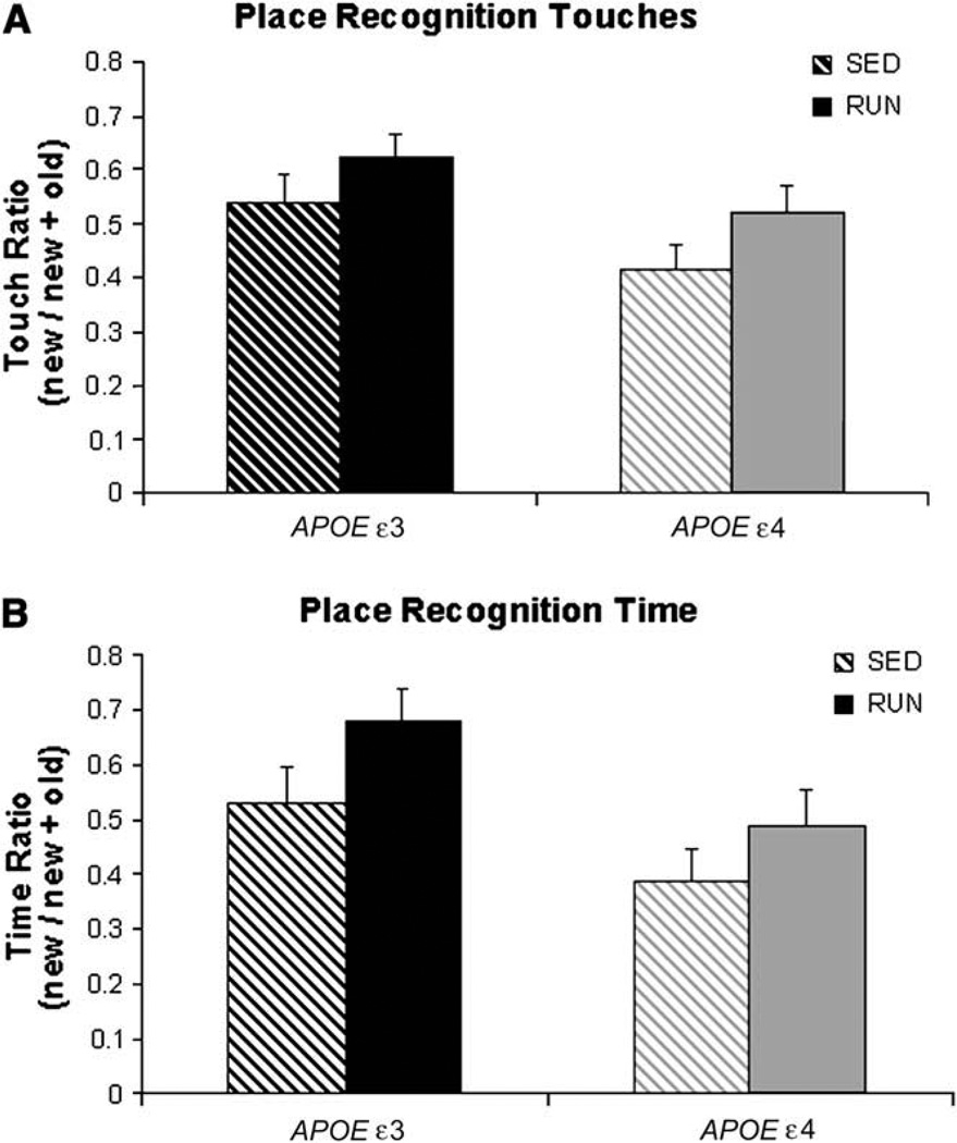
Place recognition is a single-day task, but is dependent on hippocampal function. The APOE ε4 mice were impaired in this task relative to APOE ε3 mice for both touches of the new object (F(1,26) = 6.0, P = 0.021) and time exploring the new object (F(1,26) = 7.2, P = 0.013). Exercise improved performance in both genotypes for touches (F(1,26) = 4.31, P = 0.048; Fig. 1A) and time (F(1,26) = 4.2, P = 0.051; Fig. 1B).
Fig. 1.
ε4 mice were impaired on place recognition relative to ε3 mice. Exercise improved performance for both genotypes in context of both touches (A) and time (B).
3.4. RAWM
Water-maze testing is dependent on hippocampal function, and spans several days. It is a greater challenge to hippocampal function than simple recognition tasks, and was shown to be an excellent discriminator of hippocampal dysfunction in transgenic mice [5, 25, 30]. Swim speed was not significantly affected by genotype or exercise condition, but females of both genotypes swam significantly faster than males (F(1,23) = 11.05, P = 0.003). No interactions were found between gender and exercise, or between gender and genotype. To correct for the influence of gender on error and latency outcomes, we included gender as a covariate in all analyses of RAWM. In measures of error, the effect of gender as a covariate neared significance (P = 0.06). In measures of latency, gender was a significant covariate (P = 0.04). This study was not powered to detect genotype and exercise effects within separate genders.
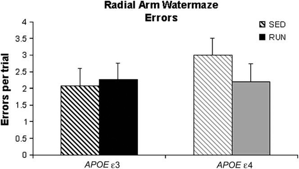
When we evaluated number of incorrect arm entries (errors), a significant interaction emerged for overall errors on the RAWM between genotype (ε3 versus ε4) and exercise condition (SED versus RUN) (F(1,26) = 4.60, P = 0.042). Although all mice eventually learned the escape platform location by day 3, ε4 SED mice made more overall errors than the ε3 SED mice during learning. The ε4 RUN mice improved such that they were indistinguishable from ε3 mice on error measures (Fig. 2). The ε3 mice showed modest improvement with exercise.
Fig. 2.
Sedentary ε4 mice made more errors than did ε3 mice on RAWM task, whereas exercised ε4 mice performed the same as ε3 mice.
Compared with APOE ε3 mice, APOE ε4 mice exhibited a delayed latency to locate the escape platform, regardless of exercise condition (F(1,26) = 8.34, P = 0.008). Exercise did not significantly affect latency measures in either genotype, nor was an interaction of genotype and exercise present.
3.5. Molecular markers
Average daily running in the RUN group correlated positively with BDNF levels (R = 0.49, P < 0.05) and with citrate synthase (R = 0.34, P < 0.05). The correlation between BDNF and citrate synthase levels approached significance (P = 0.059).
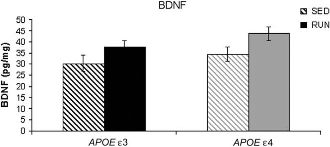
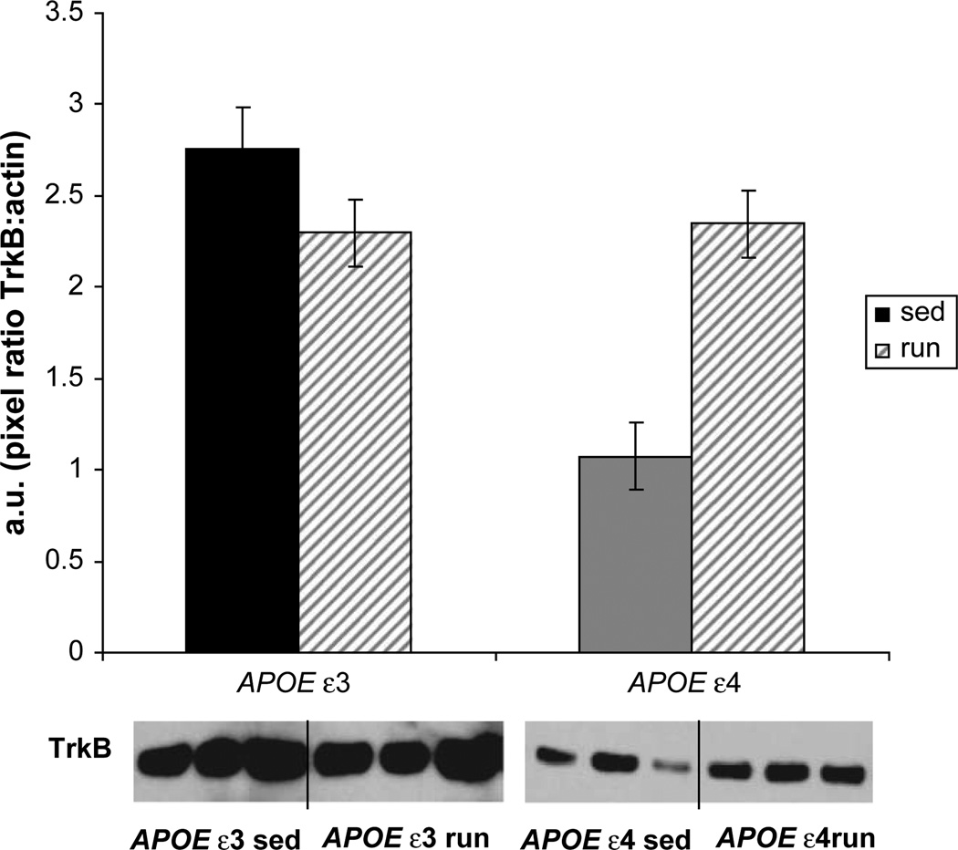
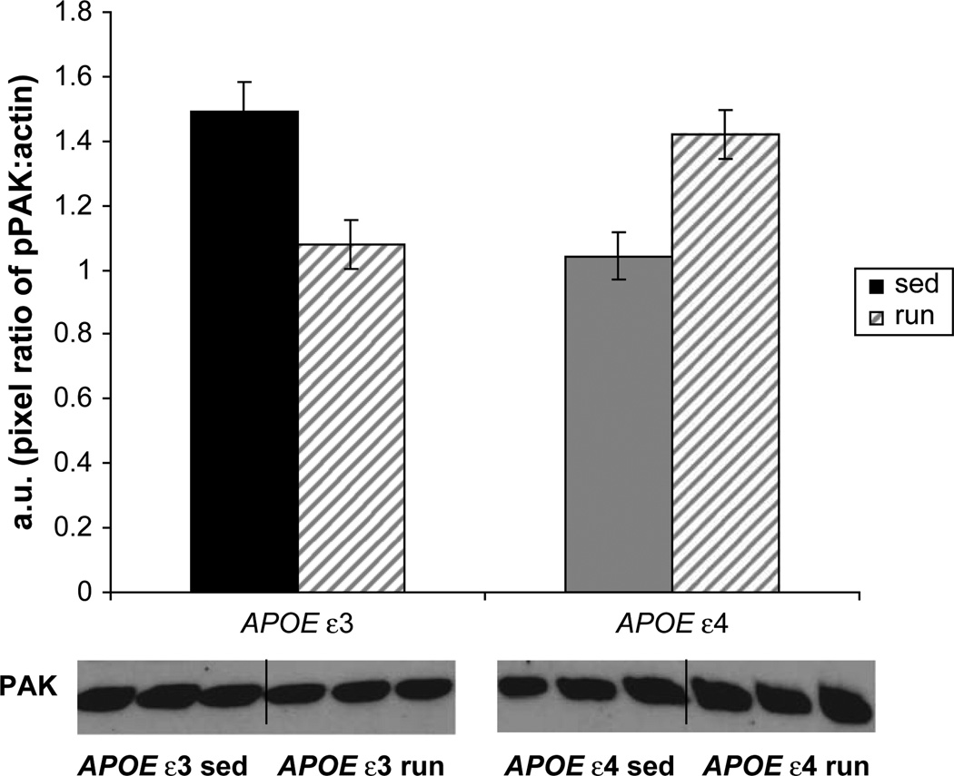
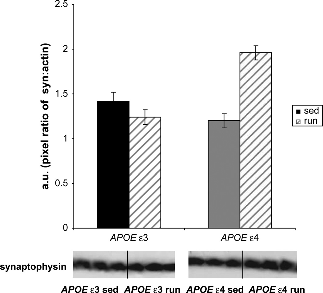
ELISA also revealed significantly greater BDNF in the RUN group relative to the SED group (Fig. 3), irrespective of genotype (F(1,25) = 6.47, P < 0.05). Although both genotypes equally increased BDNF in response to exercise, significant genotype × exercise interactions were observed for TrkB (Fig. 4), PAK (Fig. 5), and synaptophysin (Fig. 6) (TrkB, F(1,18) = 19.5, P < 0.01; PAK, F(1,18) = 24.7, P < 0.01; synaptophysin, F(1,18) = 29.6, P < 0.01). Post hoc tests revealed that for TrkB, PAK, and synaptophysin, running-induced increases in protein levels were largely restricted to the APOE ε4 genotype (TrkB, F(1,10) = 16.0, P < 0.01; PAK, F(1,10) = 11.6, P < 0.01; synaptophysin, F(1,10) = 31.5, P < 0.01). A significant decrease in PAK was evident in the APOE ε3 genotype with exercise (P = 0.006).
Fig. 3.
BDNF levels significantly increased in both APOE ε3 and APOE ε4 animals with 6 weeks of running-wheel access. There were no significant differences between the two genotypes.
Fig. 4.
The BDNF receptor, TrkB, demonstrated a significant interaction of genotype and condition (P < 0.01). Post hoc analysis revealed that APOE ε4 sedentary animals had significantly less TrkB than did sedentary APOE ε3 animals, but experienced a significant increase after 6 weeks of running-wheel exposure. No such increase was evident in APOE ε3 animals. The TrkB blots were normalized to β-actin.
Fig. 5.
A significant interaction of genotype and condition existed for phosphorylated p21 activated kinase (P < 0.01). The ε4 mice increased their levels of PAK with 6 weeks of wheel access.
Fig. 6.
Synaptophysin levels demonstrated an interaction between genotype × condition (P < 0.01), such that exercised ε4 mice exhibited increased synaptophysin, but ε3 mice did not.
4. Discussion
Exercise improved accuracy in ε4 mice on the radial-arm water maze, a task dependent on the hippocampus, and resulted in dramatic changes in plasticity in the hippocampus. Improvements with exercise on the RAWM task were specific to ε4 mice. The ε4 SED mice exhibited impairment on the RAWM task relative to ε3 mice, whereas ε4 RUN mice performed the same as ε3 mice. Importantly, differences in RAWM performance between ε3 and ε4 mice were not attributable to the extent of exercise performed, because ε3 and ε4 mice showed similar amounts of wheel-running and similar aerobic changes. Only scores of accuracy (errors) improved with exercise. Interestingly, the speed ofRAWM-solving (latency) did not change with exercise in either genotype. Place recognition, another hippocampal-dependent task, showed modest improvements in both ε3 and ε4 mice after exercise. Interestingly, no impairment of ε4 mice was evident in the hippocampus-independent task of object recognition, nor did exercise improve this task. These data show that exercise improves hippocampal function, and in the context of RAWM, restores the capacity of ε4 animals to that of ε3 animals.
The present study was not powered to analyze genders separately. However, we observed significant differences in swim speed and latency between genders, as well as trends toward a gender effect on error scores. Future investigation should analyze genders independently, to determine if exercise-induced improvements in cognition are more predominant in females or in males.
Hippocampal learning depends on BDNF signaling as well as the engagement of other downstream pathways [19]. The BDNF system differed between ε3 mice and ε4 mice in several ways. Although BDNF increased to similar levels with running in both ε3 and ε4 animals, the levels of TrkB in SED ε4 were approximately 50% lower than in ε3. Reduced TrkB, caused by a partial knockout of the receptor or administration of antibodies against TrkB, will compromise synaptic function and learning [19, 32–34]. Running increased TrkB in ε4 animals to levels indistinguishable from those in ε3 animals. Moreover, in response to exercise, phosphorylated PAK and synaptophysin, i.e., markers indicative of synaptogenesis [19, 35, 36], increased in ε4 mice. Levels of PAK actually decreased in APOE ε3 animals, despite increases in BDNF. Although these results are difficult to interpret, they may explain our failure to observe improvements in exercised ε3 animals in RAWM. We suggest that the differences evident in APOE ε4 animals with running may be essential in enhancing synaptic plasticity and the improved performance on the RAWM task. Our data are supported by previous findings where TrkB knockout mice performed well on simple avoidance learning tasks, but were impaired on radial-arm maze-learning [19].
We observed a similar induction of BDNF in both genotypes. Brain-derived neurotrophic growth factor was well-demonstrated to increase with exercise, and to improve cognitive function. It can act acutely to increase LTP and synaptic vesicle-docking [37]. Such acute effects of exercise-induced increases in hippocampal BDNF may have contributed to the improvements in place recognition observed in both genotypes. Brain-derived neurotrophic growth factor was also shown to increase in the hippocampus as a result of cognitive stimulation from RAWM itself [35, 36]. Thus it is also possible that increases in BDNF/TrkB because of exercise may have been supplemented by the activation of neurons involved in learning the platform location. This is reflected by the robust improvement observed in RAWM in exercised ε4 mice.
This study supports the human-related literature indicating that sedentary APOE ε4 carriers are particularly at risk for cognitive decline, and as such, may exhibit a more robust benefit from physical activity than noncarriers [6–9]. In the human-related literature, ε4 carriers were shown to exhibit a slower speed of processing during executive function tasks [6, 38], a longer latency of event-related potentials [6, 39, 40], and altered cortical activation during cognitive challenges [6, 41–44]. The exercise-related benefits on cognitive function and speed of processing reported to date in the human-related literature have been cross-sectional or epidemiological in nature [6–9]. In these studies, physically active participants had a history of a prolonged lifestyle of activity. However, the mice in the present study were predominantly sedentary until they were introduced to the running wheel at around age 9 months, an age at which ε4 mice already exhibit cognitive deficits. Hippocampal TrkB was significantly lower in age-matched sedentary APOE ε4 mice, as were cognitive deficits. An exercise-induced increase in BDNF and perhaps other pathways only resulted in increases in markers of synaptogenesis (PAK and synaptophysin) in APOE ε4 mice. Perhaps by virtue of having less synaptic markers in the sedentary condition, these animals had more to gain from exercise, or perhaps the APOE ε4 mice recruited greater neuronal resources to learn the RAWM than did the ε3 mice, thereby boosting their overall plastic response. It is also possible (although not explored in the present study) that exercise decreases the synaptotoxicity associated with AD. Sedentary APOE ε4 animals did not show lower levels of synaptophysin that APOE ε3 animals, a potential indication for such toxicity observed in other models [45]. However, because no previous work established the presence or absence of synaptotoxicity in this model, we cannot rule out a contribution. Overall, our studies support the hypothesis that ε4 carriers show improved cognition and perhaps less hippocampal dysfunction when they engage in an exercise program.
Acknowledgments
Funding was provided by National Institutes of Health grants AR47752-06 and AG000096-26. We would also like to thank Mr. Rick Muth for his generous support.
References
- 1.Saunders AM, Schmader K, Breitner JC, Benson MD, Brown WT, Goldfarb L, et al. Apolipoprotein E epsilon 4 allele distributions in late-onset Alzheimer’s disease and in other amyloid-forming diseases. Lancet. 1993;342:710–711. doi: 10.1016/0140-6736(93)91709-u. [DOI] [PubMed] [Google Scholar]
- 2.Larson EB, Wang L, Bowen JD, McCormick WC, Teri L, Crane P, et al. Exercise is associated with reduced risk for incident dementia among persons 65 years of age and older. Ann Intern Med. 2006;144:73–81. doi: 10.7326/0003-4819-144-2-200601170-00004. [DOI] [PubMed] [Google Scholar]
- 3.Laurin D, Verreault R, Lindsay J, MacPherson K, Rockwood K. Physical activity and risk of cognitive impairment and dementia in elderly persons. Arch Neurol. 2001;58:498–504. doi: 10.1001/archneur.58.3.498. [DOI] [PubMed] [Google Scholar]
- 4.Adlard PA, Perreau VM, Pop V, Cotman CW. Voluntary exercise decreases amyloid load in a transgenic model of Alzheimer’s disease. J Neurosci. 2005;25:4217–4221. doi: 10.1523/JNEUROSCI.0496-05.2005. [DOI] [PMC free article] [PubMed] [Google Scholar]
- 5.Nichol KE, Parachikova AI, Cotman CW. Three weeks of running wheel exposure improves cognitive performance in the aged Tg2576 mouse. Behav Brain Res. 2007;184:124–132. doi: 10.1016/j.bbr.2007.06.027. [DOI] [PMC free article] [PubMed] [Google Scholar]
- 6.Deeny SP, Poeppel D, Zimmerman JB, Roth SM, Brandauer J, Witkowski S, et al. Exercise, APOE, and working memory: MEG and behavioral evidence for benefit of exercise in epsilon4 carriers. Biol Psychol. 2008;78:179–187. doi: 10.1016/j.biopsycho.2008.02.007. [DOI] [PMC free article] [PubMed] [Google Scholar]
- 7.Etnier JL, Caselli RJ, Reiman EM, Alexander GE, Sibley BA, Tessier D, et al. Cognitive performance in older women relative to ApoE-epsilon4 genotype and aerobic fitness. Med Sci Sports Exerc. 2007;39:199–207. doi: 10.1249/01.mss.0000239399.85955.5e. [DOI] [PubMed] [Google Scholar]
- 8.Rovio S, Kareholt I, Helkala EL, Viitanen M, Winblad B, Tuomilehto J, et al. Leisure-time physical activity at midlife and the risk of dementia and Alzheimer’s disease. Lancet Neurol. 2005;4:705–711. doi: 10.1016/S1474-4422(05)70198-8. [DOI] [PubMed] [Google Scholar]
- 9.Schuit AJ, Feskens EJ, Launer LJ, Kromhout D. Physical activity and cognitive decline, the role of the apolipoprotein e4 allele. Med Sci Sports Exerc. 2001;33:772–777. doi: 10.1097/00005768-200105000-00015. [DOI] [PubMed] [Google Scholar]
- 10.Podewils LJ, Guallar E, Kuller LH, Fried LP, Lopez OL, Carlson M, et al. Physical activity, APOE genotype, and dementia risk: findings from the Cardiovascular Health Cognition Study. Am J Epidemiol. 2005;161:639–651. doi: 10.1093/aje/kwi092. [DOI] [PubMed] [Google Scholar]
- 11.Levi O, Michaelson DM. Environmental enrichment stimulates neurogenesis in apolipoprotein E3 and neuronal apoptosis in apolipoprotein E4 transgenic mice. J Neurochem. 2007;100:202–210. doi: 10.1111/j.1471-4159.2006.04189.x. [DOI] [PubMed] [Google Scholar]
- 12.Zhong N, Scearce-Levie K, Ramaswamy G, Weisgraber K. Apolipoprotein E4 domain interaction: synaptic and cognitive deficits in mice. Alzheimers Dement. 2008;4:179–192. doi: 10.1016/j.jalz.2008.01.006. [DOI] [PMC free article] [PubMed] [Google Scholar]
- 13.Jankowsky JL, Xu G, Fromholt D, Gonzales V, Borchelt DR. Environmental enrichment exacerbates amyloid plaque formation in a transgenic mouse model of Alzheimer disease. J Neuropathol Exp Neurol. 2003;62:1220–1227. doi: 10.1093/jnen/62.12.1220. [DOI] [PubMed] [Google Scholar]
- 14.Cotman CW, Berchtold NC, Christie LA. Exercise builds brain health: key roles of growth factor cascades and inflammation. Trends Neurosci. 2007;30:464–472. doi: 10.1016/j.tins.2007.06.011. [DOI] [PubMed] [Google Scholar]
- 15.Cotman CW, Berchtold NC. Exercise: a behavioral intervention to enhance brain health and plasticity. Trends Neurosci. 2002;25:295–301. doi: 10.1016/s0166-2236(02)02143-4. [DOI] [PubMed] [Google Scholar]
- 16.Seil FJ, Drake-Baumann R. TrkB receptor ligands promote activitydependent inhibitory synaptogenesis. J Neurosci. 2000;20:5367–5373. doi: 10.1523/JNEUROSCI.20-14-05367.2000. [DOI] [PMC free article] [PubMed] [Google Scholar]
- 17.Rex CS, Lin CY, Kramar EA, Chen LY, Gall CM, Lynch G. Brainderived neurotrophic factor promotes long-term potentiation-related cytoskeletal changes in adult hippocampus. J Neurosci. 2007;27:3017–3029. doi: 10.1523/JNEUROSCI.4037-06.2007. [DOI] [PMC free article] [PubMed] [Google Scholar]
- 18.Luikart BW, Parada LF. Receptor tyrosine kinase B-mediated excitatory synaptogenesis. Prog Brain Res. 2006;157:15–24. doi: 10.1016/s0079-6123(06)57002-5. [DOI] [PubMed] [Google Scholar]
- 19.Minichiello L, Korte M, Wolfer D, Kuhn R, Unsicker K, Cestari V, et al. Essential role for TrkB receptors in hippocampus-mediated learning. Neuron. 1999;24:401–414. doi: 10.1016/s0896-6273(00)80853-3. [DOI] [PubMed] [Google Scholar]
- 20.Chen LY, Rex C, Casale MS, Gall CM, Lynch G. Changes in synaptic morphology accompany actin signaling during LTP. J Neurosci. 2007;27:5363–5372. doi: 10.1523/JNEUROSCI.0164-07.2007. [DOI] [PMC free article] [PubMed] [Google Scholar]
- 21.Wang C, Wilson WA, Moore SD, Mace BE, Maeda N, Schmechel DE, et al. Human ApoE4-targeted replacement mice display synaptic deficits in the absence of neuropathology. Neurobiol Dis. 2005;18:390–398. doi: 10.1016/j.nbd.2004.10.013. [DOI] [PubMed] [Google Scholar]
- 22.Xu PT, Schmechel D, Rothrock-Christian T, Burkhart DS, Qiu HL, Popko B, et al. Human apolipoprotein E2, E3, and E4 isoform-specific transgenic mice: human-like pattern of glial and neuronal immunoreactivity in central nervous system not observed in wild-type mice. Neurobiol Dis. 1996;3:229–245. doi: 10.1006/nbdi.1996.0023. [DOI] [PubMed] [Google Scholar]
- 23.Srere PA, Brooks GC. The circular dichroism of glucagon solutions. Arch Biochem Biophys. 1969;129:708–710. doi: 10.1016/0003-9861(69)90231-8. [DOI] [PubMed] [Google Scholar]
- 24.Mumby DG, Gaskin S, Glenn MJ, Schramek TE, Lehmann H. Hippocampal damage and exploratory preferences in rats: memory for objects, places, and contexts. Learn Mem. 2002;9:49–57. doi: 10.1101/lm.41302. [DOI] [PMC free article] [PubMed] [Google Scholar]
- 25.Alamed J, Wilcock DM, Diamond DM, Gordon MN, Morgan D. Twoday radial-arm water maze learning and memory task: robust resolution of amyloid-related memory deficits in transgenic mice. Nat Protoc. 2006;1:1671–1679. doi: 10.1038/nprot.2006.275. [DOI] [PubMed] [Google Scholar]
- 26.Arendash GW, Gordon MN, Diamond DM, Austin LA, Hatcher JM, Jantzen P, et al. Behavioral assessment of Alzheimer’s transgenic mice following long-term Abeta vaccination: task specificity and correlations between Abeta deposition and spatial memory. DNA Cell Biol. 2001;20:737–744. doi: 10.1089/10445490152717604. [DOI] [PubMed] [Google Scholar]
- 27.Hodges H. Maze procedures: the radial-arm and water maze compared. Brain Res Cogn Brain Res. 1996;3:167–181. doi: 10.1016/0926-6410(96)00004-3. [DOI] [PubMed] [Google Scholar]
- 28.Park CR, Zoladz PR, Conrad CD, Fleshner M, Diamond DM. Acute predator stress impairs the consolidation and retrieval of hippocampus-dependent memory in male and female rats. Learn Mem. 2008;15:271–280. doi: 10.1101/lm.721108. [DOI] [PMC free article] [PubMed] [Google Scholar]
- 29.Gong B, Vitolo OV, Trinchese F, Liu S, Shelanski M, Arancio O. Persistent improvement in synaptic and cognitive functions in an Alzheimer mouse model after rolipram treatment. J Clin Invest. 2004;114:1624–1634. doi: 10.1172/JCI22831. [DOI] [PMC free article] [PubMed] [Google Scholar]
- 30.Wilcock DM, Alamed J, Gottschall PE, Grimm J, Rosenthal A, Pons J, et al. Deglycosylated anti-amyloid-beta antibodies eliminate cognitive deficits and reduce parenchymal amyloid with minimal vascular consequences in aged amyloid precursor protein transgenic mice. J Neurosci. 2006;26:5340–5346. doi: 10.1523/JNEUROSCI.0695-06.2006. [DOI] [PMC free article] [PubMed] [Google Scholar]
- 31.Hulse RE, Kunkler PE, Fedynyshyn JP, Kraig RP. Optimization of multiplexed bead-based cytokine immunoassays for rat serum and brain tissue. J Neurosci Methods. 2004;136:87–98. doi: 10.1016/j.jneumeth.2003.12.023. [DOI] [PMC free article] [PubMed] [Google Scholar]
- 32.von Bohlen und Halbach O, Krause S, Medina D, Sciarretta C, Minichiello L, Unsicker K. Regional- and age-dependent reduction in TrkB receptor expression in the hippocampus is associated with altered spine morphologies. Biol Psychiatry. 2006;59:793–800. doi: 10.1016/j.biopsych.2005.08.025. [DOI] [PubMed] [Google Scholar]
- 33.Danzer SC, Kotloski RJ, Walter C, Hughes M, McNamara JO. Altered morphology of hippocampal dentate granule cell presynaptic and postsynaptic terminals following conditional deletion of TrkB. Hippocampus. 2008;18:668–678. doi: 10.1002/hipo.20426. [DOI] [PMC free article] [PubMed] [Google Scholar]
- 34.Vaynman S, Ying Z, Gomez-Pinilla F. Interplay between brain-derived neurotrophic factor and signal transduction modulators in the regulation of the effects of exercise on synaptic-plasticity. Neuroscience. 2003;122:647–657. doi: 10.1016/j.neuroscience.2003.08.001. [DOI] [PubMed] [Google Scholar]
- 35.Mizuno M, Yamada K, Olariu A, Nawa H, Nabeshima T. Involvement of brain-derived neurotrophic factor in spatial memory formation and maintenance in a radial arm maze test in rats. J Neurosci. 2000;20:7116–7121. doi: 10.1523/JNEUROSCI.20-18-07116.2000. [DOI] [PMC free article] [PubMed] [Google Scholar]
- 36.Mizuno M, Yamada K, He J, Nakajima A, Nabeshima T. Involvement of BDNF receptor TrkB in spatial memory formation. Learn Mem. 2003;10:108–115. doi: 10.1101/lm.56003. [DOI] [PMC free article] [PubMed] [Google Scholar]
- 37.Jovanovic JN, Czernik AJ, Fienberg AA, Greengard P, Sihra TS. Synapsins as mediators of BDNF-enhanced neurotransmitter release. Nat Neurosci. 2000;3:323–329. doi: 10.1038/73888. [DOI] [PubMed] [Google Scholar]
- 38.O’Hara R, Sommer B, Way N, Kraemer HC, Taylor J, Murphy G. Slower speed-of-processing of cognitive tasks is associated with presence of the apolipoprotein epsilon4 allele. J Psychiatr Res. 2008;42:199–204. doi: 10.1016/j.jpsychires.2006.12.001. [DOI] [PubMed] [Google Scholar]
- 39.Green J, Levey AI. Event-related potential changes in groups at increased risk for Alzheimer disease. Arch Neurol. 1999;56:1398–1403. doi: 10.1001/archneur.56.11.1398. [DOI] [PubMed] [Google Scholar]
- 40.Wetter S, Murphy C. Apolipoprotein E epsilon4 positive individuals demonstrate delayed olfactory event-related potentials. Neurobiol Aging. 2001;22:439–447. doi: 10.1016/s0197-4580(01)00215-9. [DOI] [PubMed] [Google Scholar]
- 41.Bookheimer SY, Strojwas MH, Cohen MS, Saunders AM, Pericak-Vance MA, Mazziotta JC, et al. Patterns of brain activation in people at risk for Alzheimer’s disease. N Engl J Med. 2000;343:450–456. doi: 10.1056/NEJM200008173430701. [DOI] [PMC free article] [PubMed] [Google Scholar]
- 42.Bondi MW, Houston WS, Eyler LT, Brown GG. fMRI evidence of compensatory mechanisms in older adults at genetic risk for Alzheimer disease. Neurology. 2005;64:501–508. doi: 10.1212/01.WNL.0000150885.00929.7E. [DOI] [PMC free article] [PubMed] [Google Scholar]
- 43.Smith JD, Sikes J, Levin JA. Human apolipoprotein E allele-specific brain expressing transgenic mice. Neurobiol Aging. 1998;19:407–413. doi: 10.1016/s0197-4580(98)00076-1. [DOI] [PubMed] [Google Scholar]
- 44.Trivedi MA, Schmitz TW, Ries ML, Torgerson BM, Sager MA, Hermann BP, et al. Reduced hippocampal activation during episodic encoding in middle-aged individuals at genetic risk of Alzheimer’s disease: a cross-sectional study. BMC Med. 2006;4:1. doi: 10.1186/1741-7015-4-1. [DOI] [PMC free article] [PubMed] [Google Scholar]
- 45.Mucke L, Masliah E, Yu GQ, Mallory M, Rockenstein EM, Tatsuno G, et al. High-level neuronal expression of Abeta 1-42 in wild-type human amyloid protein precursor transgenic mice: synaptotoxicity without plaque formation. J Neurosci. 2000;20:4050–4058. doi: 10.1523/JNEUROSCI.20-11-04050.2000. [DOI] [PMC free article] [PubMed] [Google Scholar]


