Abstract
Background
Cleft lip and palate (CL/P) is one of the most common malformations in humans. Transforming growth factor alpha (TGFA) is a well characterized mammalian growth factor which might contribute to the development of CL/P. This meta-analysis aimed to summarize the association between the TGFA Taq I polymorphisms and CL/P.
Methods
We retrieved the relevant articles from PubMed, EMBASE, ISI Web of Science and SCOPUS databases. Studies were selected using specific inclusion and exclusion criteria. The odds ratios (ORs) and their 95% confidence intervals (95% CIs) were calculated to assess the association between TGFA Taq I polymorphism and CL/P risk. Meta-analyses were performed on the total data set and separately for the major ethnic groups, disease type and source of control. All analyses were performed using the Stata software.
Results
Twenty articles were included in the present analysis. There is a significant association between the TGFA Taq I polymorphism and CL/P (C1C2 vs C1C1: OR = 1.67, 95% CI = 1.23-2.25, C2C2 + C1C2 vs C1C1C1: OR = 1.52, 95% CI = 1.15-2.01; C2 vs C1:OR = 1.41, 95% CI = 1.12-1.78). Stratified analyses suggested that the TGFA Taq I polymorphism was significantly associated with CL/P in Caucasians (C1C2 vs C1C1: OR = 1.95, 95% CI = 1.34-2.86; C2C2 + C1C2 vs C1C1: OR = 1.68, 95% CI = 1.18-2.38; C2 vs V1: OR = 1.52, 95% CI = 1.14 -2.02).
Conclusion
TGFA Taq I polymorphism may be associated with the risk of CL/P.
Keywords: Cleft Lip and Palate, Clip lip, Clip palate, Transforming growth factor alpha, Single nucleotide polymorphism, Meta-analysis
Background
Facial clefting is one of the most common malformations in humans. Significant differences between populations in the prevalence of cleft lip or palate (CL/P) have been reported, with higher rates found in Asians and American Indians than those observed in Caucasians and Africans. Palate formation is complex, and there are numerous potential untoward possibilities, the most common being delayed shelf horizontalization and inadequate shelf growth [1].
Epidemiologic studies suggest that a number of environmental factors have been examined as risk factors for CL/P, including maternal smoking, exposure to antiepileptic drugs, antiemetic agents and vitamin use during the periconceptual period, maternal metabolic factors, alcohol consumption and exposure to agricultural chemicals [2]. Several studies have suggested that maternal cigarette smoking increased the risk of delivering infants with orofacial clefts [3-6]. It has previously been shown that maternal periconceptional intake of multivitamins containing folic acid decreased the occurrence of CL/P [6-8]. However, there is a study showing the different results [9]. A case–control study showed that CL/P was associated with maternal alcohol consumption [10]. However, Christensen and colleagues found that before the pregnancy there were fewer case mothers drinking alcohol than control mothers [11].
The epidemiologic characteristics and risk factors of CL/P are not clear. There is also a strong genetic component to oral clefts. The host susceptibility factors may play an important role in the development of CL/P. Ardinger and colleagues performed the first study to use a case–control design to test candidate genes [12]. They found a significantly statistical association between CL/P and two of 12 markers in five genes, with an intronic Taq1 marker in the transforming growth factor alpha (TGFA) gene showing the strongest association. TGFA encoded by a gene mapped at 2p13, is a secretion protein that binds to the epidermal growth factor receptor (EGFR) and is situated at the palate epithelium during palate closing [13]. TGFA may function as a normal embryonic version of EGF-related growth factor [14]. EGF/TGFA and glucocorticoids are believed to regulate the proliferation and differentiation of palatal epithelial cells both in vitro and in vivo. Moreover, the continued presence of EGF inhibits the fusion process; TGFA is likely to have similar effects. These biological studies suggest that mutations in the TGFA gene might contribute to the development of CL/P, especially for those mutations that affect the timing of the tissue-specific expression of this gene.
The TGFA gene shows a restriction fragment length polymorphism when treated with Taq I restriction enzyme. The mutant allele shows a four-base (TAAT) deletion. In this case, it shows a 178-base pair (bp) C1 allele and a 174-bp C2 allele [15]. TGFA Taq I polymorphism is located at intron 5 and has 602 bp in the 59 direction of the acceptor site of exon 6 [16]. For this polymorphism, C1C1 is wild genotype, C1C2 is heterozygote genotype, and C2C2 is homozygote mutation genotype. In most studies, there are different forms of comparisons such as heterozygote comparison (C1C2 vs. C1C1), homozygote comparison (C2C2 vs C1C1), dominant model (C1C2 + C2C2 vs C1C1), recessive model (C2C2 vs C1C2 + C1C1) and allelic model (C2 vs C1). Ardinger and colleagues first reported association between the Taq I polymorphisms at the TGFA locus and CL/P susceptibility in a case–control study [12]. This finding has since been replicated in some studies [6,15,17-23]. However, there are still controversies of the effect of TGFA polymorphism on the predisposition of this malformation [24-35].
The above inconsistent conclusions in the findings of the studies may be attributed to the size of the samples, the ethnic of the sample population and other reasons. In order to contribute to a better understanding of the role of this gene in the occurrence of cleft lip, cleft lip, or cleft lip and palate, we perform an updated meta-analysis on all available case–control studies, combining data together to reach a larger sample size, to get more statistical power to evaluate the association between CL/P susceptibility and TGFA Taq I polymorphism. Understanding the genetic background and etiology of CL/P is essential for both the risk assessment and findings of effective methods for prevention and treatment.
Methods
Data sources
We retrieved the articles using the following terms “Transforming growth factor alpha or TGFA” and “cleft lip or cleft palate or cleft lip and palate” from PubMed, Embase, ISI Web of Science and SCOPUS (Last search was updated on October 2013). There was no any language restriction and the age of participants was not considered as selection criteria. We evaluated potentially relevant publications by examining their titles and abstracts and all studies matching the eligible criteria were retrieved.
Study selection and data extraction
Eligible studies were selected according to the following explicit inclusion criteria: (a) evaluation of the TGFA Taq I polymorphism and CL/P risks, (b) using the methodology of a case–control study to keep the homogeneity between included studies in the meta-analysis.
Duplicate and obviously unrelated articles were eliminated by a single author (C.F.). Abstracts of the remaining articles were examined independently by two authors (C.F. and E.Z.) to determine whether the full-text article should be sought. When there were disagreements between CF and EZ in selecting papers, the third author (L.L.) would assess the articles and make the final decision with CF and EZ. A four-phase flow diagram according to Systematic Reviews (http://www.prisma-statement.org/) was shown in Figure 1. We have used the Newcastle-Ottawa Scale (NOS), suggested by Cochrane Collaboration, for assessing the quality of each included study in the present meta-analysis. The following information was obtained from each publication: first author’s name, publication year, country origin, ethnicity, case characteristics, total number of cases and controls, numbers of each group with TGFA Taq I genotypes, respectively.
Figure 1.
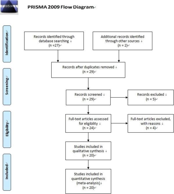
Flow chart of the study selection process.
Statistical methods
The odds ratios (ORs) and their 95% confidence intervals (95% CIs) were calculated to assess the association between TGFA Taq I polymorphism and CL/P. Pooled ORs were obtained from combination of single study by heterozygote comparison (C1C2 vs. C1C1), homozygote comparison (C2C2 vs C1C1), dominant model (C1C2 + C2C2 vs C1C1), recessive model (C2C2 vs C1C2 + C1C1) and allelic model (C2 vs C1), respectively. These comparisons were used to provide more information about the relationship between the polymorphism and the disease, assess the association in different point of view and validate the association by many ways, as well as offer the data for further study on the gene expression. Meta-analyses were performed on the total data set and separately for the source of control and disease type. We investigated the between-study heterogeneity by the Cochran’s Q test and quantified by I2. To obtain summary statistics for ORs of the polymorphism and disease, we performed initial analyses with a fixed-effect model and confirmatory analyses with a random-effect model if there was significant heterogeneity. If there is no heterogeneity, the fixed and random effects models produce similar results, and, if not, the random-effect model usually produces wider CIs than the fixed-effect model. If the P value is >0.05 of the Q test, the summary OR estimate of each study was calculated by the fixed-effect model. Otherwise, the random-effect model was used.
We assessed potential publication bias by examining funnel plots and using Egger’s test [36,37]. A funnel plot is a graph designed to check for the existence of publication bias in systematic reviews and meta-analyses. In the absence of publication bias, it assumes that the largest studies will be plotted near the average, and smaller studies will be spread evenly on both sides of the average, creating a roughly funnel-shaped distribution. Deviation from this shape can indicate publication bias. This approach is very simple to use but, sometimes, we may have doubts about the funnel asymmetry, especially if the number of studies is small. In addition, the funnel may be asymmetric due to a deficient quality of studies or because we are dealing with interventions whose effect varies with the sample of each study. For these situations, Egger’s linear regression could be used. Egger’s test plots the regression line between precision of the studies (independent variable) and the standardized effect (dependent variable). When there isn’t publication bias the regression line originates in the Y-axis zero. So much further away from zero, further evidence of publication bias. The significance of the intercept was determined by the t-test as suggested by Egger’s test. All of P values were two-sided and all analyses were performed using the Stata software version 11.0 (Stata Corp, College station, TX).
Results
Detailed characteristics of each study are summarized in Table 1. A total of 20 case–control studies including 3824 cases and 7710 controls contributed to the analysis. The subjects in the study were population of Caucasian, African, Hispanic and Asian. There are some studies including more than one ethnic population. There were 14 studies containing Europeans, 4 studies including Hispanics, 3 studies involving Africans and 2 studies comprising Asians. For the Europeans, Hispanics, Africans and Asians, sample sizes ranged from 25 to 1525, 90 to 921, 17 to 69 and 12 to 100, respectively. The total sample size were 2897 cases and 6806 controls for the Europeans, 853 cases and 753 controls for the Hispanics, 41 cases and 72 controls for the Africans and 33 cases and 79 controls for the Asians. The types of controls included population-based, hospital-based and unrelated family members.
Table 1.
Characteristics of the studies included in the meta-analysis
|
Author, year |
Country |
Ethnicity |
Clip type |
Control characteristics |
No. (case/control) |
Case |
Control |
||||||
|---|---|---|---|---|---|---|---|---|---|---|---|---|---|
| C1C1 | C1C2 | C2C2 | C1/C2 | C1C1 | C1C2 | C2C2 | C1/C2 | ||||||
| BEATY [31] |
USA |
Caucasian |
CP |
HB |
42/135 |
- |
- |
- |
78/6 |
- |
- |
- |
248/22 |
| |
|
Caucasian |
CL/P |
HB |
86/135 |
- |
- |
- |
163/9 |
- |
- |
- |
248/22 |
| |
|
African |
CP |
HB |
13/135 |
- |
- |
- |
24/2 |
- |
- |
- |
248/22 |
| |
|
African |
CL/P |
HB |
11/135 |
- |
- |
- |
22/0 |
- |
- |
- |
248/22 |
| TANABE [15] |
Japan |
Asian |
CL/P |
HB |
28/73 |
- |
- |
- |
49/7 |
- |
- |
- |
129/17 |
|
Lilian Jara[21] |
Chile |
Hispanic |
CL/P |
HB |
39/51 |
33 |
6 |
0 |
72/6 |
44 |
6 |
1 |
94/8 |
| Sassani [19] |
USA |
Caucasian |
CL/P |
HB |
81/84 |
54 |
26 |
1 |
134/28 |
70 |
13 |
1 |
153/15 |
| |
|
Asian |
CL/P |
HB |
6/6 |
4 |
2 |
0 |
10/2 |
4 |
2 |
0 |
10/2 |
| |
|
African |
CL/P |
HB |
10/7 |
4 |
5 |
1 |
13/7 |
4 |
3 |
0 |
11/3 |
| Ardinger [12] |
USA |
Caucasian |
CL/P |
HB |
78/98 |
59 |
17 |
2 |
135/21 |
89 |
8 |
1 |
186/10 |
| Shiang [20] |
USA |
Caucasian |
CP |
HB |
43/170 |
- |
- |
- |
69/17 |
- |
- |
- |
311/29 |
| Hwang [22] |
USA |
Caucasian |
CP |
HB |
69/284 |
49 |
20 |
0 |
118/20 |
239 |
44 |
1 |
522/46 |
| |
|
Caucasian |
CL/P |
HB |
114/284 |
93 |
19 |
2 |
205/23 |
239 |
44 |
1 |
522/46 |
| ROMITTI [30] |
USA |
Caucasian |
CP |
PB |
51/295 |
41 |
10* |
- |
- |
235 |
60* |
- |
- |
| |
|
Caucasian |
CL/P |
PB |
118/295 |
96 |
22* |
- |
- |
235 |
60* |
- |
- |
| Hecht [24] |
USA |
Caucasian |
CL/P |
UFM |
12/13 |
11 |
1 |
0 |
23/1 |
10 |
2 |
1 |
22/4 |
| Chenevix [18] |
Australia |
Caucasian |
CL/P |
HB |
117/113 |
84 |
30 |
3 |
198/36 |
94 |
17 |
2 |
205/21 |
| Holder [17] |
UK |
Caucasian |
CL/P |
HB |
60/60 |
36 |
14 |
5 |
86/24 |
55 |
5 |
0 |
115/5 |
|
CHENEVIX[29] |
Australia |
Caucasian |
CL/P |
HB |
96/100 |
66 |
27 |
3 |
159/33 |
90 |
9 |
1 |
189/11 |
| Stoll [25] |
France |
Caucasian |
CL/P |
HB |
98/99 |
- |
- |
- |
187/10 |
- |
- |
- |
184/14 |
| |
|
Caucasian |
CP |
HB |
57/99 |
- |
- |
- |
104/10 |
- |
- |
- |
184/14 |
| Christensen [11] |
Denmark |
Caucasian |
CP |
PB |
65/457 |
49 |
15 |
1 |
113/17 |
344 |
102 |
11 |
790/124 |
| |
|
Caucasian |
CL/P |
PB |
191/457 |
145 |
45 |
1 |
335/47 |
344 |
102 |
11 |
790/124 |
| SHAW [6] |
USA |
Caucasian |
CP |
PB |
114/379 |
87 |
27* |
- |
- |
321 |
58* |
- |
- |
| |
|
Hispanic |
CP |
PB |
35/175 |
34 |
1* |
- |
- |
164 |
11* |
- |
- |
| |
|
African |
CP |
PB |
7/20 |
6 |
1* |
- |
- |
18 |
2* |
- |
- |
| |
|
Caucasian |
CL/P |
PB |
245/379 |
212 |
33* |
- |
- |
321 |
58* |
- |
- |
| |
|
Hispanic |
CL/P |
PB |
103/175 |
94 |
9* |
- |
- |
164 |
11* |
- |
- |
| |
|
African |
CL/P |
PB |
12/20 |
11 |
1* |
- |
- |
18 |
2* |
- |
- |
| Beaty [26] |
USA |
Caucasian |
CP |
HB |
51/87 |
44 |
6 |
1 |
94/8 |
79 |
8 |
0 |
166/8 |
| |
|
Caucasian |
CL |
HB |
26/87 |
21 |
5 |
0 |
47/5 |
79 |
8 |
0 |
166/8 |
| |
|
Caucasian |
CL/P |
HB |
53/87 |
48 |
5 |
0 |
101/5 |
79 |
8 |
0 |
166/8 |
| |
|
African |
CP |
HB |
12/45 |
10 |
2 |
0 |
22/2 |
43 |
2 |
0 |
88/2 |
| |
|
African |
CL |
HB |
2/45 |
2 |
0 |
0 |
4/0 |
43 |
2 |
0 |
88/2 |
| |
|
African |
CL/P |
HB |
10/45 |
9 |
1 |
0 |
19/1 |
43 |
2 |
0 |
88/2 |
| Bertoja [34] |
Brazil |
Hispanic |
CL/P |
HB |
140/142 |
114 |
25 |
1 |
253/27 |
121 |
21 |
0 |
263/21 |
| PASSOS-BUENO [32] |
Brazil |
Hispanic |
CL/P |
HB |
536/385 |
484 |
51 |
1 |
1019/53 |
344 |
41 |
0 |
729/41 |
| Lidral [28] |
USA |
Caucasian |
CL/P |
PB |
182/251 |
- |
- |
- |
327/37 |
- |
- |
- |
449/53 |
| |
|
Caucasian |
CP |
PB |
62/251 |
- |
- |
- |
109/15 |
- |
- |
- |
449/53 |
| Lidral [27] |
USA |
Caucasian |
CL/P |
PB |
652/776 |
- |
- |
- |
1204/100 |
- |
- |
- |
1436/116 |
| Caucasian | CP | PB | 97/776 | - | - | - | 176/18 | - | - | - | 1436/116 | ||
CL: clip lip, CL/P: clip lip and palate, CP: clip palate, PB: population-based control group, HB: hospital-based control group, UFM: unrelated family members control group.
C1C1, C1C2, C2C2: genotype, C1/C2: allele frequency.
*The sum of C1C2 and C2C2.
NOS results suggested that all of the included studies are high level quality with the score >6.
Association between the genotypes of TGFA Taq1 and CL/P risk
A summary of the meta-analysis findings of the association between TGFA Taq I and CL/P risk is provided in Table 2. Meta-analysis showed statistically significant association between TGFA Taq I polymorphism and CL/P risk in heterozygote comparison, dominant and allelic model (C1C2 vs C1C1: OR = 1.67, 95% CI = 1.23-2.25, P = 0.009 for heterogeneity, I2 = 55.8%; C2C2 + C1C2 vs C1C1: OR = 1.52, 95% CI = 1.15-2.01, P < 0.001 for heterogeneity, I2 = 64.7%; C2 vs C1: OR = 1.41, 95% CI = 1.12-1.78, P < 0.001 for heterogeneity, I2 = 65.2%), but not in the homozygote and recessive model (C2C2 vs C1C1: OR = 1.57, 95% CI = 0.87-2.83, P = 0.525 for heterogeneity, I2 = 0.0%; C2C2 vs C1C2 + C1C1: OR = 1.43, 95% CI = 0.79-2.59, P = 0.634 for heterogeneity, I2 = 0.0%). Meta-analysis results of the association between TGFA Taq I polymorphism and CL/P risk under the heterozygote comparison model (C1C2 versus C1C1), the dominant model (C1C2 + C2C2 versus C1C1), and the allelic model (C2 versus C1) were also shown in Figure 2, Figure 3 and Figure 4, respectively.
Table 2.
Association between TGFA Taq1 polymorphism and CL/P risk
| Model | Number of studies | Fixed effect | Random effect | Phet | I-squared (%) |
|---|---|---|---|---|---|
| C1C2 vs. C1C1 |
12 |
1.46 [1.22,1.75] |
1.67 [1.23,2.25] |
0.009 |
55.8 |
| C2C2 vs. C1C1 |
12 |
1.57 [0.87,2.83] |
1.56 [0.78,3.16] |
0.525 |
0.0 |
| C1C2 + C2C2 vs. C1C1 |
14 |
1.33 [1.14,1.54] |
1.52 [1.15,2.01] |
0.000 |
64.7 |
| C2C2 vs. C1C2 + C1C1 |
12 |
1.43 [0.79,2.59] |
1.42 [0.70,2.85] |
0.634 |
0.0 |
| C2 vs. C1 | 18 | 1.26 [1.12,1.43] | 1.41 [1.12,1.78] | 0.000 | 65.2 |
Figure 2.
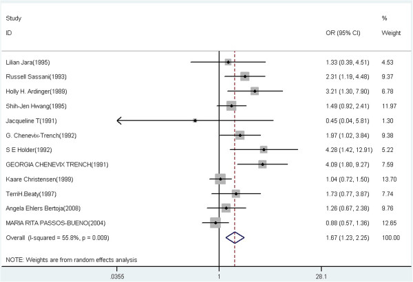
Forest plot of cancer risk associated with TGFA Taq I polymorphism under the heterozygote comparison model (C1C2 versus C1C1).
Figure 3.
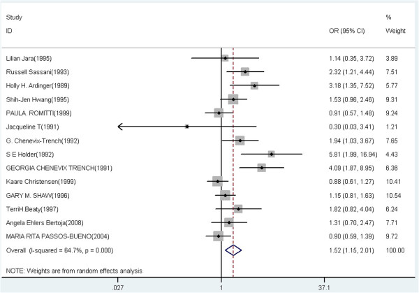
Forest plot of cancer risk associated with TGFA Taq I polymorphism under the dominant model (C1C2 + C2C2 versus C1C1).
Figure 4.
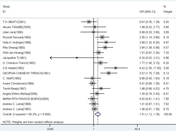
Forest plot of cancer risk associated with TGFA Taq I polymorphism under the allelic model (C2 versus C1).
Stratified meta-analysis
Stratified analyses were conducted by ethnicity, source of control and disease type. Pooled ORs and 95% CIs of stratified meta-analysis are showed in Table 3. In the subgroup analysis by ethnicity, significantly increased CL/P risks were found among Caucasian (C1C2 vs C1C1: OR = 1.95, 95% CI = 1.34-2.86; C2C2 + C1C2 vs C1C1: OR = 1.68, 95% CI = 1.18-2.38; C2 vs C1: OR = 1.52, 95% CI = 1.14-2.02). No significantly evaluated risk was found among African and Hispanic population in any of the genetic models.
Table 3.
Pooled ORs and 95% CIs of stratified meta-analysis
| Subgroup | Genotype | No of studies |
Test of association |
Test of heterogeneity |
||||
|---|---|---|---|---|---|---|---|---|
| OR (95% CI) | Z | P-value | Model | P-value | I 2 (%) | |||
|
Ethnicity | ||||||||
| African | ||||||||
| |
C1C2 + C2C2 vs. C1C1 |
3 |
1.92 [0.63,1.90] |
1.14 |
0.253 |
F |
0.754 |
0.0 |
| |
C2 vs. C1 |
3 |
1.15 [0.50,2.66] |
0.33 |
0.741 |
F |
0.254 |
26.9 |
| Caucasian | ||||||||
| |
C1C2 vs. C1C1 |
9 |
1.95 [1.34,2.86]* |
3.48 |
0.001 |
R |
0.016 |
57.3 |
| |
C2C2 vs. C1C1 |
9 |
1.50 [0.79,2.84] |
1.25 |
0.211 |
F |
0.341 |
11.2 |
| |
C1C2 + C2C2 vs. C1C1 |
9 |
1.68 [1.18,2.38]* |
2.87 |
0.004 |
R |
0.000 |
69.9 |
| |
C2C2 vs. C1C2 + C1C1 |
9 |
1.36 [0.71,2.58] |
0.93 |
0.354 |
F |
0.443 |
0.0 |
| |
C2 vs. C1 |
14 |
1.52 [1.14,2.02]* |
2.87 |
0.004 |
R |
0.000 |
74.8 |
| Hispanic | ||||||||
| |
C1C2 vs. C1C1 |
3 |
1.02 [0.72,1.43] |
0.08 |
0.935 |
F |
0.594 |
0.0 |
| |
C2C2 vs. C1C1 |
3 |
1.44 [0.27,7.67] |
0.42 |
0.672 |
F |
0.669 |
0.0 |
| |
C1C2 + C2C2 vs. C1C1 |
4 |
1.04 [0.76,1.44] |
0.27 |
0.789 |
F |
0.792 |
0.0 |
| |
C2C2 vs. C1C2 + C1C1 |
3 |
1.40 [0.26,7.46] |
0.4 |
0.692 |
F |
0.663 |
0.0 |
| |
C2 vs. C1 |
3 |
1.04 [0.75,1.44] |
0.23 |
0.816 |
F |
0.608 |
0.0 |
|
Disease | ||||||||
| CP | ||||||||
| |
C1C2 vs. C1C1 |
3 |
1.54 [1.04,2.27]* |
2.14 |
0.032 |
F |
0.215 |
34.9 |
| |
C2C2 vs. C1C1 |
3 |
1.32 [0.35,5.00] |
0.40 |
0.687 |
F |
0.478 |
0.0 |
| |
C1C2 + C2C2 vs. C1C1 |
5 |
1.45 [1.10,1.91] |
2.61 |
0.009 |
F |
0.281 |
21.0 |
| |
C2C2 vs. C1C2 + C1C1 |
3 |
1.26 [0.33,4.78] |
0.33 |
0.738 |
F |
0.496 |
0.0 |
| |
C2 vs. C1 |
8 |
1.38 [1.10,1.73]* |
2.82 |
0.005 |
F |
0.226 |
25.4 |
| CL/P | ||||||||
| |
C1C2 vs. C1C1 |
12 |
1.60 [1.16,2.20]* |
2.89 |
0.004 |
R |
0.010 |
55.7 |
| |
C2C2 vs. C1C1 |
11 |
1.64 [0.88,3.04] |
1.57 |
0.116 |
F |
0.457 |
0.0 |
| |
C1C2 + C2C2 vs. C1C1 |
11 |
1.46 [1.09,1.95]* |
2.55 |
0.011 |
R |
0.001 |
62.7 |
| |
C2C2 vs. C1C2 + C1C1 |
11 |
1.45 [0.82,2.78] |
1.25 |
0.211 |
F |
0.545 |
0.0 |
| |
C2 vs. C1 |
17 |
1.29 [1.01,1.66]* |
2.03 |
0.042 |
R |
0.000 |
63.1 |
|
Source of control | ||||||||
| HB | ||||||||
| |
C1C2 vs. C1C1 |
10 |
1.84 [1.32,2.56]* |
3.58 |
0.000 |
R |
0.019 |
54.7 |
| |
C2C2 vs. C1C1 |
10 |
2.96 [1.35,2.70]* |
4.70 |
0.000 |
F |
0.965 |
0.0 |
| |
C1C2 + C2C2 vs. C1C1 |
10 |
1.99 [1.35,2.70]* |
3.66 |
0.000 |
R |
0.007 |
60.1 |
| |
C2C2 vs. C1C2 + C1C1 |
10 |
2.38 [1.06,5.55]* |
5.17 |
0.000 |
F |
0.968 |
0.0 |
| |
C2 vs. C1 |
14 |
1.63 [1.22,2.18]* |
3.28 |
0.001 |
R |
0.001 |
63.0 |
| PB | ||||||||
| |
C1C2 + C2C2 vs. C1C1 |
3 |
0.99 [0.79,1.24] |
0.10 |
0.917 |
F |
0.549 |
0.0 |
| C2 vs. C1 | 3 | 1.00 [0.83,1.20] | 0.02 | 0.986 | F | 0.778 | 0.0 | |
*OR had statistical significance with corresponding 95% CI not including 1. F: Fixed effect model; R: Random effect model.
In the subgroup analysis according to disease type, the ORs of the heterozygote comparison, dominant and allelic model with CL/P are statistically significant (C1C2 vs C1C1: OR = 1.60, 95% CI = 1.16-2.20; C2C2 + C1C2 vs C1C1: OR = 1.46, 95% CI = 1.09-1.95; C2 vs C1: OR = 1.29, 95% CI = 1.01-1.66). For the CP, the significant results were observed in heterozygote comparison, dominant and allelic model (C1C2 vs C1C1: OR = 1.54, 95% CI = 1.04-2.27; C2C2 + C1C2 vs C1C1: OR = 1.45, 95% CI = 1.10-1.19; C2 vs C1: OR = 1.38, 95% CI = 1.10-1.73).
In the subgroup analysis by control characteristics, the ORs of the heterozygote comparison, homozygote, dominant, recessive and allelic model for the hospital-based control are statistically significant (C1C2 vs C1C1: OR = 1.84, 95% CI = 1.32-2.56; C2C2 vs C1C1: OR = 2.96, 95% CI = 1.35-2.70; C2C2 + C1C2 vs C1C1: OR = 1.99, 95% CI = 1.35-2.70; C2C2 vs C1C2 + C1C1: OR = 2.38, 95% CI = 1.06-5.55; C2 vs C1: OR = 1.63, 95% CI = 1.22-2.18). While, no statistically significant association was found in population-based controls.
Heterogeneity between studies was observed in overall comparisons and also subgroup analyses. Thus, meta-analyses were performed using random-effect models (Table 2).
To explore potential sources of heterogeneity in this meta-analysis, meta-regression analyses were implemented. The covariates included ethnicity and source of control. In all of the heterozygote comparison, homozygote, dominant, recessive and allelic models, above potential factors were probably not the major sources of heterogeneity (P-values were all >0.05 or near 0.05). The heterogeneity might attribute to other factors, the insufficient data limited to identify their sources only using meta-regression.No publication bias was detected by either the inverted funnel plot or Egger’s test. The shapes of the funnel plots seemed approximately symmetrical and P values of the Egger’ tests were not statistical significant (P values were all >0.05). Figure 5 and Figure 6 showed the funnel plot under the heterozygote comparison model (C1C2 versus C1C1) and the allelic model (C2 versus C1).
Figure 5.
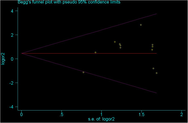
Funnel plot under the heterozygote comparison model (C1C2 versus C1C1).
Figure 6.
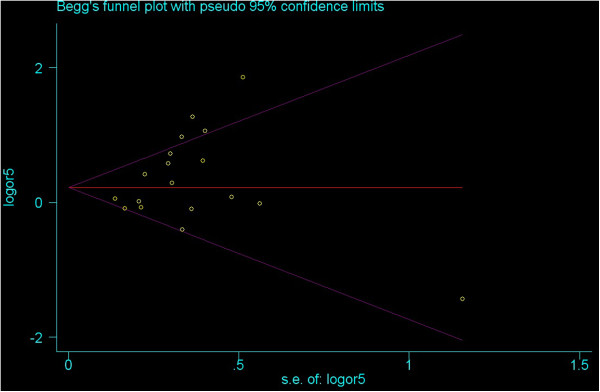
Funnel plot under the allelic model (C2 versus C1).
Discussion
The individual susceptibility plays an important role in the development of most diseases. Therefore, the studies of genetic susceptibility are helpful for disease prevention, diagnosis, and treatment.
TGFA is a well characterized mammalian growth factor. Previous studies found that although TGFA is expressed in mice during palatogenesis and mice with a null mutation of the TGFA gene have abnormal skin, hair, and eyes, but they do not have CL/P [38,39]. TGFA is a likely ligand for epidermal growth factor receptor and newborn epidermal growth factor receptor-negative/-negative mice have high incidence of CL/P which may explain the association between TGFA polymorphisms and CL/P [40]. In 1989, it was firstly published that the TGFA Taq I polymorphism contributed to the development CL/P in humans. From then on, a growing number of studies have been done to examine the relationship between TGFA Taq I polymorphism and the risks of CL/P. However, the results are not consistent. In order to better understand the association, we subjected previously published data to meta-analysis to evaluate genetic associations between the TGFA Taq I polymorphism and CL/P susceptibility.
Through this meta-analysis we found that TGFA Taq I polymorphism increased the risk of CL/P. Mitchell adopted a reappraisal and showed that there was a statistically significant association between TGFA and CL/P, However, there was evidence of significant heterogeneity from different studies, regarding an association between genetic variation at the TGFA locus and CL/P remains inconclusive [41]. A meta-analysis of gene-environment interaction showed the suggestive evidence for gene-environment interaction between the infant’s genotype at the Taq1 marker in TGFA and maternal smoking was limited to CP [42]. Meta-review of thyroid cancer gene expression profiling studies identifies important diagnostic biomarkers, including relatively novel or uncharacterized genes such as TGFA [43]. These results of systematic reviews in some extent support the findings in the present meta-analysis.
In order to reduce heterogeneity, we performed the stratified analyses. It is well know that the incidence of most genetic polymorphisms could vary between different ethnic populations and the wide range in TGFA Taq I allele frequencies across different studies suggests the heterogeneity between populations may exist [28]. In the subgroup analysis by ethnicity, we found that TGFA Taq I polymorphism increased the risk of CL/P in Caucasian population, which agreed with Ardinger and colleagues [12], Shiang and colleagues [20], Hwang and colleagues [22], Chenevix-Trench and colleagues [18], Holder and colleagues [17], and disagreed with Lidral and colleagues [27], [28], Stoll and colleagues [25] and Christensen and colleagues [11]. In the subgroup analysis by disease type, we found the ORs of different disease type were statistically significant, which suggested disease type was not mainly result the heterogeneity. In the subgroup analyses of control characteristics, only the ORs of hospital-based control groups were statistically significant. Therefore, subgroup analyses suggested that ethnicity and control characteristics might contribute to the heterogeneity in this meta-analysis. The meta-regression analyses suggested that ethnicity or control characteristics were probably not the major sources of heterogeneity. The heterogeneity might attribute to other factors. The insufficient data are limited to identify the source of heterogeneity only using meta-regression.
Despite trying our best to perform a comprehensive meta-analysis, some limitations exist in our study. Although the results for publication bias in our study were not statistically significant, our analysis used published international studies, which could arose publication bias. Lack of the original data of available studies limited our further evaluation of potential interactions, such as age, gender, family history, environmental factors and lifestyle. There were not significant results in stratified analyses among African and Hispanic population, because there were too few studies after stratifying. Therefore, more studies are needed to provide more evidence on the association between TGFA polymorphism and CL/P in different ethnic populations.
Although CL/P is a complex disease, our study provided evidence showing the important role of TGFA genes polymorphisms in the development of CL/P, genetic factors determining disease susceptibility and severity may facilitate personalized medicine. Further understanding of the interactions between genetic regulatory mechanisms is critical for discovering new therapies for managing human CL/P. Specifically, targeted therapy about the polymorphisms of related genes might be a promising avenue for future CL/P diagnosis and treatment.
In summary, our meta-analysis supports that the TGFA Taq I polymorphism is more likely to contribute to the susceptibility of CL/P, especially in the subgroup of Caucasian population.
Conclusion
TGFA Taq I polymorphism may be associated with the risk of CL/P.
Competing interests
The authors declare that they have no competing interests.
Authors’ contributions
CF participated in selecting the study, extracting the data, performing the statistical analysis and drafting the manuscript. EZ and WD participated in study selection, data extraction and manuscript drafting. ZX collected and extracted the data. YZ and LL participated in study selection and the statistical analysis. All authors read and approved the final manuscript.
Pre-publication history
The pre-publication history for this paper can be accessed here:
Contributor Information
Cuijuan Feng, Email: fcjxll@outlook.com.
Enjiao Zhang, Email: zej980980@hotmail.com.
Weiyi Duan, Email: duanwy2013@163.com.
Zhongfei Xu, Email: xzf7090@hotmail.com.
Yang Zhang, Email: kp3666@126.com.
Li Lu, Email: luli@mail.cmu.edu.cn.
Acknowledgements
This research was supported by the Public Welfare Fund Project for Science of Liaoning Province (No: 2012002015) and a grant from the Science and Technology Project of Shenyang (Grant no.: F13-220-9-73).
References
- Melnick M, Jaskoll T, Marazita M. Localization of H-2K in developing mouse palates using monoclonal antibody. J Embryol Exp Morphol. 1982;70:45–60. [PubMed] [Google Scholar]
- Wyszynski DF, Beaty TH. Review of the role of potential teratogens in the origin of human nonsyndromic oral clefts. Teratology. 1996;53:309–317. doi: 10.1002/(SICI)1096-9926(199605)53:5<309::AID-TERA5>3.0.CO;2-W. [DOI] [PubMed] [Google Scholar]
- Czeizel A, Nagy E. A recent aetiological study on facial clefting in Hungary. Acta Pediatr Hung. 1986;27:145–166. [PubMed] [Google Scholar]
- Khoury MJ, Gomez-Farias M, Mulinare J. Does maternal cigarette smoking during pregnancy cause cleft lip and palate in offspring? Am J Dis Child. 1989;143:333–337. doi: 10.1001/archpedi.1989.02150150091023. [DOI] [PubMed] [Google Scholar]
- Werler MM, Lammer EJ, Rosenberg L, Mitchell AA. Maternal cigarette smoking during pregnancy in relation to oral clefts. Am J Epidemiol. 1990;132:926–932. doi: 10.1093/oxfordjournals.aje.a115735. [DOI] [PubMed] [Google Scholar]
- Shaw GM, Wasserman CR, Lammer EJ, O’Malley CD, Murray JC, Basart AM, Tolarova MM. Orofacial clefts, parental cigarette smoking, and transforming growth factor-alpha gene variants. Am J Hum Genet. 1996;58:551–561. [PMC free article] [PubMed] [Google Scholar]
- Tolarova MM. Orofacial clefts in Czechoslovakia: incidence, genetics and prevention of cleft lip and palate over a 19-year period. Scand J Plast Reconstr Surg. 1987;21:19–25. doi: 10.3109/02844318709083574. [DOI] [PubMed] [Google Scholar]
- Tolarova MM, Harris JA. Reduced recurrence of orofacial clefts after periconceptional supplementation with high-dose folic acid and multivitamins. Teratology. 1995;51:71–78. doi: 10.1002/tera.1420510205. [DOI] [PubMed] [Google Scholar]
- Hayes C, Werler MM, Willett WC, Mitchell AA. Case–control study of periconceptional folic acid supplementation and oral clefts. Am J Epidemiol. 1996;143:1229–1234. doi: 10.1093/oxfordjournals.aje.a008710. [DOI] [PubMed] [Google Scholar]
- Munger RG, Romitti PA, Daack-Hirsch S, Burns TL, Murray JC, Hanson J. Maternal alcohol use and risk of orofacial cleft birth defects. Teratology. 1996;54:27–33. doi: 10.1002/(SICI)1096-9926(199607)54:1<27::AID-TERA4>3.0.CO;2-0. [DOI] [PubMed] [Google Scholar]
- Christensen K, Olsen J, Nørgaard-Pedersen B, Basso O, Støvring H, Milhollin-Johnson L, Murray JC. Oral clefts, transforming growth factor alpha gene variants, and maternal smoking: a population-based case–control study in Denmark, 1991–1994. Am J Epidemiol. 1999;149:248–255. doi: 10.1093/oxfordjournals.aje.a009799. [DOI] [PubMed] [Google Scholar]
- Ardinger HH, Buetow KH, Bell GI, Bardach J, Van-Demark DR, Murray JC. Association of genetic variation of the transforming growth factor-alpha gene with cleft lip and palate. Am J Hum Genet. 1989;45:348–353. [PMC free article] [PubMed] [Google Scholar]
- Dixon MJ, Garner J, Fergusson MW. Immunolocalization of epidermal growth factor (EGF), EGF receptor and transforming growth factor alpha (TGFa) during murine palatogenesis in vivo and in vitro. Anat Embryol. 1991;184:83–91. doi: 10.1007/BF01744264. [DOI] [PubMed] [Google Scholar]
- Lee DC, Rochford R, Todaro GJ, Villarreal LP. Developmental expression of rat transforming growth factor-a mRNA. Mol Cell Biol. 1985;5:3644–3646. doi: 10.1128/mcb.5.12.3644. [DOI] [PMC free article] [PubMed] [Google Scholar]
- Tanabe A, Taketani S, Endo-Ichikawa Y, Tokunaga R, Ogawa Y, Hiramoto M. Analysis of the candidate genes responsible for nonsyndromic cleft lip and palate in Japanese people. Clin Sci. 2000;99:105–111. [PubMed] [Google Scholar]
- Vieira AR. Association between the transforming growth factor alpha gene and nonsyndromic oral clefts: a HuGE review. Am J Epidemiol. 2006;163:790–810. doi: 10.1093/aje/kwj103. [DOI] [PubMed] [Google Scholar]
- Holder SE, Vintiner GM, Farren B, Malcolm S, Winter RM. Confirmation of an association between RFLPs at the transforming growth factor-alpha locus and non-syndromic cleft lip and palate. J Med Genet. 1992;29:390–392. doi: 10.1136/jmg.29.6.390. [DOI] [PMC free article] [PubMed] [Google Scholar]
- Chenevix-Trench G, Jones K, Green AC, Duffy DL, Martin NG. Cleft lip with or without cleft palate: associations with transforming growth factor alpha and retinoic acid receptor loci. Am J Hum Genet. 1992;51:1377–1385. [PMC free article] [PubMed] [Google Scholar]
- Sassani R, Bartlett SP, Feng H, Goldner-Sauve A, Haq AK, Buetow KH, Gasser DL. Association between alleles of the transforming growth factor-alpha locus and the occurrence of cleft lip. Am J Hum Genet. 1993;45:565–569. doi: 10.1002/ajmg.1320450508. [DOI] [PubMed] [Google Scholar]
- Shiang R, Lidral AC, Ardinger HH, Buetow KH, Romitti PA, Munger RG, Murray JC. Association of transforming growth-factor alpha gene polymorphisms with nonsyndromic cleft palate only (CPO) Am J Hum Genet. 1993;53:836–843. [PMC free article] [PubMed] [Google Scholar]
- Jara L, Blanco R, Chiffelle I, Palomino H, Carreño H. Association between alleles of the transforming growth factor alpha locus and cleft lip and palate in the Chilean population. Am J Hum Genet. 1995;57:548–551. doi: 10.1002/ajmg.1320570406. [DOI] [PubMed] [Google Scholar]
- Hwang SJ, Beaty TH, Panny SR, Street NA, Joseph JM, Gordon S, McIntosh I, Francomano CA. Association study of transforming growth factor alpha (TGF alpha) TaqI polymorphism and oral clefts: indication of gene-environment interaction in a population-based sample of infants with birth defects. Am J Epidemiol. 1995;141:629–636. doi: 10.1093/oxfordjournals.aje.a117478. [DOI] [PubMed] [Google Scholar]
- Shaw GM, Wasserman CR, Murray JC, Lammer EJ. Infant TGF-alpha genotype, orofacial clefts, and maternal periconceptional multivitamin Use. Cleft Palate Craniofcial J. 1998;35:366–370. doi: 10.1597/1545-1569_1998_035_0366_itagoc_2.3.co_2. [DOI] [PubMed] [Google Scholar]
- Hecht JT, Wang YP, Blanton SH, Michels VV, Daiger SP. Cleft lip and palate: no evidence of linkage to transforming growth factor alpha. Am J Hum Genet. 1991;49:682–686. [PMC free article] [PubMed] [Google Scholar]
- Stoll C, Qian JF, Feingold J, Sauvage P, May E. Genetic variation in transforming growth factor alpha: possible association of Bam HI polymorphism with bilateral sporadic cleft lip and palate. Hum Genet. 1993;92:81–82. doi: 10.1007/BF00216150. [DOI] [PubMed] [Google Scholar]
- Beaty TH, Maestri NE, Hetmanski JB, Wyszynski DF, Vanderkolk CA, Simpson JC. Testing for interaction between materrnal somking and TGFA genotype among oral cleft case born in Maryland 1992–1996. Cleft Palate Craniofcial J. 1997;34:447–454. doi: 10.1597/1545-1569_1997_034_0447_tfibms_2.3.co_2. [DOI] [PubMed] [Google Scholar]
- Lidral AC, Murray JC, Buetow KH, Basart AM, Schearer H, Shiang R, Naval A, Layda E, Magee K, Magee W. Studies of the candidate genes TGFB2, MSX1, TGFA and TGFB3 in the etiology of cleft lip and palate in the Philippines. Cleft Palate Craniofac J. 1997;34:1–6. doi: 10.1597/1545-1569_1997_034_0001_sotcgt_2.3.co_2. [DOI] [PubMed] [Google Scholar]
- Lidral AC, Romitti PA, Basart AM, Doetschman T, Leysens NJ, Daack-Hirsch S, Semina EV, Johnson LR, Machida J, Burds A, Parnell TJ, Rubenstein JL, Murray JC. Association of MSX1 and TGFB3 with nonsyndromic clefting in humans. Am J Hum Genet. 1998;63:557–568. doi: 10.1086/301956. [DOI] [PMC free article] [PubMed] [Google Scholar]
- Chenevix-Trench G, Jones K, Green A, Martin N. Further Evidence for an Association Between Genetic Variation in Transforming Growth Factor Alpha and Cleft Lip and Palate. Am J Hum Genet. 1991;48:1012–1013. [PMC free article] [PubMed] [Google Scholar]
- Romitti PA, Lidral AC, Munger RG, Daack-Hirsch S, Burns TL, Murray JC. Candidate genes for nonsyndromic cleft lip and palate and maternal cigarette smoking and alcohol consumption: evaluation of genotype-environment interactions from a population-based case–control study of orofacial clefts. Teratology. 1999;59:39–50. doi: 10.1002/(SICI)1096-9926(199901)59:1<39::AID-TERA9>3.0.CO;2-7. [DOI] [PubMed] [Google Scholar]
- Beaty TH, Wang H, Hetmanski JB, Fan YT, Zeiger JS, Liang KY, Chiu YF, Vanderkolk CA, Seifert KC, Wulfsberg EA, Raymond G, Panny SR, McIntosh I. A case–control study of nonsyndromic oral clefts in Maryland. Ann Epidemiol. 2001;11:434–442. doi: 10.1016/s1047-2797(01)00222-8. [DOI] [PubMed] [Google Scholar]
- Passos-Bueno MR, Gaspar DA, Kamiya T, Tescarollo G, Rabanéa D, Richieri-Costa A, Alonso N, Araújo B. Transforming growth factor-ɑ and nonsyndromic cleft lip with or without palate in Brazilian patients: results of a large case–control study. Cleft Palate Craniofcial J. 2004;41:387–391. doi: 10.1597/03-054.1. [DOI] [PubMed] [Google Scholar]
- Chevrier C, Bahuau M, Perret C, Iovannisci DM, Nelva A, Herman C, Vazquez MP, Francannet C, Robert-Gnansia E, Lammer EJ, Cordier S. Genetic susceptibilities in the association between maternal exposure to tobacco smoke and the risk of nonsyndromic oral cleft. Am J Med Genet A. 2008;146A:2396–2406. doi: 10.1002/ajmg.a.32505. [DOI] [PubMed] [Google Scholar]
- Ehlers Bertoja A, Sampaio Alho C, De Franca E, Menegotto B, Miriam Robinson W. TGFA/TAQ I polymorphism in nonsyndromic cleft lip and palate patients from Rio Grande Do Sul, Brazil. Cleft Palate Craniofcial J. 2008;45:539–544. doi: 10.1597/07-087.1. [DOI] [PubMed] [Google Scholar]
- Zhu J, Hao L, Li S, Bailey LB, Tian Y, Li Z. MTHFR, TGFB3, and TGFA polymorphisms and their association with the risk of non-syndromic cleft lip and cleft palate in China. Am J Med Genet A. 2010;152A:291–298. doi: 10.1002/ajmg.a.33113. [DOI] [PubMed] [Google Scholar]
- Begg CB, Mazumdar M. Operating characteristics of a rank correlation test for publication bias. Biometrics. 1994;50:1088–1101. [PubMed] [Google Scholar]
- Egger M, Davey Smith G, Schneider M, Minder C. Bias in meta-analysis detected by a simple, graphical test. BMJ. 1997;315:629–634. doi: 10.1136/bmj.315.7109.629. [DOI] [PMC free article] [PubMed] [Google Scholar]
- Mann GB, Fowler KJ, Gabriel A, Nice EC, Williams RL, Dunn AR. Mice with a null mutation of the TGFa gene have abnormal skin architecture, wavy hair, and curly whiskers and often develop corneal inflammation. Cell. 1993;73:249–261. doi: 10.1016/0092-8674(93)90227-h. [DOI] [PubMed] [Google Scholar]
- Luetteke NC, Qiu TH, Peiffer RL, Oliver P, Smithies O, Lee DC. TGFa deficiency results in hair follicle and eye abnormalities in targeted and waved-1 mice. Cell. 1993;73:263–278. doi: 10.1016/0092-8674(93)90228-i. [DOI] [PubMed] [Google Scholar]
- Miettinen PJ, Chin JR, Shum L, Slavkin HC, Shuler CF, Derynck R, Werb Z. Epidermal growth factor receptor function is necessary for normal craniofacial development and palate closure. Nat Genet. 1999;22:69–73. doi: 10.1038/8773. [DOI] [PubMed] [Google Scholar]
- Mitchell LE. Transforming growth factor alpha locus and nonsyndromic cleft lip with or without cleft palate: a reappraisal. Genet Epidemiol. 1997;14:231–240. doi: 10.1002/(SICI)1098-2272(1997)14:3<231::AID-GEPI2>3.0.CO;2-8. [DOI] [PubMed] [Google Scholar]
- Zeiger JS, Beaty TH, Liang KY. Oral clefts, maternal smoking, and TGFA: a meta-analysis of gene-environment interaction. Cleft Palate Craniofac J. 2005;42:58–63. doi: 10.1597/02-128.1. [DOI] [PubMed] [Google Scholar]
- Griffith OL, Melck A, Jones SJ, Wiseman SM. Meta-analysis and meta-review of thyroid cancer gene expression profiling studies identifies important diagnostic biomarkers. J Clin Oncol. 2006;24:5043–5051. doi: 10.1200/JCO.2006.06.7330. [DOI] [PubMed] [Google Scholar]


