Abstract
An 83-year-old woman presented with acute aphasia. Brain MRI, performed 3 hours after symptom onset, showed isolated fluid-attenuated inversion recovery vascular hyperintensities (FVH) in the left middle cerebral artery, including dot-like and serpentine hyperintensities (figure). Immediately after this first MRI (i.e., 3 hours and 15 minutes after symptom onset), aphasia resolved. A second MRI performed 15 minutes later showed FVH disappearance.
An 83-year-old woman presented with acute aphasia. Brain MRI, performed 3 hours after symptom onset, showed isolated fluid-attenuated inversion recovery vascular hyperintensities (FVH) in the left middle cerebral artery, including dot-like and serpentine hyperintensities (figure). Immediately after this first MRI (i.e., 3 hours and 15 minutes after symptom onset), aphasia resolved. A second MRI performed 15 minutes later showed FVH disappearance.
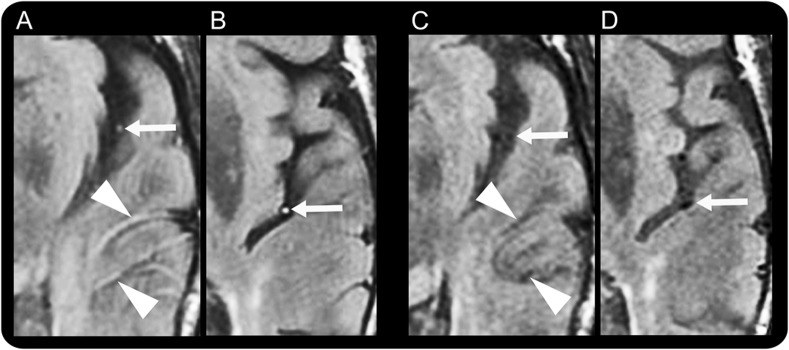
Figure. Vascular intensity changes on fluid-attenuated inversion recovery during and after resolution of symptoms in our TIA patient.

Fluid-attenuated inversion recovery vascular hyperintensities (FVH) during aphasia, including dot-like (arrows in A and B) and serpentine (arrowheads in A) hyperintensities, were seen in the middle cerebral artery branches. There were no abnormalities on diffusion-weighted or gradient echo images or magnetic resonance angiography. After aphasia resolution, the MRI showed FVH disappearance (C, D).
Only 20% of TIA patients showed FVH when MRI was performed within 24 hours.1 Because FVH in this case were transient and correlated with symptom resolution, the prior report may have underestimated the true frequency of their occurrence.
Footnotes
Author contributions: Guillaume Taieb: drafting/revising the manuscript, study concept or design, analysis or interpretation of data. Dimitri Renard: drafting/revising the manuscript, analysis or interpretation of data. Francesco Macri Gerasoli: drafting/revising the manuscript, study concept or design, analysis or interpretation of data.
Study funding: No targeted funding reported.
Disclosure: The authors report no disclosures relevant to the manuscript. Go to Neurology.org for full disclosures.
References
- 1.Kobayashi J, Uehara T, Toyoda K, et al. Clinical significance of fluid-attenuated inversion recovery vascular hyperintensities in transient ischemic attack. Stroke 2013;44:1635–1640 [DOI] [PubMed] [Google Scholar]


