Abstract
Purpose:
Skin dosimetry is important for fluoroscopically-guided interventions, as peak skin doses (PSD) that result in skin reactions can be reached during these procedures. There is no consensus as to whether or not indirect skin dosimetry is sufficiently accurate for fluoroscopically-guided interventions. However, measuring PSD with film is difficult and the decision to do so must be madea priori. The purpose of this study was to assess the accuracy of different types of indirect dose estimates and to determine if PSD can be calculated within ±50% using indirect dose metrics for embolization procedures.
Methods:
PSD were measured directly using radiochromic film for 41 consecutive embolization procedures at two sites. Indirect dose metrics from the procedures were collected, including reference air kerma. Four different estimates of PSD were calculated from the indirect dose metrics and compared along with reference air kerma to the measured PSD for each case. The four indirect estimates included a standard calculation method, the use of detailed information from the radiation dose structured report, and two simplified calculation methods based on the standard method. Indirect dosimetry results were compared with direct measurements, including an analysis of uncertainty associated with film dosimetry. Factors affecting the accuracy of the different indirect estimates were examined.
Results:
When using the standard calculation method, calculated PSD were within ±35% for all 41 procedures studied. Calculated PSD were within ±50% for a simplified method using a single source-to-patient distance for all calculations. Reference air kerma was within ±50% for all but one procedure. Cases for which reference air kerma or calculated PSD exhibited large (±35%) differences from the measured PSD were analyzed, and two main causative factors were identified: unusually small or large source-to-patient distances and large contributions to reference air kerma from cone beam computed tomography or acquisition runs acquired at large primary gantry angles. When calculated uncertainty limits [−12.8%, 10%] were applied to directly measured PSD, most indirect PSD estimates remained within ±50% of the measured PSD.
Conclusions:
Using indirect dose metrics, PSD can be determined within ±35% for embolization procedures. Reference air kerma can be used without modification to set notification limits and substantial radiation dose levels, provided the displayed reference air kerma is accurate. These results can reasonably be extended to similar procedures, including vascular and interventional oncology. Considering these results, film dosimetry is likely an unnecessary effort for these types of procedures when indirect dose metrics are available.
Keywords: fluoroscopy, peak skin dose, radiochromic film, dose calculation, skin injury
I. INTRODUCTION
Skin dosimetry is important for fluoroscopically-guided interventions as the radiation doses used to perform these procedures are on occasion high enough to cause skin reactions.1 It has been reported that doses of at least 5–10 Gy are required to cause a National Cancer Institute (NCI) Grade 2 skin reaction,2 and doses greater than 15 Gy are required to cause the most severe skin injuries.1 Skin dosimetry can be either direct or indirect,3 with indirect dosimetry being more common. An example of indirect dosimetry is reference air kerma (also referred to as cumulative dose, reference point air kerma, or cumulative air kerma); an example of direct dosimetry is the use of radiochromic film to measure the peak skin dose (PSD). Prospective direct measurement of PSD during fluoroscopically-guided interventions is a complicated and time-consuming task. A number of attempts have been made to relate indirect dose metrics and calculated quantities to direct measurements of PSD,4–11 with the authors of these works using widely varying methods for calculating PSD from indirect metrics including reference air kerma and kerma area product (KAP), and with one exception using film to directly measure PSD. It is difficult to make meaningful comparisons between these studies owing to the different calculation methods used and because film calibration methods were not always reported. The results of these studies were equally variable, including findings of a coefficient of determination of 0.8 between reference air kerma and PSD for endovascular thoracoabdominal aneurysm repair;4 that reference air kerma overestimated measured PSD for low doses and was within 50% for doses greater than 1 Gy;5 a measured PSD that was 21% lower than reference air kerma for a single patient;6 that manufacturer-reported reference air kerma was an “unreliable predictor of actual dose” when using visual comparison of a preprinted calibration tablet to film for pediatric cardiac catheterization;7 and that in about half of 16 cases, reference air kerma overestimated the PSD.8
Recently, the following statement appeared in a review of radiation effects on patients’ skin and hair: “Skin dosimetry is unlikely to be more accurate than ±50%” (Ref. 1). The goal of this study was to determine if it is possible to consistently estimate PSD within ±50% of the true PSD. The current study considered embolization procedures, a subset of vascular and oncologic interventions. We hypothesized that for these types of procedures, in most cases, indirect skin dosimetry would be within ±50% of the directly measured PSD. We also studied differences in indirect dosimetry accuracy when using only reference air kerma, when estimating PSD using accepted methods,12,13 and when using data from the radiation dose structured report (RDSR) (Ref. 14) to perform a full PSD reconstruction.
II. METHODS
This Health Insurance Portability and Accountability Act compliant study was performed with Institutional Review Board approval.
II.A. Film dosimetry
Gafchromic XR-RV3 radiochromic film (Ashland Inc., Covington, KY) was used to directly measure PSD during consecutive embolization procedures at both MD Anderson Cancer Center (Site A) and the University of Tennessee Medical Center at Knoxville (Site B). PSD was measured for a total of 41 procedures between the two sites, 21 at Site A and 20 at Site B. The radiochromic film was calibrated as described previously4–6,9,15,16 by cutting a piece of film from the lot used for direct PSD measurements into a number of small squares which were exposed, free-in-air, along with a 6 cc ionization chamber (Radcal Corporation, Monrovia, CA). A phantom consisting of 22.9–25.4 cm (9–10 in.) of acrylic was placed on the patient support of a C-arm fluoroscope of the same model (Axiom Artis/zee, Siemens, Malvern, PA) as those used for the embolization procedures. Both lots of film used, one at each site, were calibrated individually. A mix of fluoroscopy at 7.5 pulses/s and acquisition at 2–4 frames/s was used to expose the film during calibration while varying the attenuator thickness between 22.9 and 25.4 cm, resulting in exposure of the film to a similar range of beam qualities as those used during embolization procedures. During calibration, the kVp ranged from 70 to 90 and the added filtration ranged from 0.3 mm Cu to 0.1 mm Cu. A minimum of 8 points were acquired for each calibration, and the calibration films were exposed to air kermas ranging from 0 to 6.0 Gy. A four parameter logistic model Rodbard fit17,18 was applied to the calibration data to derive the calibration function. After exposure, all films were stored in a dark location for at least one week prior to scanning.
Scanning for films from both study sites was performed at Site A using a single Epson V700 flatbed scanner (Epson, Long Beach, CA) and the associated Epson Scan software (v. 3.81US) with all corrections disabled, i.e., Professional Mode. The film was scanned at a resolution of 300 dpi in 48 bit color, and the signal in the red color channel was used for all film measurements. All image analysis, including application of the Rodbard fit, was performed using ImageJ 1.45 (NIH, Bethesda, MD). All calibration films were exposed in the same orientation, with the orange side facing the x-ray source (the film has an orange side and a white side), and the radiologic technologists placing films for patient studies were instructed to place the film immediately under the patient, covered by a hospital sheet, in the same orientation. At least 4000, and typically 7000, pixels were included in the rectangular regions of interest used to measure the PSD. Visual examination while adjusting window width and level was used to identify the area of maximum darkening.
II.B. Dose calculation
Indirect PSD were calculated according to the methods published by Jones and Pasciak12,13 using varying levels of procedural detail. For all procedures, PSD was calculated using the information provided in the patient protocol produced by the fluoroscopes used in this study. Calculations were performed piecemeal12 for acquisition series, while all reference air kerma for fluoroscopy was treated as a whole. This is referred to as Method 1 throughout this paper (Table I). The patient table height was considered to be the source-to-patient distance. The patient table height was calculated by subtracting the default table-to-object distance (TOD) of 15 cm from the distance reported in Digital Imaging and Communications in Medicine (DICOM) header tag (0018,0111)-–Distance Source to Patient.19 The default TOD was verified to be correct in private tag (0023,1008) in the DICOM header produced by the fluoroscopes used in this study. The backscatter factor for fluoroscopy was calculated using published tables.20 Half value layers were determined using the measured patient anteroposterior thickness and Fig. 3 from Ref. 12, the x-ray field size at the patient surface was calculated from the ratio of KAP to reference air kerma scaled by the square of the ratio of source-to-patient distance to the interventional reference point location. KAP and reference air kerma were reported in the patient protocol, patient table height for acquisition series was extracted from the DICOM headers of the corresponding series on the Picture Archiving and Communication System, and the location of the interventional reference point was determined from the fluoroscope operator's manual. The mode, or mean when the mode was not defined, of the source-to-patient distance for each acquisition series was used as the source-to-patient distance for fluoroscopy.
TABLE I.
Description of peak skin dose calculation methods used in this study.
| Description | Information sources | Fluoroscopy | Acquisition | Additional information |
|---|---|---|---|---|
| Method 1 | Patient protocol produced by fluoroscope (reference air kerma, kerma area product, kVp, added filtration); DICOMa header (distance source to patient) | Wholesale, all reference air kerma considered as a whole | Piecemeal, each series considered individually | “Standard” method based on Refs. 12 and 13. |
| Method 2 | Patient protocol produced by fluoroscope (reference air kerma); DICOM header (distance source to patient) | Wholesale, all reference air kerma considered as a whole | Piecemeal, each series considered individually | Single backscatter factor of 1.4 used; source-to-patient distance equal to patient table height for last acquisition series. |
| Method 3 | Patient protocol produced by fluoroscope (reference air kerma) | Wholesale, all reference air kerma considered as a whole | Piecemeal, each series considered individually | Single backscatter factor of 1.4 used; location of interventional reference point used for source-to-patient distance. |
| Method 4 | RDSR | Piecemeal, each series considered individually | Piecemeal, each series considered individually | Used RDSR as data source. |
DICOM = Digital imaging and communications in medicine.
For a subset of 16 cases from Site A, PSD was calculated using data from the RDSR, with each fluoroscopy and acquisition event treated piecemeal (Method 4). The source-to-patient distance was calculated based on the primary gantry angle, patient table height, table lateral location, and the location of the interventional reference point. The secondary gantry angle was ignored, as it was never more than a few degrees.
Rotational angiography (cone beam CT) events and exposures from primary angles greater than 60° were not included in calculated PSD for any calculation method. A single broad-beam attenuation factor of 0.8 was used for the patient support, including table and pad, and a single “f-factor,” relating dose in tissue to air kerma, of 1.06 was used.13
II.C. Method simplification
In an effort to identify a simpler method for estimating PSD indirectly, two simplified indirect PSD metrics were calculated. Method 2 used a single backscatter factor of 1.4 and a single source-to-patient distance equal to the patient table height for the last acquisition series, and Method 3 used a single backscatter factor of 1.4 and a single source-to-patient distance equal to the interventional reference point (Table I).
II.D. Statistical analysis
Both Excel 2010 (Microsoft, Redmond, WA) and SAS 9.3 (SAS Institute, Inc., Cary, NC) were used to perform statistical analyses. As dose data are lognormal, all statistical analyses were performed on log-transformed data. Mixed-effects linear modeling was used to investigate the effect of indirect dose estimate type while accounting for Site, as well as to assess intersite heterogeneity and intrapatient correlation.
III. RESULTS
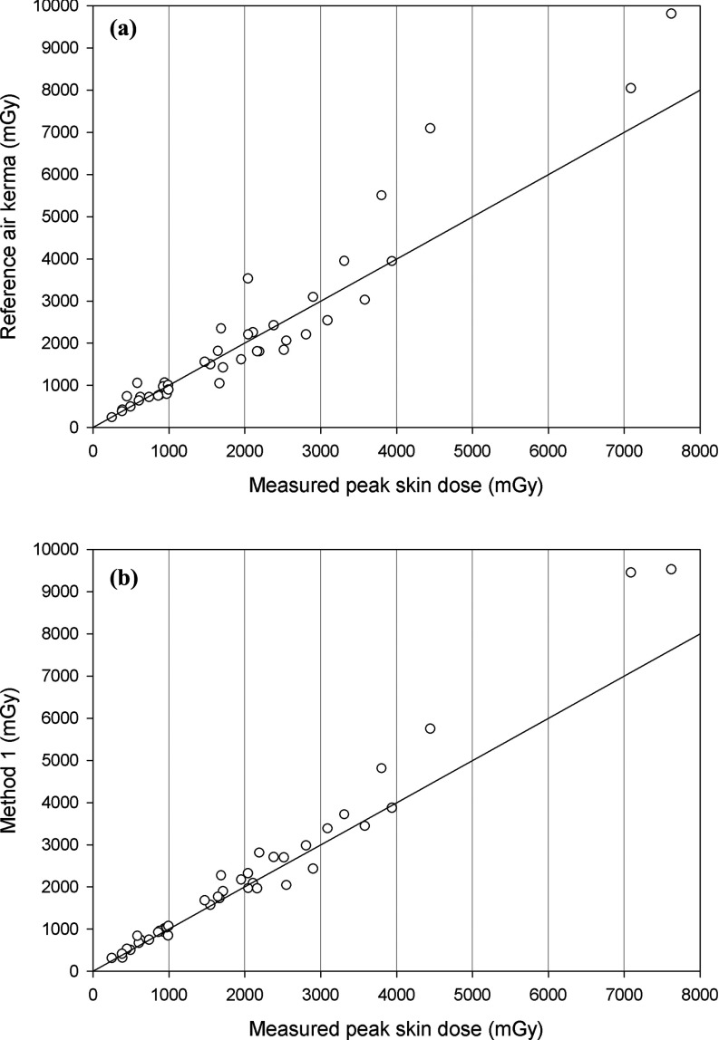
Figure 1 plots reference air kerma versus measured PSD and Method 1 versus measured PSD. Both reference air kerma and Method 1 demonstrated a strong positive correlation (Pearson's correlation coefficient [ρ] = 0.959 and ρ = 0.987, respectively) with measured PSD. Figure 2 plots the percent differences from measured PSD for reference air kerma and each calculation method for all cases. The variance at Site A and Site B as estimated by a mixed-effects linear model with heterogeneous variance structure was not significantly different (P = 0.402, Hartley's test). A mixed-effects linear model indicated that there was no significant interaction between Site and calculation method, and there was a significant effect due to calculation method (P = 0.0411). Tukey-Kramer pairwise comparisons indicated no significant difference between measured PSD and reference air kerma, and no significant difference between reference air kerma and Method 1. However, there was a significant difference between measured PSD and Method 1 (P = 0.0327), with Method 1 resulting in calculated doses that were significantly higher than measured PSD.
FIG. 1.
(a) Reference air kerma versus measured peak skin dose, (b) Method 1 versus measured peak skin dose. Identity lines are plotted for reference.
FIG. 2.
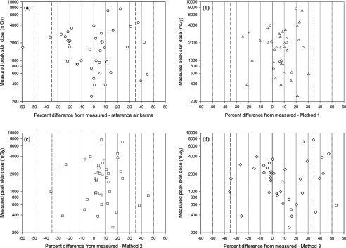
Percent difference between measured peak skin dose and (a) reference air kerma; (b) Method 1; (c) Method 2; and (d) Method 3. See Table I for descriptions of the different calculation methods.
There were no cases at either site for which the calculated PSD using Method 1 differed from the measured PSD by 35% or more (35% was chosen as it is the regulatory limit for accuracy of reference air kerma display on fluoroscopes21), the largest difference was +30.4% [Fig. 2(b)]. There were six cases (14.6% of cases), one at Site A and five at Site B, for which the difference between reference air kerma and measured PSD exceeded 35%. There was one case (2.4% of cases) at Site B for which the difference between reference air kerma and measured dose exceeded 50% (−59.3%). A mixed-effects linear model indicated that calculated PSD using Methods 1, 2, and 3 were not significantly different (P = 0.191). The averages of the absolute value of the percent differences from the measured PSD for the indirect metrics for all cases were 16.9% (reference air kerma), 11.8% (Method 1), 12.6% (Method 2), and 17.3% (Method 3). In general, Methods 1 and 2 were more accurate that reference air kerma and Method 3, albeit not significantly so, and less likely to result in large differences from measured PSD.
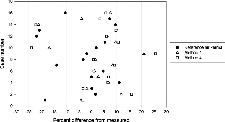
Figure 3 plots reference air kerma, Method 1, and Method 4 for the 16 cases from Site A for which the RDSR was available. A mixed-effects linear model indicated no significant differences (P = 0.184) in accuracy between reference air kerma, Method 1, and Method 4.
FIG. 3.
Comparison of Method 1 (standard method, Table I) and Method 4 (using RDSR, Table I) from Site A. Reference air kerma plotted for reference.
IV. DISCUSSION
Figures 1 and 2 demonstrate that both reference air kerma and Method 1 were more likely to overestimate than underestimate the measured PSD, particularly at higher doses. The most likely causes of this positive bias were the calibrations of the reference air kerma displays on the fluoroscopes used to perform the procedures studied, and uncertainties related to film dosimetry. A review of historical quality control records revealed that the percent difference in the displayed reference air kerma compared to the measured reference air kerma ranged from −5% to +20% across the range of possible operating conditions (e.g., field of view, pulse rate, mode of operation, patient thickness) of the fluoroscopes. Uncertainties related to film dosimetry are discussed later in this section. Calculated PSD using Method 1 were significantly higher than measured PSD, but less likely to result in large differences (±35%) from measured PSD compared to reference air kerma. Also notable is that in only 1 case out of 41 embolization procedures did the reported reference air kerma differ from the measured PSD by more than 50%, and in no cases did Method 1 differ from the measured PSD by 35% or more. This is in contrast to the assertion that skin dosimetry is unlikely to be more accurate than ±50%.1 This also implies that assigning a patient experiencing a skin dose exceeding the recommended substantial radiation dose level (SRDL) of 5 Gy (Ref. 22) to the correct dose “band” in Table I of Ref. 1 is possible in most cases.
The use of the more detailed information contained in the RDSR (Method 4) did not significantly improve the accuracy of the calculated PSD, and it can be seen from Fig. 3 that the differences between Methods 1 and 4 were usually small.
IV.A. Factors contributing to large differences
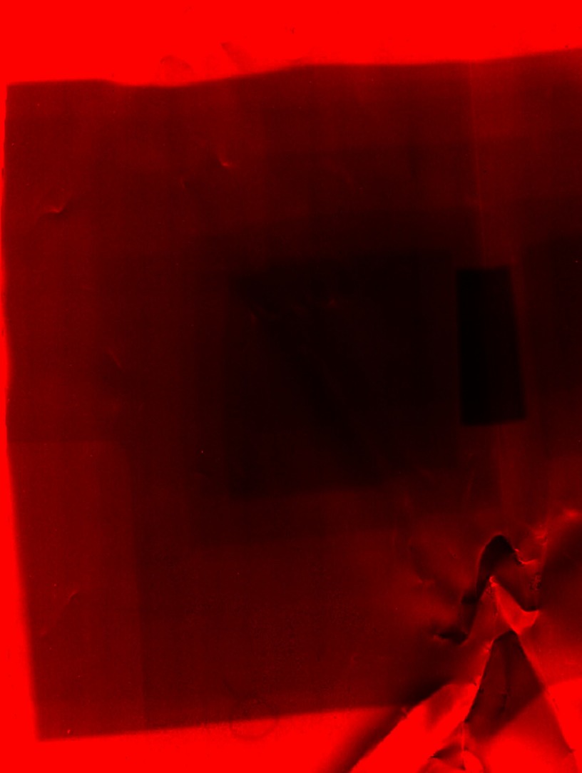
An analysis of cases for which differences between either reference air kerma or Method 1 and measured PSD exceeded 35% revealed two key factors that contributed to these differences. These factors were unusually small or large source-to-patient distances and the use of cone beam CT when only a few acquisition series were acquired. In the two cases from Site B for which reference air kerma underestimated the measured PSD by more than 35%, the source-to-patient distance ranged from 544 to 579 mm from the x-ray source, resulting in PSD that were much higher than indicated by reference air kerma alone. These are source-to-patient distances that would more commonly be seen for interventional cardiology procedures, a source-to-patient distance of 635 mm corresponds to the skin surface of the patient being located at the IRP. For these two cases, Method 1, which accounted for the source-to-patient distance, was within 10% of the measured PSD. In the two cases from Site B for which reference air kerma overestimated the measured PSD by more than 35%, the source-to-patient distance ranged from 781 to 787 mm, resulting in PSD that were much lower than indicated by reference air kerma. A source-to-patient distance of 785 mm corresponds to the skin surface of the patient being located at isocenter, 150 mm further from the x-ray source than the IRP. Method 1 improved the accuracy for both cases, however, for one case Method 1 was still 30.4% higher than the measured PSD. A review of the data and film from this case revealed that a very small x-ray field (73 cm2) was used for fluoroscopy, which contributed 75% of the total reference air kerma to the case. This fluoroscopy was performed at a different primary gantry angle than the bulk of the acquisition imaging, resulting in multiple distinct irradiation sites on the film (Fig. 4). Therefore, both reference air kerma and Method 1 overestimated the measured PSD, as Method 1 did not account for radiation dose being distributed between multiple distinct skin sites, which seldom occurs in these types of procedures. In the one case from Site A for which reference air kerma overestimated the measured PSD by more than 35%, one cone beam CT acquisition contributed 20% of the total reference air kerma to the procedure, and when combined, cone beam CT and acquisition series acquired at primary angles greater than 30° contributed 29% of the total reference air kerma for the procedure. The source-to-patient distance also ranged from 739 to 750 mm during most of the procedure. These two factors combined to cause the mismatch between reference air kerma and measured PSD. Method 1, which accounted for source-to-patient distance, reduced the difference to 15.3%.
FIG. 4.
Red channel from scanned radiochromic film for case B17, demonstrating multiple distinct x-ray fields.
IV.B. Uncertainties associated with film dosimetry
As anyone who has attempted film dosimetry can attest, there are numerous potential pitfalls and sources of error associated with the process. Therefore, it is prudent to re-examine the results presented here in terms of the ranges of actual PSD that might be expected based on the uncertainties inherent in film dosimetry.
McCabe et al. have published an authoritative review of radiochromic film calibration for dosimetry in medical imaging.16 Understanding that several of their recommendations are impractical for the average user of radiochromic film to implement, they included in this review a discussion of the types of uncertainty that are to be expected, their associated magnitudes, and a quadratic sum of the individual sources of uncertainty. These sources of uncertainty include scanner nonuniformity, which depends on the level of film darkening; the influence of backscatter on calibration; and film-to-film variation in response. The energy dependence of the response of radiochromic film is another potential source of uncertainty. Reports of the energy dependence of Gafchromic film are consistent, with earlier generations reported to have energy dependence from 0% to 5% from 80 to 125 kVp,15,23,24 and a slightly lower response (7%) at 60 kVp.23 McCabe et al. reported an energy dependence for orange-facing XR-RV3 film of approximately 5% from 100 to 120 kVp (half-value layer of 4.98–6.96 mm Al), with the energy dependence decreasing slightly as air kerma increased. McCabe et al. also reported an 8% overprediction at 120 kVp by XR-RV3 film that is calibrated free-in-air and used in backscatter conditions, as done in this study. A final source of uncertainty is polarization effects, which result from the layered construction of Gafchromic film and the nature of the light used to scan the film. Butson et al. reported a maximum uncertainty of 3% for XRQA film exposed to 2000 mGy and scanned with an Epson V700 scanner.25 Table II lists these sources of uncertainty and presents limits for the measured PSD in this study, which can be considered a “worst-case” scenario.
TABLE II.
Sources of uncertainty in Gafchromic film dosimetry and their associated magnitudes.
| Uncertainty source | Magnitude (%) |
|---|---|
| Calibration uncertainty (95% CIa) (Ref. 16) | ±6.4 |
| Polarization effects (Ref. 25) | ±3 |
| Energy dependence (Ref. 16) | ±5 |
| Free-in-air calibration used in backscatter conditions (Ref. 16) | −8 |
| Scanner nonuniformity (Ref. 16) | ±5 |
| Limits on measured peak skin dose in this studyb | [−12.8, 10.0] |
CI = Confidence interval.
Quadratic sum of individual uncertainty sources.
The results of this study were largely unchanged when the limits presented in Table II were applied to the data (Fig. 5). There were still no procedures for which either the lower or upper bounds of the percent difference between Method 1 and measured PSD was 50% or more. For two additional procedures (total 3 of 41, 7.3% of cases), the limits of reference air kerma differed from measured PSD by 50% or more. Both were procedures that were discussed previously in Sec. IV A. The limits of PSD calculated using Method 2 differed from the measured PSD by more than 50% for one procedure. A review of the data from this case revealed that four acquisition series were acquired, with patient table heights ranging from 629 to 870 mm. The use of the patient table height for the last acquisition series, equal to 629 mm, as the source-to-patient distance for the entire procedure led to the large difference. The percent difference for the limits of PSD calculated using Method 3 exceeded 50% for three additional cases (total 4 of 41, 9.8% of cases).
FIG. 5.
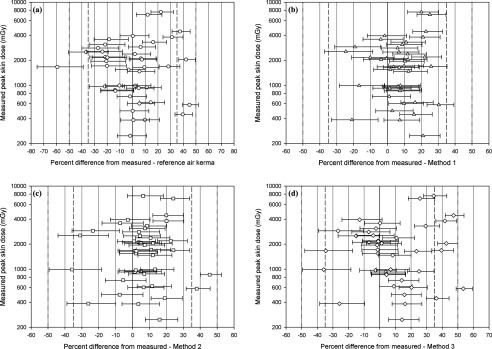
Impact of Gafchromic film calibration and measurement uncertainties on results. Percent difference between measured peak skin dose and (a) reference air kerma; (b) Method 1; (c) Method 2; and (d) Method 3. See Table I for descriptions of the different calculation methods. The horizontal error bars represent the limits calculated in Table II [−12.8%, 10.0%]. Note the different x-axis scale in part a compared to parts (b), (c), and (d).
The results of this study indicate that while reference air kerma is in most cases within ±50% of the actual PSD, there is additional value to be gained by performing a PSD reconstruction for embolization procedures. The “hybrid” simplified method using the patient table height for the last acquisition series (Method 2) matched the measured PSD to within ±50% in most cases. It is reasonable to use a single backscatter factor for all embolization procedures performed using modern C-arm fluoroscopes, as the backscatter factor varies linearly within a relatively narrow range for these procedures, by approximately 15% from 1.3 to 1.5. The source-to-patient distance, on the other hand, can vary from approximately 550 to 785 mm, and the associated quadratic inverse square law correction can span a range of 100% or more. The impact of this variation in source-to-patient distance is observed in the differences between Methods 2 and 3 (Fig. 2).
These results can be extended to other procedures using similar geometries, e.g., vascular interventions and other interventional oncology procedures. The reference air kerma alone is a reasonably accurate indicator of the true PSD for these types of procedures. Reported reference air kerma levels can be used without modification to set notification levels and SRDL for these procedures, provided the calibration of the displayed reference air kerma is accurate. For example, based on a fit to the plot of reference air kerma versus measured PSD from Site A, a reference air kerma of 4977 mGy would correspond to a PSD of 5000 mGy. One additional step that may be required, both when setting notification levels or SRDL and calculating PSD, is to apply a measured single-point correction factor to the displayed reference air kerma. This would be required in labs where the reported reference air kerma is known to differ from the true reference air kerma by a substantial amount, e.g., ±20% or more. Modern fluoroscopes and KAP meters are capable of reporting reference air kerma more accurately than currently required by regulation (±35%),21 however, some fluoroscopes still report reference air kerma values that are close to the regulatory accuracy limits. We recommend that the accuracy of the displayed reference air kerma be verified at acceptance testing and on a regular basis afterwards. All of the reference air kerma displays used in this study were verified on an annual basis to be accurate to within ±20%. Because the PSD can be estimated accurately using easily accessible data, film dosimetry is likely unnecessary for most vascular interventions and interventional oncology procedures when indirect dose metrics are available.
There are situations where a more detailed PSD reconstruction may still be indicated. These include cases where steep gantry angles are used for a substantial fraction of the case, and cases where an unexpected skin reaction occurs given the reported reference air kerma. However, accurately estimating or measuring PSD can be difficult when steep gantry angles are used. When attempting to measure PSD, the limited area of the film may not intercept the x-ray beam from certain angles. Indirect estimation of PSD can account for irradiation from any angle, however, it can be difficult to accurately determine if individual radiation fields overlap on the skin of the patient. Doing so requires an accurate model of the patient that is correctly registered to an accurate model of the fluoroscopic system. Future developments, including automated systems that calculate and monitor PSD in real-time, may address this problem, provided they handle the procedural and patient geometry well.
Limitations of this study included the uncertainty in film calibration and dose measurement, analyzed previously; the exclusion of certain contributions to reference air kerma from dose calculations, including fluoroscopy or acquisition performed at primary gantry angles greater than 60° and cone beam CT; and the limited range of air kerma to which the film was exposed during calibration. The first two are usually related, as fluoroscopy at large gantry angles is often performed to position the patient for a cone beam CT acquisition. These exclusions seldom impact the accuracy of calculated PSD, as demonstrated in this study. However, the accuracy of calculated PSD is likely to be affected for procedures for which a variety of large gantry angles are routinely used, including interventional cardiology procedures. It is for these types of procedures that real-time calculation software is likely to offer the largest benefit. Failure to account for the distribution of dose on the skin as the gantry is rotated and small x-ray fields are used can lead to an overestimation of skin dose and perhaps early termination of a procedure, potentially exposing the patient to risks associated with a repeated procedure and/or risks related to incomplete therapy. In two procedures at Site B, the measured film doses, 7.6 and 7.1 Gy, exceeded the maximum air kerma to which the film was exposed during calibration, 6.0 Gy. We refitted the calibration data from Site B after deleting the measurement at 6.0 Gy, then calculated the dose for this point using the measured film darkening. The calculated dose was 5.8 Gy, a difference of −3%. We concluded that the measured PSD for the two aforementioned procedures most likely slightly underestimated the true PSD.
V. CONCLUSIONS
The results of this study have demonstrated that it is possible to determine PSD using indirect methods with better than 50% accuracy for most, if not all, vascular and interventional oncology procedures. A corollary to these results is that in most cases, film dosimetry is unnecessary when indirect dose metrics are available. A simple “hybrid” method using the last source-to-patient distance recorded is usually sufficiently accurate. In most cases, reference air kerma alone is well within ±50%, and can be used without modification to set notification levels and substantial radiation dose levels, provided the accuracy of the displayed reference air kerma has been verified. If the calculated peak skin dose is greater than or equal to 5 Gy, patient follow-up should be initiated.22,26 The results presented here likely do not apply to procedures performed using different geometries, such as interventional cardiology and neuroradiology procedures, and these procedures must be studied separately.
ACKNOWLEDGMENTS
The authors would like to thank the technologists in their respective interventional radiology departments for their assistance in handling the films. MD Anderson Cancer Center is supported by Cancer Center Support Grant No. P30CA016672.
REFERENCES
- 1.Balter S., Hopewell J. W., Miller D. L., Wagner L. K., and Zelefsky M. J., “Fluoroscopically guided interventional procedures: A review of radiation effects on patients’ skin and hair,” Radiology 254, 326–341 (2010). 10.1148/radiol.2542082312 [DOI] [PubMed] [Google Scholar]
- 2. National Cancer Institute, Cancer Therapy Evaluation Program (available URL: http://ctep.cancer.gov/protocolDevelopment/electronic_applications/ctc.htm). Last accessed April 2014.
- 3.Balter S., “Methods for measuring fluoroscopic skin dose,” Pediatr. Radiol. 36(Suppl 2), 136–140 (2006). 10.1007/s00247-006-0193-3 [DOI] [PMC free article] [PubMed] [Google Scholar]
- 4.Panuccio G., Greenberg R. K., Wunderle K., Mastracci T. M., Eagleton M. G., and Davros W., “Comparison of indirect radiation dose estimates with directly measured radiation dose for patients and operators during complex endovascular procedures,” J. Vasc. Surg. 53, 885–894 (2011). 10.1016/j.jvs.2010.10.106 [DOI] [PubMed] [Google Scholar]
- 5.Sukupova L., Novak L., Kala P., Cervinka P., and Stasek J., “Patient skin dosimetry in interventional cardiology in the Czech Republic,” Radiat. Prot. Dosim. 147, 106–110 (2011). 10.1093/rpd/ncr284 [DOI] [PubMed] [Google Scholar]
- 6.Tsai H.-Y., Lai P.-L., Li Y.-Y., and Tyan Y.-S., “Establishing an individual dosing system for patients undergoing interventional transcatheter arterial embolization: Radiochromic film and Monte Carlo simulation,” Radiat. Meas. 46, 2107–2110 (2011). 10.1016/j.radmeas.2011.10.016 [DOI] [Google Scholar]
- 7.Herron B., Strain J., Fagan T., Wright L., and Shockley H., “X-ray dose from pediatric cardiac catheterization: A comparison of materials and methods for measurement or calculation,” Pediatr. Cardiol. 31, 1157–1161 (2010). 10.1007/s00246-010-9770-1 [DOI] [PubMed] [Google Scholar]
- 8.Zontar D., Kuhelj D., Skrk D., and Zdesar U., “Patient peak skin doses from cardiac interventional procedures,” Radiat. Prot. Dosim. 139, 262–265 (2010). 10.1093/rpd/ncq013 [DOI] [PubMed] [Google Scholar]
- 9.Bor D., Olgar T., Toklu T., Caglan A., Onal E., and Padovani R., “Patient doses and dosimetric evaluations in interventional cardiology,” Phys. Med. 25, 31–42 (2009). 10.1016/j.ejmp.2008.03.002 [DOI] [PubMed] [Google Scholar]
- 10.Domienik J., Papierz S., Jankowski J., Peruga J. Z., Werduch A., and Religa W., “Correlation of patient maximum skin doses in cardiac procedures with various dose indicators,” Radiat. Prot. Dosim. 132, 18–24 (2008). 10.1093/rpd/ncn247 [DOI] [PubMed] [Google Scholar]
- 11.Kwon D., Little M. P., and Miller D. L., “Reference air kerma and kerma-area product as estimators of peak skin dose for fluoroscopically guided interventions,” Med. Phys. 38, 4196–4204 (2011). 10.1118/1.3590358 [DOI] [PMC free article] [PubMed] [Google Scholar]
- 12.Jones A. K. and Pasciak A. S., “Calculating the peak skin dose resulting from fluoroscopically guided interventions. Part I. Methods,” J. Appl. Clin. Med. Phys. 12, 231–244 (2011). 10.1120/jacmp.v12i4.3670 [DOI] [PMC free article] [PubMed] [Google Scholar]
- 13.Jones A. K. and Pasciak A. S., “Calculating the peak skin dose resulting from fluoroscopically-guided interventions. Part II. Case studies,” J. Appl. Clin. Med. Phys. 13, 174–186 (2012). 10.1120/jacmp.v13i1.3693 [DOI] [PMC free article] [PubMed] [Google Scholar]
- 14. National Electrical Manufacturer's Association, Digital Imaging and Communications in Medicine (DICOM) Supplement 94: Diagnostic X-Ray Radiation Dose Reporting (Dose SR) (NEMA, Rosslyn, VA, 2005) (available URL: ftp://medical.nema.org/medical/dicom/final/sup94_ft.pdf). Last accessed April 2014. [Google Scholar]
- 15.Giles E. R. and Murphy P. H., “Measuring skin dose with radiochromic dosimetry film in the cardiac catheterization laboratory,” Health Phys. 82, 875–880 (2002). 10.1097/00004032-200206000-00017 [DOI] [PubMed] [Google Scholar]
- 16.McCabe B. P., Speidel M. A., Pike T. L., and Van Lysel M. S., “Calibration of GafChromic XR-RV3 radiochromic film for skin dose measurement using standardized x-ray spectra and a commercial flatbed scanner,” Med. Phys. 38, 1919–1930 (2011). 10.1118/1.3560422 [DOI] [PMC free article] [PubMed] [Google Scholar]
- 17.Rodbard D., Munson P., and De Lean A., “Improved curve-fitting, parallelism testing, characterization of sensitivity and specificity, validation, and optimization for radioligand assay,” in Radioimmunoassay and Related Procedures in Medicine (International Atomic Energy Agency, Vienna, Austria, 1978), pp. 469–504 [Google Scholar]
- 18. National Institutes of Health, ImageJ Documentation (available URL: http://imagej.sourceforge.net/docs/menus/analyze.html). Last accessed April 2014.
- 19. National Electrical Manufacturer's Association, Digital Imaging and Communications in Medicine (DICOM) Base Standard PS 3.3: Information Object Definitions (NEMA, Rosslyn, VA, 2011) (available URL: ftp://medical.nema.org/medical/dicom/2011/11_03pu.pdf). Last accessed April 2014. [Google Scholar]
- 20.Petoussi-Henss N., Zankl M., Drexler G., Panzer W., and Regulla D., “Calculation of backscatter factors for diagnostic radiology using Monte Carlo methods,” Phys. Med. Biol. 43, 2237–2250 (1998). 10.1088/0031-9155/43/8/017 [DOI] [PubMed] [Google Scholar]
- 21. United States Food and Drug Administration, 21 CFR Part 1020 - Electronic Products; Performance Standard for Diagnostic X-Ray Systems and Their Major Components; Final Rule,2006. [PubMed]
- 22. National Council on Radiation Protection and Measurements, Radiation Dose Management for Fluoroscopically-Guided Interventional Medical Procedures, NCRP Report No. 168 (NCRP, Bethesda, MD, 2011).
- 23.Butson M. J., Yu P. K. N., Cheung T., and Metcalfe P., “Radiochromic film for medical radiation dosimetry,” Mat. Sci. Eng. R 41, 61–120 (2003). 10.1016/S0927-796X(03)00034-2 [DOI] [Google Scholar]
- 24.Delle Canne S., Carosi A., Bufacchi A., Malatesta T., Capperella R., Fragomeni R., Adorante N., Bianchi S., and Begnozzi L., “Use of GAFCHROMIC XR type R films for skin-dose measurements in interventional radiology: Validation of a dosimetric procedure on a sample of patients undergone interventional cardiology,” Phys. Med. 22, 105–110 (2006). 10.1016/S1120-1797(06)80004-9 [DOI] [PubMed] [Google Scholar]
- 25.Alnawaf H., Butson M. J., Cheung T., and Yu P. K. N., “Scanning orientation and polarization effects for XRQA radiochromic film,” Phys. Med. 26, 216–219 (2010). 10.1016/j.ejmp.2010.01.003 [DOI] [PubMed] [Google Scholar]
- 26.Steele J. R., Jones A. K., and Ninan E. P., “Quality intiatives: Establishing an interventional radiology patient radiation safety program,” Radiographics 32, 277–287 (2012). 10.1148/rg.321115002 [DOI] [PubMed] [Google Scholar]


