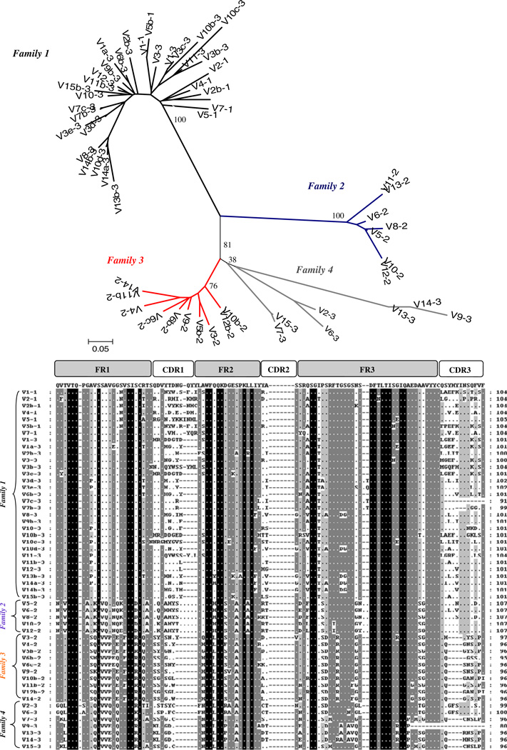
Fig. 5.
Comparisons of medaka genomic VL segments. The upper por-tion of this diagram depicts an unrooted phylogenetic tree based on amino acid sequences. Both the phylogenetic tree and corresponding alignment (lower portion of diagram agree with a classification of medaka VL into four different families). Alignments were performed and scored based on recommendations of the IMGT (http://imgt.cines.fr)

