
Figure 3.
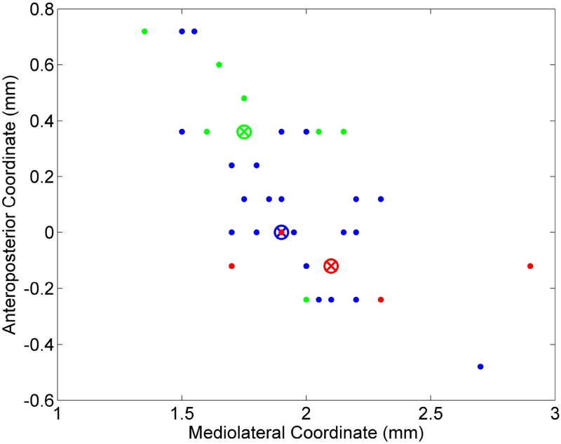
Microwire positions of recorded slow phasic neurons within the right hemisphere VP. X-axis refers to mediolateral position and Y-axis refers to anteroposterior position. Green dots indicate early reversal pattern, blue dots indicate progressive reversal pattern, and red dots indicate late reversal pattern without regard to directionality. Colored circles with X inside indicate k-means centroid of their respective reversal clusters. Each centroid represents the geometric median of the points in that cluster.