Abstract
Background
Treadmill gait analysis was more advantageous than over-ground walking because it allowed continuous measurements of the gait parameters. The purpose of this study was to investigate the concurrent validity and the test-retest reliability of the OPTOGait photoelectric cell system against the treadmill-based gait analysis system by assessing spatio-temporal gait parameters.
Material/Methods
Twenty-six stroke patients and 18 healthy adults were asked to walk on the treadmill at their preferred speed. The concurrent validity was assessed by comparing data obtained from the 2 systems, and the test-retest reliability was determined by comparing data obtained from the 1st and the 2nd session of the OPTOGait system.
Results
The concurrent validity, identified by the intra-class correlation coefficients (ICC [2, 1]), coefficients of variation (CVME), and 95% limits of agreement (LOA) for the spatial-temporal gait parameters, were excellent but the temporal parameters expressed as a percentage of the gait cycle were poor. The test-retest reliability of the OPTOGait System, identified by ICC (3, 1), CVME, 95% LOA, standard error of measurement (SEM), and minimum detectable change (MDC95%) for the spatio-temporal gait parameters, was high.
Conclusions
These findings indicated that the treadmill-based OPTOGait System had strong concurrent validity and test-retest reliability. This portable system could be useful for clinical assessments.
MeSH Keywords: Exercise Test, Gait, Spatio-Temporal Analysis, Walking
Background
Gait analysis equipment using a computer has been used increasingly by clinicians to obtain objective and accurate measurements of spatio-temporal gait patterns [1,2]. In particular, the advent of modern and portable instrument walkway systems induced rapid determination of the spatio-temporal gait parameters during over-ground walking [3–5]. On the other hand, assessing the gait pattern during over-ground walking is limited by the instrumented walkway length and limited working space.
Treadmill gait analysis has been considered more advantageous than over-ground walking because it required less space and allowed continuous measurements of the gait parameters. Moreover, intervention effects could be compared under equivalent conditions by the constant walking speed at pre- and post-test [6–8]. Treadmill walking encourages repetitive and intensive gait training and facilitates a normal gait pattern. Therefore, it is considered a viable intervention for treating gait impairments associated with neurological disorders such as Parkinson’s disease [9–11] and after stroke [12–14].
Instrumented treadmills could be good measurement tools available to clinicians for monitoring the progress of training, and quantifying spatial and temporal gait parameters [15,16]. For example, Kiss reported a strong correlation (r, 0.948–0.974) between instrumented treadmills and ultrasound-based measuring system [15]. Wearing et al. reported a high correlation (r, 0.79–0.95) between the 2 measuring systems (instrumented treadmill vs. conventional instrumented walkway) despite systematic differences [17]. One study using an instrument treadmill with healthy seniors reported high test-retest reliability [18].
The OPTOGait system, which uses high-density photoelectric cells, was recently introduced. The system allows quantification of spatial and temporal gait parameters on essentially all flat surfaces. The OPTOGait system uses transmitting and receiving bars placed parallel to each other. When a subject passes between the transmitting bar and the receiving bar, the system detects any interruption in the light signal due to the presence of feet within the recording area, and automatically calculates the spatial and temporal gait parameters. The method has high concurrent validity and test-retest reliability compared to those of conventional instrumented walkway systems for over-ground walking [19]. On the other hand, despite the availability in a clinical setting, the concurrent validity and reliability of treadmill-based gait analysis are unknown.
This study examined the concurrent validity and test-retest reliability of the OPTOGait system against the instrumented treadmill system by assessing the spatial and temporal gait parameters of stroke patients and healthy adults measured while walking at a comfortable speed.
Material and Methods
Subjects
Twenty-six stroke patients (age 65±4.6 years; 34 females, 20 males; height 164.9±5.8 cm; weight 64.3±8.4 kg) and 18 healthy young adults (age 28±.35 years; 9 females, 8 males; height 170.9±8.8 cm; weight 62.3±12.4 kg) were enrolled in this study. The stroke patients could walk independently on a treadmill for 10 min without gait aids or orthosis. The potential candidates needed to understand and act in accordance with simple spoken instructions. The healthy young adults had no medical history of cardiovascular, neurological, or orthopedic problems likely to affect their ability to walk on a treadmill. All protocols and procedures were approved by the Institutional Review Board of Sahmyook University and all subjects signed a statement of informed consent.
Instruments
The spatial and temporal gait parameters were collected using 2 commercially available systems: the OPTOGait photoelectric cells system (OPTOGait, Microgate S.r.I, Italy, 2010), and the Zebris instrumented gait analysis system (Zebris Medical GmbH, FDM-T system, Isny, Germany). The OPTOGait system used in this study consisted of a single transmitting bar and receiving bar positioned on the side bars of a treadmill (APSUN Inc, Korea) (Figure 1). Each single bar was 100×8 cm and contained 96 light diodes that were located 3 mm above floor level and approximately 1 cm apart. The data was extracted at 1000Hz and saved in a PC using OPTOGait Version 1.6.4.0 software (Microgate S.r.I, Italy).
Figure 1.
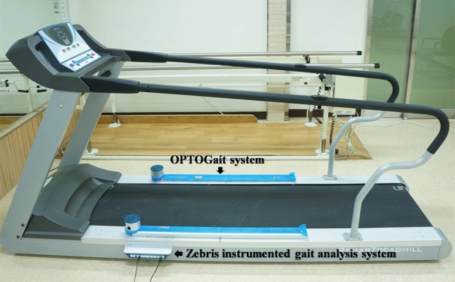
Lateral view of the 2 measuring systems. The transmitting bar and receiving bar of the OPTOGait system were placed on the side bars of the Zebris treadmill.
The Zebris instrumented gait analysis system consisted of a capacitance-based foot pressure platform housed within a treadmill. The pressure platform had a sensing area of 112×49 cm and incorporated 3432 sensors. The treadmill had a contact surface of 210×70 cm, and its speed could be adjusted between 0.1 and 6 km/h at 0.1 km/h intervals. The data were obtained at a sampling rate of 100 Hz and saved in a PC using Win FDM-T Version 2.5.1 software (Zebris Medical GmbH, Germany).
Procedure
Two researchers were responsible for system software and collecting data. General information, such as height and weight, was recorded. The subjects participated in the experiment wearing light-weight, comfortable clothing. The subjects were instructed to walk barefoot on the treadmill at their preferred walking speed. Prior to gait analysis, the subjects were allowed 10 min to become acquainted with treadmill walking [20]. During the acclimatization session, the individual’s preferred walking speed was measured and entered into the OPTOGait software. The participants were asked to walk on the treadmill again, and the treadmill speed was adjusted to match the self-selected walking speed determined during the acclimatization session. The spatio-temporal gait parameters were measured by placing the OPTOGait system on the Zebris treadmill system and operating both simultaneously. Once the participants were comfortable, a 60-s data capture period was used. Testing was repeated 30 min after the initial trial to evaluate the test-retest reliability.
Statistical analysis
The spatio-temporal variables analyzed included the speed, cadence, gait cycle, step length, step time, stride length, duration of single and double limb support, and swing and stance phase duration. Statistical analysis was conducted using SPSS version 19.0 (SPSS Inc., Chicago, IL, USA). All measurements are expressed as the mean and standard deviation (SD). The differences between measurements systems with respect to the gait parameters were determined using a paired t-test. Paired t-tests were performed to determine the mean difference in the measurements for each gait variable recorded using the OPTOGait and Zebris treadmill system. The level of agreement between the OPTOGait and Zebris treadmill system was analyzed using the intra-class correlation coefficient (ICC [2, 1]) [21]. For absolute comparisons of the parameters obtained in the 2 sessions, the coefficients of variation of the method errors (CVME) [22] and the 95% limits of agreement (LOA) described by Bland and Altman were calculated [23]. The CVME values were converted to percentages by calculating the coefficients of variation of the method errors obtained using the standard deviations of the differences scores (Sd) between the results obtained using the 2 systems (ME=Sd/✓2, CVME=2ME/(X1+X2)×100%).
The differences between the first and second sessions with respect to the gait parameters were determined using a paired t-test. The test-retest reliabilities of the gait parameters measured using the OPTOGait system are expressed as ICC (3, 1), and were described by Shrout and Flessiss. The CVME and 95% LOA [23] were calculated for absolute comparisons of the parameters obtained during the 2 sessions. To estimate the absolute variability, the standard errors of measurement (SEMs) were calculated by measuring the range of errors for each gait parameter. The SEMs were calculated as (SD)×(1–ICC [2, 1])1/2 using the higher of the 2 SD measurements. For the convenience of interpretation, the SEMs are expressed as the percentage of the mean values (SEM%). In addition, the minimum detectable changes (MDC) at the confidence level of 95% were calculated to determine the smallest change needed to indicate a change as real and beyond the bounds of the measurement error. This was achieved by converting MDC to the percentage of the mean (MDC95%) after calculating it using the formula, ✓2×1.96×SEM [24]. The absolute variability data was analyzed using Microsoft Excel®. The statistical significance was set at p<0.05 for all procedures.
Results
Concurrent validity
Table 1 lists the mean and SD of the spatial and temporal gait parameters. A paired t-test was used to determine the systematic differences between the gait parameters obtained using the 2 systems. As a result, the step length and stride length of the OPTOGait system were significantly higher than those of the Zebris treadmill system in the stroke patients group. When expressed as a percentage of the gait cycle, the swing phase duration recorded by the OPTOGait system was significantly shorter than that of the Zebris treadmill system, whereas the stance phase of the OPTOGait system was significantly higher than that of the Zebris treadmill system. Furthermore, the period of the single limb support (SLS) phase of the OPTOGait system was significantly shorter than that of the Zebris treadmill system, whereas the total double limb support (TDLS) phase of the OPTOGait system was significantly greater than that of the Zebris treadmill system in both groups.
Table 1.
Mean (S.D.) and level of agreement of the gait parameters for subjects using the OPTOGait system and Zebris treadmill system.
| Gait parameters | Young subjects | Stroke patients | ||||||||
|---|---|---|---|---|---|---|---|---|---|---|
| OPTOGait | Zebris treadmill | ICC (95%CI) | CV% | 95%LOA | OPTOGait | Zebris treadmill | ICC (95%CI) | CV% | 95%LOA | |
| Speed (m/s) | 0.85 (0.08) | 0.85 (0.08) | 0.995 (0.990~0.998) | 0.63 | −0.012~0.014 | 0.71 (0.09) | 0.72 (0.08) | 0.908 (0.844~0.946) | 3.83 | −0.033~0.026 |
| Cadence (steps/m) | 94.93 (8.67) | 94.75 (8.75) | 0.997 (0.994~0.998) | 0.50 | −1.087~0.665 | 97.60 (10.98) | 97.63 (11.27) | 0.988 (0.980~0.993) | 1.23 | −1.987~1.820 |
| Gait cycle (s) | 1.28 (0.12) | 1.28 (0.12) | 0.999 (0.998~1.000) | 0.27 | −0.006~0.012 | 1.24 (0.14) | 1.25 (0.15) | 0.990 (0.983~0.995) | 1.15 | −0.023~0.039 |
| Lt. step (cm) | 53.42 (3.24) | 53.53 (3.33) | 0.958 (0.920~0.978) | 1.25 | −1.362~1.807 | 44.76 (5.73) | 44.50 (5.58) | 0.975 (0.957~0.986) | 1.99 | −2.542~1.772 |
| Rt. step (cm) | 53.66 (3.26) | 53.58 (3.25) | 0.932 (0.870~0.965) | 1.59 | −2.406~1.962 | 45.90 (5.18) | 45.53 (4.86) | 0.947 (0.910~0.969) | 2.42 | −3.193~1.943 |
| Lt. step time (s) | 0.64 (0.06) | 0.64 (0.06) | 0.990 (0.981~0.995) | 0.96 | −0.008~0.018 | 0.61 (0.08) | 0.62 (0.08) | 0.958 (0.928~0.976) | 2.67 | −0.019~0.027 |
| Rt. step time (s) | 0.64 (0.06) | 0.64 (0.06) | 0.985 (0.971~0.992) | 1.14 | −0.020~0.015 | 0.61 (0.08) | 0.62 (0.08) | 0.852 (0.756~0.912) | 5.01 | −0.012~0.027 |
| Stride (cm) | 106.97 (5.54) | 107.00 (6.12) | 0.969 (0.939~0.984) | 0.97 | −1.986~2.319 | 90.64 (9.01) | 89.98 (8.69) | 0.976 (0.959~0.986) | 1.51 | −5.017~2.575 |
| Lt. SLS (%) | 31.39 (1.69) | 34.82 (1.50)*** | 0.370 (0.052~0.620) | 3.83 | −0.165~7.143 | 29.79 (2.25) | 33.20 (2.00)*** | 0.297 (0.029~0.525) | 5.68 | −1.378~7.987 |
| Rt. SLS (%) | 31.44 (1.64) | 34.76 (1.36)*** | 0.238 (−0.099~0.526) | 3.92 | 0.026~7.074 | 29.63 (2.27) | 33.00 (2.39)*** | 0.098 (−0.177~0.359) | 7.07 | −2.811~9.386 |
| TDLS (%) | 37.19 (3.21) | 30.39 (2.60)*** | 0.310 (−0.016~0.577) | 7.18 | −13.975~−0.158 | 40.64 (3.91) | 33.80 (2.41)*** | −0.294 (−0.523~−0.026) | 10.05 | −16.894~3.571 |
| Lt. swing phase (%) | 31.44 (1.62) | 34.84 (1.44)*** | 0.243 (−0.088~0.526) | 4.02 | −0.047~7.147 | 29.98 (2.35) | 32.92 (2.31)*** | 0.042 (−0.232~0.309) | 7.20 | −3.596~9.061 |
| Rt. swing phase (%) | 31.38 (1.70) | 34.77 (1.43)*** | 0.378 (0.061~0.626) | 3.75 | −0.097~7.109 | 30.07 (2.32) | 33.31 (2.06)*** | 0.315 (0.048~0.539) | 5.78 | −1.358~8.206 |
| Lt. stance phase (%) | 68.56 (1.62) | 65.23 (1.37)*** | 0.239 (−0.093~0.523) | 1.96 | −7.165~0.077 | 70.02 (2.35) | 67.09 (2.32)*** | 0.041 (−0.232~0.308) | 3.32 | −9.063~3.605 |
| Rt. stance phase (%) | 68.36 (2.15) | 65.18 (1.49)*** | 0.468 (0.169~0.688) | 2.02 | −7.139~0.627 | 69.93 (2.32) | 66.68 (2.05)*** | 0.319 (0.053~0.543) | 2.66 | −8.000~1.344 |
ICC– intra correlation coefficient; CVME – coefficients of variation of method error; LOA – limits of agreement; SLS – single limb support; TDLS – total double limb support. Significant difference between the two measuring instruments (***, p≤0.001).
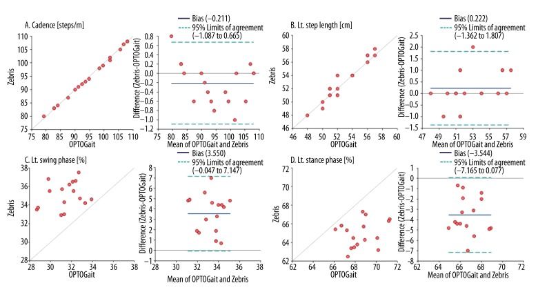
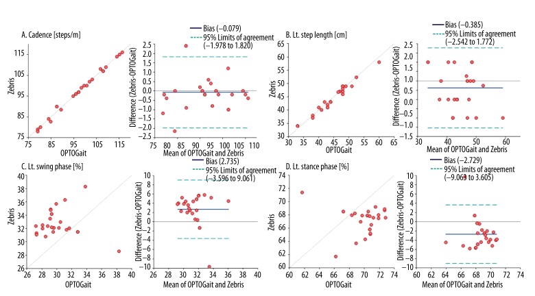
Table 1 lists the level of agreement between the OPTOGait and the Zebris treadmill system for each gait variable. In the young subjects, the ICCs for the spatial and temporal gait parameters were excellent (ICC [2, 1], 0.932~0.999) but the temporal parameters expressed as a percentage of the gait cycle were poor (ICC [2, 1], 0.238~0.468). The CVME values were small for all gait parameters (0.27~7.18%). At 95% LOA, the spatial and temporal gait parameters were distributed in a symmetrical manner but the temporal parameters expressed as a percentage of the gait cycle were skewed to 1 side, except for the TDLS. In the stroke patients group, the ICCs were excellent for speed, cadence, gait cycle, step length, step time, and stride length (ICC [2, 1], 0.852~0.990) and poor for the temporal parameters expressed as a percentage of the gait cycle (ICC [2, 1], 0.294~0.319). The CVME values were relatively small for all gait parameters (1.15~7.07%), except for TDLS% (10.05%). Scatter plots and Bland-Altman plots of the main spatial and temporal gait parameters provided by the OPTOGait system against the Zebris treadmill system are presented in Figures 2 and 3.
Figure 2.
Relationship between OPTOGait system and Zebris treadmill system for main spatio-temporal gait parameters in healthy young adults.
Figure 3.
Relationship between OPTOGait system and Zebris treadmill system for main spatio-temporal gait parameters in stroke patients.
Test-retest reliability
Tables 2 and 3 show the mean (SD) and test-retest reliability of the spatio-temporal gait parameters across the 2 measurement sessions obtained using the OPTOGait system in young and stroke subjects, respectively.
Table 2.
Mean (S.D.) and test-retest reliability of the gait parameters for the two measurement sessions in healthy young adults.
| Gait parameters | Session 1 | Session 2 | ICC (95% CI) | 95% LOA | CV % | SEM % | MDC % |
|---|---|---|---|---|---|---|---|
| Speed (m/s) | 0.84 (0.07) | 0.85 (0.07) | 0.978 (0.943–0.992) | −0.026~0.037 | 1.35 | 2.03 | 5.63 |
| Cadence (steps/m) | 95.13 (8.35) | 94.73 (9.21) | 0.962 (0.901–0.948) | −5.173~4.373 | 1.81 | 0.37 | 1.02 |
| Gait cycle (s) | 1.27 (0.11) | 1.27 (0.13) | 0.961 (0.900–0.985) | −0.057~0.073 | 1.84 | 0.40 | 1.11 |
| Lt. step (cm) | 53.44 (2.91) | 53.39 (3.42) | 0.805 (0.668–0.929) | −5.101~4.324 | 3.19 | 1.89 | 5.24 |
| Rt. step (cm) | 54.22 (3.84) | 54.55 (3.61) | 0.835 (0.613–0.935) | −3.865~4.532 | 2.78 | 1.17 | 3.23 |
| Lt. step time (s) | 0.64 (0.06) | 0.64 (0.07) | 0.920 (0.799–0.969) | −0.049~0.050 | 2.79 | 0.88 | 2.43 |
| Rt. step time (s) | 0.64 (0.07) | 0.64 (0.06) | 0.940 (0.848–0.977) | −0.050~0.043 | 2.58 | 0.66 | 1.82 |
| Stride (cm) | 106.28 (4.46) | 107.66 (6.49) | 0.812 (0.566–0.925) | −5.035~8.083 | 2.25 | 1.14 | 3.16 |
| Lt. SLS (%) | 31.31 (1.52) | 31.47 (1.89) | 0.873 (0.694–0.951) | −1.527~1.861 | 1.94 | 0.76 | 2.12 |
| Rt. SLS (%) | 31.15 (1.50) | 31.74 (1.71) | 0.857 (0.658–0.944) | −1.100~2.277 | 1.93 | 0.78 | 2.15 |
| TDLS (%) | 37.56 (2.93) | 36.81 (3.52) | 0.911 (0.778–0.966) | −3.430~1.930 | 2.60 | 0.84 | 2.34 |
| Lt. swing phase (%) | 31.17 (1.51) | 31.72 (1.72) | 0.855 (0.655–0.943) | −1.151~2.262 | 1.95 | 0.79 | 2.20 |
| Rt. swing phase (%) | 31.29 (1.53) | 31.46 (1.89) | 0.872 (0.691–0.950) | −1.548~1.870 | 1.96 | 0.77 | 2.14 |
| Lt. stance phase (%) | 68.84 (1.52) | 68.28 (1.71) | 0.856 (0.657–0.944) | −2.265~1.142 | 0.89 | 0.36 | 1.00 |
| Rt. stance phase (%) | 68.72 (1.52) | 68.28 (2.31) | 0.774 (0.492–0.909) | −3.018~2.140 | 1.35 | 0.76 | 2.11 |
ICC – intra correlation coefficient; LOA – limits of agreement; CVME – coefficients of variation of method error; SEM – standard error of measurement; MDC – minimum detectable change; SLS – single limb support; TDLS – total double limb support.
Table 3.
Mean (S.D.) and test-retest reliability of the gait parameters for the two measurement sessions in stroke patients.
| Gait parameters | Session 1 | Session 2 | ICC (95% CI) | 95% LOA | CV % | SEM % | MDC % |
|---|---|---|---|---|---|---|---|
| Speed (m/s) | 0.71 (0.10) | 0.71 (0.10) | 0.966 (0.926–0.985) | −0.049~0.051 | 2.97 | 0.47 | 1.31 |
| Cadence (steps/m) | 97.65 (11.36) | 97.56 (10.81) | 0.970 (0.935–0.986) | −5.408~5.224 | 2.27 | 0.35 | 0.97 |
| Gait cycle (s) | 1.23 (0.15) | 1.23 (0.14) | 0.962 (0.918–0.983) | −0.080~0.076 | 2.52 | 0.46 | 1.28 |
| Lt. step (cm) | 44.56 (5.76) | 44.96 (5.80) | 0.985 (0.968–0.996) | −1.537~2.344 | 1.63 | 0.19 | 0.54 |
| Rt. step (cm) | 45.99 (5.35) | 45.81 (5.11) | 0.929 (0.848–0.967) | −4.060~3.694 | 3.18 | 0.83 | 2.29 |
| Lt. step time (s) | 0.61 (0.07) | 0.61 (0.08) | 0.911 (0.812–0.959) | −0.067~0.066 | 3.54 | 1.17 | 3.24 |
| Rt. step time (s) | 0.61 (0.08) | 0.61 (0.08) | 0.926 (0.843–0.966) | −0.063~0.064 | 3.56 | 0.97 | 2.69 |
| Stride (cm) | 90.45 (9.28) | 90.83 (8.90) | 0.973 (0.941–0.988) | −3.750~4.520 | 1.86 | 0.28 | 0.77 |
| Lt. SLS (%) | 29.73 (2.34) | 29.85 (2.20) | 0.901 (0.792–0.954) | −1.855~2.117 | 2.61 | 0.78 | 2.16 |
| Rt. SLS (%) | 29.55 (2.26) | 29.71 (2.31) | 0.949 (0.890–0.977) | −1.279~1.586 | 1.78 | 0.40 | 1.10 |
| TDLS (%) | 40.74 (4.12) | 40.52 (3.89) | 0.957 (0.907–0.980) | −2.529~2.083 | 2.49 | 0.44 | 1.21 |
| Lt. swing phase (%) | 30.01 (2.23) | 30.12 (2.44) | 0.920 (0.830–0.963) | −1.730~1.940 | 2.21 | 0.65 | 1.80 |
| Rt. swing phase (%) | 29.91 (2.35) | 30.04 (2.39) | 0.854 (0.701–0.932) | −2.378~2.647 | 3.25 | 1.16 | 3.23 |
| Lt. stance phase (%) | 69.98 (2.23) | 69.87 (2.44) | 0.920 (0.830–0.963) | −1.940~1.730 | 0.97 | 0.28 | 0.77 |
| Rt. stance phase (%) | 70.09 (2.34) | 69.95 (2.39) | 0.854 (0.701–0.932) | −2.647~2.378 | 1.41 | 0.50 | 1.38 |
ICC – intra correlation coefficient; LOA – limits of agreement; CVME – coefficients of variation of method error; SEM – standard error of measurement; MDC – minimum detectable change; SLS – single limb support; TDLS – total double limb support.
The paired t-test indicated that there were no significant systematic differences in any gait parameters between the 2 sessions in each group. In the young subjects, the ICCs for all the gait parameters were excellent (ICC [3, 1], 0.7745~0.978). This was also reflected in the 95% LOA. All the gait parameters were distributed symmetrically. The CVME values were small for all gait parameters (0.89~3.19%). For the 2 sessions, all the parameters showed a low level of SEM (0.36~0.03%), indicating strong and absolute reliability. The MDC values (1.02~5.63%) for all gait parameters indicated a low level of variation between the 2 sessions (Table 2).
In the stroke patients group, the ICCs for all gait parameters were also excellent (ICC [3, 1], 0.854~0.985). At 95% LOA, all spatial and temporal gait parameters were distributed in a symmetrical manner. The CVME values were small for all gait parameters (0.97~3.56%). The SEM (0.19a~1.17%) and the MDC (0.54~3.24%) showed an absolutely strong reliability and a low level of variation between the 2 sessions, respectively (Table 3).
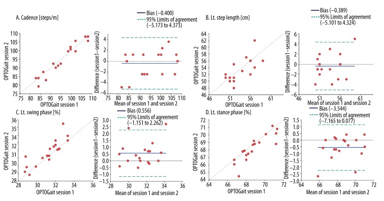
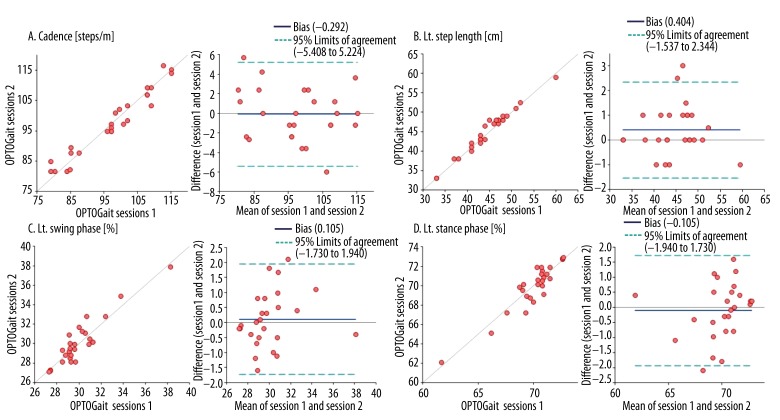
Scatter plots and Bland-Altman plots of the main spatial and temporal gait parameters provided by OPTOGait session 1 and session 2 are presented in Figures 4 and 5.
Figure 4.
Agreement for the main spatial and temporal gait parameters between session 1 and session 2 of OPTOGait system in healthy young adults.
Figure 5.
Agreement for the main spatial and temporal gait parameters between session 1 and session 2 of OPTOGait system in stroke patients.
Discussion
Treadmill-based gait analysis was available to clinicians for monitoring the progress of training and quantifying the spatial and temporal gait parameters. However, there were many restrictions (e.g., space, experimental process, price, and portability) on the use of the equipment. On the other hand, the OPTOGait photoelectric cell system was the equipment available in both over-ground based and treadmill-based gait analysis, but the agreement was not clear with other equipment. Therefore, we investigated the concurrent validity and the test-retest reliability of the OPTOGait photoelectric cell system against the treadmill-based gait analysis system by assessing the spatio-temporal gait parameters in healthy young adults and stroke patients.
Systematic differences were observed in the temporal parameter expressed as a percentage of the gait cycle measured by the 2 systems in both groups. In the healthy young adults groups, the SLS phase (Left: 3.43%, Right: 3.32%) and swing phase duration (Left: 3.4%, Right 3.39%) recorded by the OPTOGait were shorter than the Zebris treadmill system, whereas the TDLS phase (6.8%) and stance phase duration (Left: 3.33%, Right: 3.18%) were longer. Similarly, in the stroke patients group, the SLS phase (Left: 3.41%, Right: 3.37%) and swing phase duration (Left: 2.94%, Right: 3.24%) recorded by the OPTOGait system were shorter than the Zebris treadmill system, whereas the TDLS phase (6.84%) and stance phase duration (Left: 2.94%, Right: 3.25%) were longer. The temporal differences were attributed to the LED diodes of the OPTOGait system being raised 3 mm higher from the floor. Therefore, the sensing of heel contact occurred earlier, and the sensing of toe lift-off occurred later than in the Zebris treadmill system in the gait cycle. In addition, there was a 3-mm gap between the Zebris pressure sensors under the treadmill belt and the OPTOGait diodes sensor on the treadmill sidebars. Another reason for the temporal difference was the difference in the measuring principle. The OPTOGait system detects the blocked segment and the time interval by sensing interruptions of the progression of light between the photoelectric diodes, whereas the Zebris pressure sensors identify the precise pressure threshold induced by a load. These temporal differences were reported in a comparative study of the OPTOGait system and force plate for an assessment of the vertical jump height, and of the OPTOGait system and portable walkway system for floor-based gait analysis [25].
No significant differences in the spatial and temporal gait parameters (e.g., speed, cadence, gait cycle, step time, step length, and stride length) were observed. To assess the gait variables using the treadmill-based OPTOGait system, the walking speed must be entered into the software. In the present study, the walking speed was already calculated using the Zebris treadmill system during the acclimatization session and the same walking speed was also entered into the OPTOGait system.
The step length calculation process during treadmill walking was similar between the 2 systems. During continuous stepping on the treadmill belt at a constant speed, the OPTOGait system quantified the respective 1-dimensional data using the photoelectric cells, and the Zebris system was also quantified using a 1-dimensional ground reaction force measuring system. Measuring step length should be considered the anterio-posterior and the medio-lateral components according to the line of progression. Lienhard et al. reported that the weak point of a 1 dimensional gait evaluation could result in a substantial underestimation of the step length compared to the ground gait analysis systems that consider the line of progression, particularly in patients presenting large step widths [19]. However, both systems did not consider the medio-lateral component of the gait in the measuring principle, so they quantified the value of the only anterio-posterior component of the step length parallel to the bars regardless of the individual walking direction.
The temporal parameters expressed as a percentage of the gait cycle in both healthy young adult group (ICC <0.468) and stroke patient group (ICC <0.319) showed a low correlation between the 2 systems. The CV% values were relatively higher (1.96~7.18 in healthy young adult group, and 2.66~10.05 in stroke patient group), and the 95% LOA values were skewed to 1 side compared to the other spatial and temporal parameters, indicating the presence of a systematic difference. On the other hand, the spatial and temporal gait parameters, such as speed, cadence, gait cycle, step time, step length, and stride length in both the healthy young group (ICC >0.932) and the stroke patient group (ICC >0.852) showed a strong correlation between the 2 systems. The CV% values were relatively lower (<1.59% in the healthy young adult group, and <3.83% in the stroke patient group respectively), and the 95% LOA values (including zero) were within a narrow range, with a symmetric distribution. These findings showed excellent agreement between the 2 measuring systems, with little systematic bias.
This study revealed high test-retest reliability and consistency with respect to the derivation of the spatial and temporal gait parameters of the healthy young adults group (ICC >0.774) and the stroke patients group (ICC >0.854) using the OPTOGait system. For all parameters, the ICCs indicated excellent test-retest reliability. Moreover, the CV% values for all parameters were 0.89~2.79% and 0.97~3.56%, and the 95% LOA values (including zero) were within a narrow range, with a symmetric distribution. These findings indicated that only slight differences were as important as the relative reliability. SEM is a quantitative expression of the range of errors that occur whenever the same participant repeated certain tests [26]. In this study, SEM for the test-retest were converted to percentages of the mean values (SEM%), and showed a low measurement error (<2.03% in he healthy young adult group, and <1.17% in stroke patient group), indicating strong absolute reliability. The MDC was defined as the minimum change that could occur during the measurement, not due to accidental changes. Because it represents the degree of sensitivity to change, the MDC was needed to determine if the actual change occurred during the performance of the 2 sessions [27]. This value could be a criterion for assessing the changes in a performed process. The MDC values in this study were relatively low (<5.63% in the healthy young adult group, and <3.24% in the stroke patient group) when expressed as a percentage of the means. Therefore, the measurements were sufficiently sensitive to the changes, suggesting that all spatial and temporal parameters measured using the OPTOGait system could be useful for sensing the changes that occurred in the gait process.
Unlike the over-ground gait, the treadmill gait encouraged the symmetry of steps [28,29], allowed walking at a constant speed, and could collect many gait cycles and gait parameters. As a result of this study, the OPTOGait system and Zebris system showed excellent concurrent validity, and the treadmill-based OPTOGait had high test-retest reliability.
Conclusions
This study compared the spatio-temporal gait parameters measured using the treadmill-based OPTOGait photoelectric cells system and the Zebris instrumented gait analysis system and examined the test-retest reliability of the spatial and temporal gait parameters using the OPTOGait system during treadmill walking with healthy young adults and stroke patients. Before measuring gait parameters using treadmill-based equipment, clinicians should first understand the characteristics of both systems and use each system only under the appropriate circumstances.
Footnotes
Source of support: This study was supported by Sahmyook University
References
- 1.Steinwender G, Saraph V, Scheiber S, et al. Intrasubject repeatability of gait analysis data in normal and spastic children. Clin Biomech. 2000;15:134–39. doi: 10.1016/s0268-0033(99)00057-1. [DOI] [PubMed] [Google Scholar]
- 2.Maynard V, Bakheit AM, Oldham J, Freeman J. Intra-rater and inter-rater reliability of gait measurements with CODA mpx30 motion analysis system. Gait Posture. 2003;17:59–67. doi: 10.1016/s0966-6362(02)00051-6. [DOI] [PubMed] [Google Scholar]
- 3.Stokic DS, Horn TS, Ramshur JM, Chow JW. Agreement between temporospatial gait parameters of an electronic walkway and a motion capture system in healthy and chronic stroke populations. Am J Phys Med Rehabil. 2009;88:437–44. doi: 10.1097/PHM.0b013e3181a5b1ec. [DOI] [PubMed] [Google Scholar]
- 4.McDonough AL, Batavia M, Chen FC, et al. The validity and reliability of the GAITRite system’s measurements: A preliminary evaluation. Arch Phys Med Rehabil. 2001;82:419–25. doi: 10.1053/apmr.2001.19778. [DOI] [PubMed] [Google Scholar]
- 5.Webster KE, Wittwer JE, Feller JA. Validity of the GAITRite walkway system for the measurement of averaged and individual step parameters of gait. Gait Posture. 2005;22:317–21. doi: 10.1016/j.gaitpost.2004.10.005. [DOI] [PubMed] [Google Scholar]
- 6.Parvataneni K, Ploeg L, Olney SJ, Brouwer B. Kinematic, kinetic and metabolic parameters of treadmill versus overground walking in healthy older adults. Clin Biomech. 2009;24:95–100. doi: 10.1016/j.clinbiomech.2008.07.002. [DOI] [PubMed] [Google Scholar]
- 7.Riley PO, Paolini G, Della Croce U, et al. A kinematic and kinetic comparison of overground and treadmill walking in healthy subjects. Gait Posture. 2007;26:17–24. doi: 10.1016/j.gaitpost.2006.07.003. [DOI] [PubMed] [Google Scholar]
- 8.Watt JR, Franz JR, Jackson K, et al. A three-dimensional kinematic and kinetic comparison of overground and treadmill walking in healthy elderly subjects. Clin Biomech. 2010;25:444–49. doi: 10.1016/j.clinbiomech.2009.09.002. [DOI] [PubMed] [Google Scholar]
- 9.Earhart GM, Williams AJ. Treadmill training for individuals with Parkinson disease. Phys Ther. 2012;92:893–97. doi: 10.2522/ptj.20110471. [DOI] [PMC free article] [PubMed] [Google Scholar]
- 10.Nadeau A, Pourcher E, Corbeil P. Effects of 24 Weeks of Treadmill Training on Gait Performance in Parkinson Disease. Med Sci Sports Exerc. 2014;46(4):645–55. doi: 10.1249/MSS.0000000000000144. [DOI] [PubMed] [Google Scholar]
- 11.Yang YR, Tseng CY, Chiou SY, et al. Combination of rTMS and treadmill training modulates corticomotor inhibition and improves walking in Parkinson disease: a randomized trial. Neurorehabil Neural Repair. 2013;27:79–86. doi: 10.1177/1545968312451915. [DOI] [PubMed] [Google Scholar]
- 12.Moore JL, Roth EJ, Killian C, Hornby TG. Locomotor training improves daily stepping activity and gait efficiency in individuals poststroke who have reached a “plateau” in recovery. Stroke. 2010;41:129–35. doi: 10.1161/STROKEAHA.109.563247. [DOI] [PubMed] [Google Scholar]
- 13.Tyrell CM, Roos MA, Rudolph KS, Reisman DS. Influence of systematic increases in treadmill walking speed on gait kinematics after stroke. Phys Ther. 2011;91:392–403. doi: 10.2522/ptj.20090425. [DOI] [PMC free article] [PubMed] [Google Scholar]
- 14.Harris-Love ML, Forrester LW, et al. Hemiparetic gait parameters in overground versus treadmill walking. Neurorehabil Neural Repair. 2001;15:105–12. doi: 10.1177/154596830101500204. [DOI] [PubMed] [Google Scholar]
- 15.Kiss RM. Comparison between kinematic and ground reaction force techniques for determining gait events during treadmill walking at different walking speeds. Med Eng Phys. 2010;32:662–67. doi: 10.1016/j.medengphy.2010.02.012. [DOI] [PubMed] [Google Scholar]
- 16.Squadrone R, Gallozzi C. Biomechanical and physiological comparison of barefoot and two shod conditions in experienced barefoot runners. J Sports Med Phys Fitness. 2009;49:6–13. [PubMed] [Google Scholar]
- 17.Wearing SC, Reed LF, Urry SR. Agreement between temporal and spatial gait parameters from an instrumented walkway and treadmill system at matched walking speed. Gait Posture. 2013;38:380–84. doi: 10.1016/j.gaitpost.2012.12.017. [DOI] [PubMed] [Google Scholar]
- 18.Faude O, Donath L, Roth R, et al. Reliability of gait parameters during treadmill walking in community-dwelling healthy seniors. Gait Posture. 2012;36:444–48. doi: 10.1016/j.gaitpost.2012.04.003. [DOI] [PubMed] [Google Scholar]
- 19.Lienhard K, Schneider D, Maffiuletti NA. Validity of the Optogait photoelectric system for the assessment of spatiotemporal gait parameters. Med Eng Phys. 2013;35:500–4. doi: 10.1016/j.medengphy.2012.06.015. [DOI] [PubMed] [Google Scholar]
- 20.Van de Putte M, Hagemeister N, St-Onge N, et al. Habituation to treadmill walking. Biomed Mater Eng. 2006;16:43–52. [PubMed] [Google Scholar]
- 21.Shrout PE, Fleiss JL. Intraclass correlations: uses in assessing rater reliability. Psychol Bull. 1979;86:420–28. doi: 10.1037//0033-2909.86.2.420. [DOI] [PubMed] [Google Scholar]
- 22.Portney LG, Watkins MP. Foundations of Clinical Research: Applications to Practice. 3rd ed. Prentice Hall Publisher; 2009. [Google Scholar]
- 23.Bland JM, Altman DG. Statistical methods for assessing agreement between two methods of clinical measurement. Lancet. 1986;1:307–10. [PubMed] [Google Scholar]
- 24.Jacobson NS, Truax P. Clinical significance: a statistical approach to defining meaningful change in psychotherapy research. J Consult Clin Psychol. 1991;59:12–19. doi: 10.1037//0022-006x.59.1.12. [DOI] [PubMed] [Google Scholar]
- 25.Glatthorn JF, Gouge S, Nussbaumer S, et al. Validity and reliability of Optojump photoelectric cells for estimating vertical jump height. J Strength Cond Res. 2011;25:556–60. doi: 10.1519/JSC.0b013e3181ccb18d. [DOI] [PubMed] [Google Scholar]
- 26.Stratford PW, Goldsmith CH. Use of the standard error as a reliability index of interest: an applied example using elbow flexor strength data. Phys Ther. 1997;77:745–50. doi: 10.1093/ptj/77.7.745. [DOI] [PubMed] [Google Scholar]
- 27.Haley SM, Fragala-Pinkham MA. Interpreting change scores of tests and measures used in physical therapy. Phys Ther. 2006;86:735–43. [PubMed] [Google Scholar]
- 28.Pohl M, Mehrholz J, Ritschel C, Ruckriem S. Speed-dependent treadmill training in ambulatory hemiparetic stroke patients: a randomized controlled trial. Stroke. 2002;33:553–58. doi: 10.1161/hs0202.102365. [DOI] [PubMed] [Google Scholar]
- 29.Silver KH, Macko RF, Forrester LW, et al. Effects of aerobic treadmill training on gait velocity, cadence, and gait symmetry in chronic hemiparetic stroke: a preliminary report. Neurorehabil Neural Repair. 2000;14:65–71. doi: 10.1177/154596830001400108. [DOI] [PubMed] [Google Scholar]


