Abstract
Forkhead box P3 (Foxp3) is the major transcription factor controlling the development and function of regulatory T (Treg) cells. Previous studies have indicated epigenetic regulation of Foxp3 expression. Here, we investigated whether the deoxyribonucleic acid (DNA) methyltransferase inhibitor 5-aza-2′-deoxycytidine (5-Aza) applied peripherally could modulate central nervous system (CNS) inflammation, by using a mouse experimental autoimmune encephalomyelitis (EAE) model. We found that disease activity was inhibited in a myelin oligodendrocyte glycoprotein (MOG) peptide–induced EAE mouse briefly pretreated with low-dose (0.15 mg/kg) 5-Aza, ameliorating significant CNS inflammatory responses, as indicated by greatly decreased proinflammatory cytokines. On the contrary, control EAE mice expressed high levels of IFN-γ and interleukin (IL)-17. In addition, 5-Aza treatment in vitro increased GFP expression in CD4+GFP− T cells isolated from GFP knock-in Foxp3 transgenic mice. Importantly, 5-Aza treatment increased Treg cell numbers, in EAE mice, at both disease onset and peak. However, Treg inhibition assays showed 5-Aza treatment did not enhance per-cell Treg inhibitory function, but did maintain a lower activation threshold for effector cells in EAE mice. In conclusion, 5-Aza treatment prevented EAE development and suppressed CNS inflammation, by increasing the number of Treg cells and inhibiting effector cells in the periphery.
INTRODUCTION
Multiple sclerosis (MS) is a chronic inflammatory, demyelinating, autoimmune disease of the central nervous system (CNS) that leads to progressive neurological damage and disability (1). The pathogenesis of MS is probably due to a failure of immune tolerance triggered by environmental factors in genetically susceptible individuals. The disease mechanisms of the rodent model experimental autoimmune encephalomyelitis (EAE) are similar to the clinical, immunological and histopathological characteristics of MS (2,3). In this model, immunization with neuroantigens leads to EAE with characteristic inflammatory cell infiltration and demyelination of the CNS (4). Induction of EAE leads to significant CNS changes, including trafficking and expansion of neuroantigen-specific Th1 and Th17 cells, localized inflammation, demyelination and nerve cell damage. Th1, Th2, Th17 and regulatory T (Treg) cell subpopulations are highly correlated with EAE disease onset, progression and relapse (4–7).
Treg cells constitute about 1–2% of human peripheral blood mononuclear cells (PBMCs) and about 5–10% of CD4+ T lymphocytes (8). The bone marrow produces naive T cells that then develop into mature Treg cells in the thymus. Treg cells can also be induced in the periphery by transforming growth factor (TGF)-β, tolerogenic dendritic cells or continuous stimulation with specific antigens (9–11). In healthy individuals, Treg cells are essential for the suppression of pathological responses to autoantigens, recovery from infection by foreign pathogens and maintenance of homeostasis. Treg cell–mediated suppression, possibly due to direct cell contact and/or induction of inhibitory cytokines, decreases effector T-cell proliferation and inflammatory cytokine release, leading to reduced inflammation and the maintenance of self-tolerance (12). Because several autoimmune diseases are characterized by a decreased population of Treg cells, and disruption of Treg cell function, these processes may be related to the breakdown of self-tolerance and the pathogenesis of autoimmune diseases (12). In EAE, an increase in the population of in situ Treg cells or the passive transfer of in vitro–generated, antigen-specific Treg cells can attenuate disease induction and progression (13).
Forkhead box P3 (Foxp3) is a specific cell marker that regulates T-cell function by controlling the transcription of several genes, including GITR (glucocorticoid-induced TNFR family–related gene), CTLA4 (cytotoxic T-lymphocyte antigen 4), IL2 (interleukin 2) and IFNG (interferon gamma) (11), that regulate the function and fate of Treg cells (14). Whereas transforming growth factor (TGF)-β stimulation of peripheral CD4+ T cells transiently upregulates Foxp3, constitutive Foxp3 expression is required for the stable suppressive function of Treg cells (15,16). Loss of Foxp3 can lead to autoimmune diseases, such as the scurfy phenotype in mice and immune dysregulation, polyendocrinopathy, enteropathy and X-linked syndrome (IPEX) in humans (17).
Epigenetic mechanisms, such as histone modification and deoxyribonucleic acid (DNA) methylation, are known to regulate Foxp3 gene expression (18,19). One previous study demonstrated that 5-aza-2′-deoxycytidine (5-Aza), an inhibitor of DNA nucleotide methylation transferase (DNMT) (20), blocks DNA methylation over the 5′-untranslated regions of the Foxp3 promoter and increases Foxp3 expression in CD4+Foxp3− cells (21). Other preclinical studies indicated that in vitro 5-Aza administration increases the population and immunosup-pressive function of Foxp3+ regulatory T cells (22). The therapeutic potential of demethylating agents for the treatment of autoimmune diseases in vivo has also been investigated recently. For example, in mouse models of autoimmune diabetes and colitis, 5-Aza treatment increased Treg cell populations in vivo, decreased autoimmune responses, modulated disease severity and prolonged survival (23,24). However, the effect of 5-Aza on demyelination, during the pathogenesis of EAE, has not yet been investigated.
Previous studies indicated that in vitro 5-Aza treatment converted CD4+CD25−Foxp3− effector T cells into CD4+CD25−Foxp3+ regulatory T cells, thus increasing their capacity to inhibit immune responses (25). However, increased expression of interleukin (IL)-17 in effector T cells, following in vitro treatment with 5-Aza, was also reported (26). Because both Foxp3− and IL-17–expressing T cells have important roles in the pathogenesis of EAE (5), we sought here to evaluate the precise effect of 5-Aza on the pathogenesis of EAE.
MATERIALS AND METHODS
Mice and Induction of EAE
C57BL/6 mice were purchased from the National Laboratory Animal Center in Taiwan, and C.Cg-FOXP3tm2Tch/J FOXP3-EGFP reporter mice in BALB/c background were provided by Jau-Ling Suen (Kaohsiung Medical University, Kaohsiung, Taiwan). The Foxp3-EGFP reporter mice used in this study were generated by backcrossing Foxp3-EGFP BALB/c reporter mice with C57BL/6 mice for at least 10 generations, to generate a background similar to wild-type C57BL/6 mice. All mice were housed in a specific pathogen-free facility in microisolator cages that contained sterilized food, autoclaved bedding and water. Animal procedures and experimental protocols were approved by the Institute of Animal Care and Use Committee of the National Chung Cheng University (Chiayi, Taiwan).
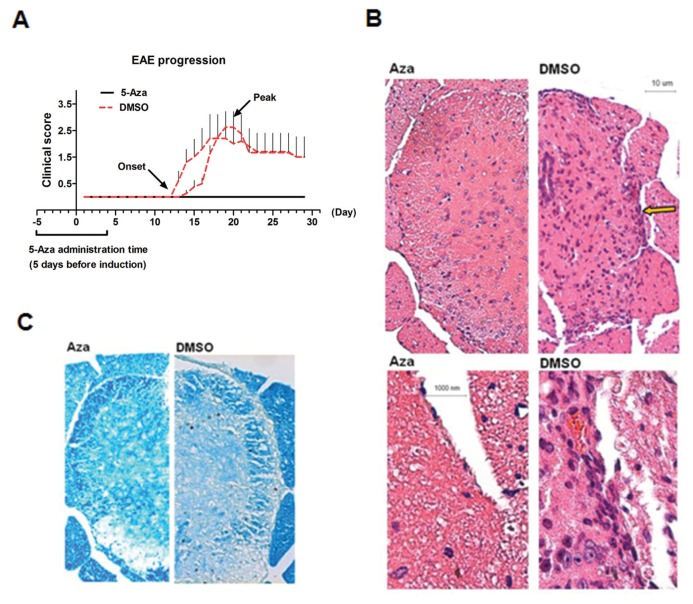
EAE was induced as previously described with some modifications (27). On EAE d 0, 8- to 12-wk-old C57BL/6 female mice (n = 5/group) were subcutaneously injected with 150 μL incomplete Freund adjuvant emulsion containing 100 μg of a myelin oligodendrocyte glycoprotein (MOG) peptide, MOG35-55 (MEVGWYRSPFSRVVHLYRNGK), and 400 μg of Mycobacterium tuberculosis extract (H37Ra). On d 0 and d 2 after immunization, mice were given 250 ng pertussis toxin in 150 μL phosphate-buffered saline (PBS) by intraperitoneal (IP) injection. Mice were monitored daily and clinical signs of disease progression were recorded, as described previously (27). Wild type C57BL/6 and Foxp3-EGFP transgenic mice were given IP 5-Aza in 0.1 mol/L DMSO at a dose of 0.15 mg/kg/d, 5 d before EAE induction, for 10 d (Figure 1A). Control mice were treated with 0.1 mol/L DMSO alone for ten days.
Figure 1.
Treatment of mice with 5-Aza inhibits the development of experimental auto-immune encephalomyelitis (EAE). (A) Low-dose 5-Aza (0.15 mg/kg/d) or control DMSO treatments of C57BL/6 mice were administrated at 5 d before induction of EAE for a total of 10 d. Each line represents an independent experiment. Two independent experiments were performed (n = 5 mice/group). The time of disease onset and peak is also indicated. (B) Spinal cords of mice treated with 5-Aza or DMSO were stained with H&E at the peak of EAE pathogenesis (upper panels: 40×, lower panels: 100×); arrows show inflammatory cell infiltration. Histology was performed in three different mice for each treatment, and representative images are shown. (C) Luxol fast blue stain of spinal cords at the peak of EAE in mice pretreated with 5-Aza or DMSO (40×). 5-Aza-treated tissue showed intact myelin, whereas DMSO-treated mice showed attacked myelin. Histology was performed in three different mice for each treatment. Representative images are shown.
Growth Medium, Buffers and Reagents
RPMI-1640–based medium supplemented with 1% l-glutamine, 1% penicillin-streptomycin, 10% fetal bovine serum (FBS), 10 mmol/L HEPES and 50 μmol/L β-mercaptoethanol was used to culture T cells. The isolation buffer was PBS supplemented with 2 mmol/L EDTA and 2% FBS. The hypotonic red blood cell (RBC) lysis buffer was 0.15 mol/L NH4Cl, 10 mmol/L KHCO3 and 0.1 mmol/L Na2EDTA (pH 7.2). Solutions were sterilized by passage through a 0.22-μm filter and were stored at room temperature. 5-Aza was purchased from Sigma (St. Louis, MO, USA). A solution of 5-Aza was prepared in dimethyl sulfoxide (DMSO; Sigma) and stored at −80°C.
Flow Cytometry
Flow cytometry was performed with a FACSCalibur flow cytometer by using CellQuest software (BD Biosciences [BD, Franklin Lakes, NJ, USA]). Cell staining was performed with specific antibodies at 4°C for 30 min. Anti-mouse CD4−FITC (clone GK1.5), anti-mouse CD25-PE Cy5 (clone PC61.5) and anti-mouse Foxp3-PE (NRRF-30) were purchased from eBio-science (San Diego, CA, USA). Anti-mouse IFN-γ-PE (XMG1.2), anti-mouse IL-17A-PE (TC11-18H10), anti-mouse CD3-PE (145-2C11), anti-mouse CD3-PerCP (145-2C11), anti-mouse CD3-PE Cy5 (145-2C11) and anti-mouse CD4−APC (RM4-5) were purchased from BD.
RNA Isolation, cDNA Synthesis and Real-Time Polymerase Chain Reaction
Total ribonucleic acid (RNA) was isolated from spinal cords, and cDNA was synthesized from 1 μg RNA by using oligo dt. Real-time polymerase chain reaction (PCR) with a SYBR Green on StepOne cycler (Applied Biosystems/Life Technologies, Carlsbad, CA, USA) was used for amplification of cytokine genes. Expression of each gene was determined relative to that of β-actin.
Histopathology
Spinal cords were dissected from mice after heparin-PBS perfusion and fixed in 10% paraformaldehyde overnight. Paraffin-embedded sections (6–10 μm thickness) were then stained with hema-toxylin and eosin (H&E) or Luxol Fast Blue and examined by light microscopy.
Isolation of CNS-Infiltrating Lymphocytes
Mice were perfused with 30 mL heparinized PBS before isolation of infiltrating mononuclear cells from pooled spinal cord and brain tissues. Single-cell suspensions were prepared and subjected to Percoll gradient (70%/30%) centrifugation. The isolated mononu-clear cells were then stained with anti-CD3, anti-CD4 or anti-CD25 antibodies for 30 min at 4°C after blocking nonspe-cific staining. Cells from GFP knock-in mice were stained only with anti-CD3 and anti-CD4 antibodies. Data were analyzed by using CellQuest software (BD Biosciences).
In Vitro 5-Aza Treatment of CD4+CD25−T Cells
Spleens were gently ground into single-cell suspensions between frosted microscope slides, and RBCs were removed by using a hypotonic RBC lysis buffer. Splenocytes were enriched for CD4+ T cells by using a CD4+ negative selection kit (BD IMag™, BD Biosciences) and were then enriched for CD4+CD25+ T cells by using anti-mouse CD25-PE and anti-PE magnetic particles (BD IMag). Splenocytes isolated from GFP knock-in mice were stained with APC anti-CD4 antibody (RM4-5; BD Biosciences). CD4+GFP− cells were sorted by using a FACSAria™ III cell sorter (BD Biosciences). Isolated CD4+CD25− T cells (4.5 × 10⁵/mL) were activated for 2 d in the presence of anti-CD3 monoclonal antibody (mAb) (1 μg/mL), anti-CD28 mAb (0.2 μg/mL) and mouse IL-2 (10 ng/mL). Cultured T cells were treated with 5-Aza (1 μmol/L) or DMSO only on d 0 and then incubated for 1–4 d. T cells were harvested and intracellular staining for Foxp3 was performed by using a Foxp3 staining kit, according to the manufacturer’s guidelines (catalog no. 00-5523; eBioscience).
In Vitro Assay for Treg Cell–Suppressive Function
T cells were isolated by a selection kit followed by FACSAria III cell sorting. Selection was performed with a CD4+ negative selection kit (BD IMag), followed by staining with anti-mouse CD25-PE, before sorting or before using GFP as a selection marker (if the mice were GFP transgenic mice). CD25− or GFP− T cells were used as responding effector cells, and T cell–depleted splenocytes were used as supporting cells after treatment with mitomycin C (20 μg/mL) at 37°C for 40 min. The supporting cells, effector cells (CD4+CD25−) and Treg cells (CD4+CD25+) were cultured at a ratio of 10:2:1 in the presence of anti-CD3 mAb (1 μg/mL) for 4.5 d.
Statistical Analysis
The differences between experimental and control groups were analyzed by unpaired t test with Welch correction, except for the comparison of EAE clinical scores, which used the Mann-Whitney U test. A p value <0.05 was considered statistically significant.
All supplementary materials are available online at www.molmed.org.
RESULTS
5-Aza Pretreatment Inhibits MOG-Induced EAE in B6 Mice
Multiple sclerosis (MS) is a Th1/Th17 CD4+ T cell-mediated chronic autoimmune disease of the central nervous system (28). Among helper T cells, regulatory T cells are pivotal in suppressing overt immune responses and maintaining homeostasis (28). Previous studies revealed that 5-Aza, a DNA-demethylating agent, upregulated regulatory T cell expression and benefited mouse models of autoimmune diabetes and colitis (23, 24). In this study, we tested whether 5-Aza administration in peripheral areas could modulate CNS inflammatory responses in the EAE mouse model.
Five days before EAE induction (that is, MOG inoculation), a 10-d regimen of low-dose (0.15 mg/kg, intraperitoneally administrated) 5-Aza was started (please refer to Materials and Methods and Figure 1A). The results indicated a significant reduction in EAE disease scores relative to DMSO-treated controls (n = 5) (Figure 1A). In particular, there were no symptoms of EAE in 5-Aza-treated mice during the entire 35-d observation period, whereas most control DMSO-treated mice showed neurological symptoms (Figure 1A and Table 1).
Table 1.
EAE clinical scores after 5-Aza treatment.
| Group | Incidence (%) | Mean day of onset | Mean maximum score |
|---|---|---|---|
| Aza | 0 | 0 ± 0 | 0 ± 0 |
| DMSO | 90 | 11.81 ± 1.31 | 2.45 ± 0.35 |
To examine the effect of 5-Aza on CNS inflammation, histological examination of spinal cord tissues was performed on each group. As expected, the spinal cords of DMSO-treated control mice showed significant leukocyte infiltration into the CNS, whereas the 5-Aza-treated mice showed no infiltrating cells (Figure 1B). To explore the demyelination status of these treated mice, luxol fast blue was used to stain spinal cord myelin, showing the myelin to be preserved with an intact margin, while control DMSO-treated mice showed demyelination, with vacuoles in the myelin (Figure 1C). Thus, 5-Aza pre-treatment strongly inhibited EAE development and CNS inflammation.
5-Aza Decreases CNS-Infiltrating Lymphocytes in EAE Mice
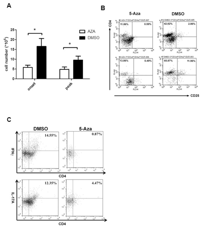
The above results showed that 5-Aza significantly inhibited disease development in EAE mice. To examine the modulatory effect of 5-Aza treatment on CNS inflammation, CNS-infiltrating lymphocytes were isolated from mice treated with 5-Aza or DMSO at EAE onset and at disease peak. As shown in Figure 2, 5-Aza-treated mice had significantly fewer infiltrating cells at both time points (Figure 2A, P < 0.05). Moreover, the percentage of CD4+CD25− effector cells among infiltrated cells was lower in 5-Aza-treated mice than in control mice at both time points (Figure 2B, CD4+CD25−upper left in dot plot), probably due to lower inflammatory immune responses in the CNS.
Figure 2.
5-Aza treatment decreases the number of CNS-infiltrating lymphocytes in EAE mice. Brains and spinal cords of MOG-induced EAE mice treated with 5-aza or DMSO were harvested at EAE disease onset or peak, and CNS-infiltrating lymphocytes were isolated by Percoll density gradient. Three independent experiments were performed. (A) Total infiltrating cell numbers were analyzed after Percoll gradient isolation. Open bars indicate 5-Aza treatment and black bars show control DMSO-treated mice (each group contained five mice, *p < 0.05). (B) CNS-infiltrating cells stained with CD3, CD4 and CD25, and CD3+ cells were gated to count CD4+CD25+ T cells from 5-Aza-treated (5-Aza, left panel) or DMSO-treated (DMSO, right panel) control EAE mice during disease onset (upper panel) and disease peak (lower panel). (C) Flow cytometry analysis of the expression of inflammatory cytokines (IFN-γ: upper panel; IL-17: lower panel) in CNS-infiltrating CD4+ T cells. A representative profile is shown. Three independent experiments were performed.
We further evaluated cytokine expression by the CNS-infiltrating cells, including IL-17 and IFN-γ, by intracellular cytokine staining. The cytokines expressed by CNS-infiltrating cells in 5-Aza-treated mice were significantly lower than those in DMSO-treated mice (Figure 2C). These data indicate that 5-Aza treatment decreases CNS pathogenic effector cells in EAE mice, thus inhibiting disease progression. Overall, this suggests that peripheral treatment with 5-Aza prevents the development of EAE and decreases Th1 and Th17 cell populations in the CNS.
5-Aza Increases Foxp3 Expression in Splenocytes and GFP Expression in CD4+GFP− T Cells In Vitro
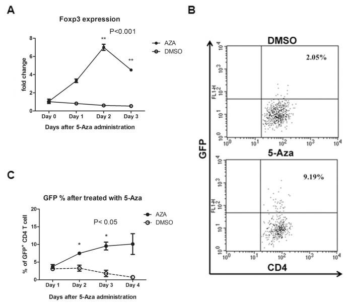
Previous studies have indicated that DNA demethylation is involved in the regulation of Foxp3 expression (18,23,25,29–31). Reduced expression of Foxp3 can affect both the number and activity of Treg cells (32). To evaluate the effect of the demethylating agent 5-Aza on the conversion of splenocytes into Treg cells, we first treated splenocytes with 5-Aza to assess its effect on Foxp3 expression. As shown in Figure 3, 5-Aza significantly upregulated Foxp3 RNA, to a maximum level on d 2 (Figure 3A, p < 0.001). We also tested the effect of 5-Aza on splenocytes from Foxp3-EGFP transgenic mice. In particular, CD3+GFP− T cells were isolated from mice treated with 5-Aza to examine possible conversion of conventional T cells into Treg cells. Flow cytometry results indicated an increased number of GFP+ cells after 5-Aza treatment (Figure 3B) in vitro, in addition to an increased percentage of GFP+ cells (Figure 3C, p < 0.05). In agreement with previous studies (33–35), our results indicate that 5-Aza can convert CD4+Foxp3− conventional T cells into Foxp3-expressing Treg cells in vitro.
Figure 3.
Treatment of isolated splenocytes with 5-Aza increases the expression of Foxp3 and GFP. (A) Isolated murine splenocytes were activated by anti-CD3 (1 μg/mL), anti-CD28 (0.2 μg/mL) and mouse IL-2 (10 ng/mL). Cells were treated with 1 μmol/L 5-Aza (solid circles) or DMSO alone (open circles) every day for 4 d. Foxp3 mRNA expression was measured by quantitative real-time PCR with normalization to β-actin (**p < 0.001). (B, C) CD3+GFP− T cells were harvested from Foxp3-EGFP C57BL/6 transgenic mice (C.Cg-FOXP3tm2Tch/J) and isolated by a BD FACSAria III cell sorter. The isolated CD3+GFP−T cells were activated as above and then treated with 1 μmol/L 5-Aza or DMSO alone. Flow cytometry was used to monitor GFP expression. A representative flow cytometry result is shown (B). Results are summarized from d 0 to d 4, with 5-Aza (solid circles) and DMSO alone (open circles) (C) (*p < 0.05). All data are representative of three independent experiments with triplicate culture wells.
5-Aza Treatment Enlarges Spleen Size and Increases Numbers of CD4+CD25+ Cells In Vivo
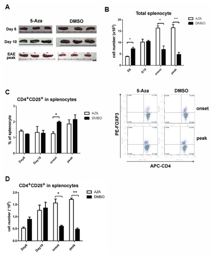
Having demonstrated that 5-Aza could convert CD4+/GFP− cells into CD4+/GFP+ cells in vitro, we then compared splenocyte populations after 5-Aza treatment in vivo. The size of the spleens in 5-Aza-treated mice was initially smaller than those of DMSO-treated mice at d 6 after MOG induction. However, the spleens enlarged until the peak of EAE in the 5-Aza-treated group, whereas the spleens in DMSO-treated mice shrank both at EAE onset (data not shown) and peak (Figure 4A). A similar pattern was observed in the number of splenocytes such that 5-Aza treatment significant lowered the number of cells, compared with control, at d 6. This trend was reversed at disease onset and peak (Figure 4B, p < 0.05).
Figure 4.
Treatment of mice with 5-Aza induces distinct changes in spleen morphology and spenocyte populations during EAE progression. Mice pretreated with 5-Aza or DMSO were MOG-induced for EAE, as described in Figure 1 (n = 3–5 mice/group, two independent experiments were performed). (A) Spleens were harvested from 5-Aza-treated (left panel) or DMSO-treated (right panel) EAE mice at d 6 and 10 and disease peak after MOG induction. Each interval between bars represents 1 cm. (B) Total cell numbers of splenocytes were counted at the indicated time points (*p < 0.05, **p < 0.01; white bar: 5-Aza, black bar: DMSO). (C) Splenocytes stained with CD3, CD4 and CD25, and CD3+ cells, were gated to analyze the percentage of CD4+CD25+ T cells from 5-Aza-treated (white bars) or DMSO-treated (black bars) EAE mice at the indicated time points (left panel). Representative results from flow cytometry are also shown (right panel) (*p < 0.05). (D) The total number of CD4+CD25+ T cells in splenocytes from 5-Aza-treated (white bars) or DMSO-treated (black bars) EAE mice were counted at the indicated time points (*p < 0.05, **p < 0.01).
Further investigation of different types of splenocytes (Supplementary Figure S1) revealed that 5-Aza treatment resulted in a similar percentage of CD4+CD25+ cells as the DMSO-treated mice at d 6 after MOG induction (Figure 4C). However, this phenomenon was reversed at disease onset and peak (Figure 4C), a result that may be attributed to the inflammatory response in DMSO-treated EAE mice. However, 5-Aza-treated mice were found to have significantly higher numbers of CD4+CD25+ cells than the DMSO-treated EAE mice at both disease onset and peak (Figure 4D), a possible explanation for 5-Aza suppression of MOG-induced EAE.
5-Aza-Treated Mice Show a Lower Activation Threshold of Effector T Cells
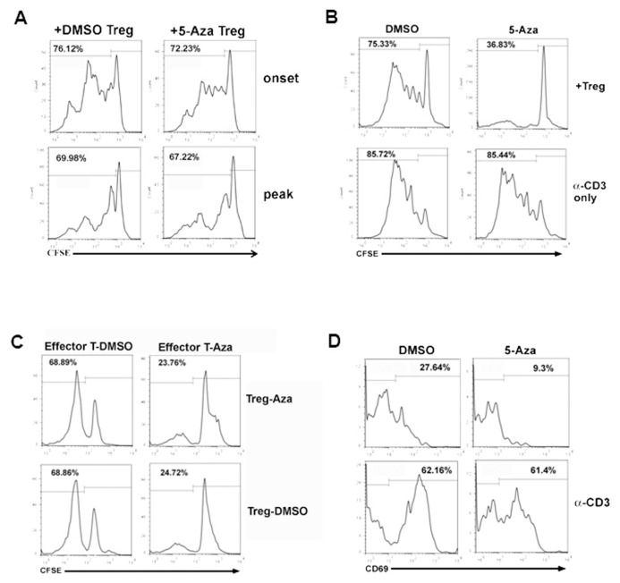
Next, we performed a Treg immune suppression assay to determine whether 5-Aza enhances the immunosuppressive activity of regulatory T cells in EAE mice. Treg cells from 5-Aza- or DMSO-treated EAE mice were isolated from the spleen at EAE onset (Figure 5A, 5-Aza-Treg, DMSO-Treg) and then cocultured with CD4+ effector T cells from naive B6 mice. Activation assays showed that these effector T cells were functionally intact (Supplementary Figure S2). Unexpectedly, however, Treg cells isolated from 5-Aza mice showed suppressive functions similar to those from the DMSO control mice (Figure 5A).
Figure 5.
Regulatory T cells have similar suppressive function, but effector T cells have a lower activation threshold in 5-Aza-treated mice (A) Effector T cells isolated from naive B6 mice were cocultured with Treg cells isolated from 5-Aza-treated (5-Aza Treg) or DMSO-treated (DMSO Treg) EAE mice at disease onset and peak. Treg inhibition assays were then performed by CFSE-labeling. (B) Effector T cells isolated from EAE mice treated with DMSO (DMSO, left panel) or 5-Aza (5-Aza, right panel) were cocultured with Treg cells isolated from 5-Aza-treated mice for Treg inhibition assay (upper panel). For control experiments, effector T cells were stimulated with anti-CD3 antibody only (lower panel). (C) CFSE-labeled CD4+CD25− effector T cells isolated from DMSO-treated (Effector T-DMSO) or 5-Aza-treated (Effector T-Aza) EAE mice were cocultured with regulatory T cells isolated from 5-Aza-treated (Treg-Aza) or DMSO-treated (Treg-DMSO) EAE mice for 5 d, and CFSE signals were detected by flow cytometry. A representative CFSE profile is shown. Three independent experiments were performed with similar trends. (D) CD4+CD25− T cells isolated from DMSO- or 5-Aza-treated EAE mice were stained with CD69 activation marker before (upper panel) or after (lower panel) stimulation with anti-CD3. Data are representative of three independent experiments with similar results.
Consequently, we wondered whether the Treg-suppressive function in 5-Aza-treated mice could inhibit effector T cells. Treg-suppressive assays of effector cells and Treg cells isolated and cocultured from the same mice showed that Treg cells from 5-Aza-treated mice significantly inhibited the proliferation of carboxyfluorescein succinimidyl ester (CFSE)-labeled effector cells from the same mice (Figure 5B, upper right). Surprisingly, this inhibitory effect was not seen in cells isolated from DMSO-treated mice (Figure 5B, upper left). Effector T cells isolated from 5-Aza-treated (Figure 5B, lower right) or DMSO-treated (Figure 5B, upper left) mice showed the same proliferation pattern after stimulation with anti-CD3 antibody, thus suggesting they were functionally intact. The above data indicate that regulatory T cells could inhibit the proliferation of effector T cells isolated from the same 5-Aza-treated mice, but not cells from (DMSO-treated) EAE-diseased mice (Figures 5A, B).
Furthermore, Treg and effector cells isolated from 5-Aza- or DMSO-treated mice were cocultured with each other, showing that Treg cells significantly inhibited the proliferation of CFSE-labeled effector cells isolated from the same 5-Aza-treated mice (Figure 5C, upper right). Surprisingly, Treg cells isolated from DMSO-treated EAE mice could also inhibit the proliferation of effector cells isolated from 5-Aza-treated mice (Figure 5C, lower right). However, neither Treg cells isolated from 5-Aza-treated mice nor from DMSO-treated mice could inhibit the proliferation of DMSO-treated mouse effector cells (Figure 5C, left panel). These results suggest that 5-Aza does not enhance Treg per-cell inhibitory function.
Finally, we wondered whether 5-Aza treatment could attenuate the activity of effector T cells. To test this hypothesis, CD69 activation marker analysis was performed to detect the activation status of the cells. Indeed, CD4+ T cells from 5-Aza-treated mice expressed less CD69 activation markers than DMSO-treated mice (Figure 5D, upper panel). However, both cells showed the same activation ability, as demonstrated by in vitro treatment with anti-CD3 antibody (Figure 5D, lower panel), thus suggesting that 5-Aza treatment maintains a lower activation threshold for the effector CD4 T cells.
DISCUSSION
In this study, we found that peripherally administrated low-dose (0.15 mg/kg/d), 10-d 5-Aza could prevent autoimmune disease in the CNS of a murine EAE model (Figure 1A). In fact, none of the short-term 5-Aza-pretreated MOG-induced mice developed EAE during our observation period (Figure 1A). Moreover, 5-Aza treatment increased both Foxp3 RNA and protein expression in vitro and in vivo and also upregulated the number of regulatory T cells in vivo. These results are consistent with previous studies of experimental autoimmune diabetes and colitis (23,25). Although the per-cell inhibitory function of Treg cells was not enhanced, 5-Aza treatment did lower the activation threshold of effector cells (Figure 5D).
Th17 cells and their cytokine, IL-17, play important roles in the pathogenesis of EAE. Our results showed that 5-Aza-treated mice had much lower expression of proinflammatory CNS cytokines compared with control mice. In particular, the expression of EAE-associated proinflammatory cytokines, such as IL-17 and IFN-γ, were lower at both EAE onset and peak in 5-Aza-treated mice (Figure 2C). A previous study showed that the Th17-specific cytokine gene Il17a, which contains six CpG dinucleotides in its 200-bp highly conserved promoter region, is regulated by promoter methylation (36). In an in vitro culture system, CD4+ cells cultured with 5-Aza expressed more IL-17A than cells grown without 5-Aza (26). However, it was also reported that in vivo IL-17A deficiency or neutralization mitigates EAE, but does not completely abrogate all symptoms (37,38). On the other hand, it has also been reported that neither T-cell–driven overexpression of IL-17A and IL-17F, nor their complete loss, had a major impact on the development of EAE (37). In particular, it has been shown that autoimmune demyelinating disease can be driven by multiple myelin-specific T cells of distinct lineages, with different extents of cell–cell interactions and dependence on cytokine production to achieve their pathogenic effects (38). According to our results, pretreatment with 5-Aza can prevent the onset of such diseases (for example, EAE).
Foxp3 is a master regulator of Treg cells. In this study, we found that both in vivo and in vitro 5-Aza treatment significantly elevated Foxp3 expression in T cells. Short-term treatment with 5-Aza significantly increased the percentage of regulatory T cells and also converted conventional T cells into Treg cells in vitro. These results are consistent with previous reports that 5-Aza treatment can convert Foxp3− cells into Foxp3+ cells (23,25), at least partly because of demethylation of the Foxp3 promoter. Thus, even if 5-Aza treatment upregulated both Foxp3 and IL17, its induction of Foxp3 might be more robust than that of IL-17. Moreover, the increased Treg cell population might also block the differentiation of pathogenic Th17 cells.
On the other hand, it was demonstrated that gene promoter hypermethylation was more prominent in cell-free DNA isolated from relapsing remitting MS patients than from healthy individuals (39). This result suggests that MS might be an epigenetic disease, and reversing epigenetic repression of the Foxp3 methylated promoter by demethylating agents may have therapeutic potential for the treatment of this auto-immune disease.
Together, these results show that short-term (10-d) low-dose 5-Aza treatment ameliorated the development of EAE and inhibited proinflammatory responses in the CNS, effects that could be fully attributed to 5-Aza-mediated DNA demethylation. Together with the results of a previous study (23), we strongly suggest that epigenetic regulation is involved in the development of Treg cells and that 5-Aza has the potential to modulate abnormal immune responses in autoimmune diseases.
We also examined the effect of 5-Aza pretreatment on populations of splenocytes from MOG-induced EAE mice at different time points. Treatment with 5-Aza showed a similar percentage of CD4+CD25+ cells as the DMSO-treated mice at d 6 after MOG induction (Figure 4C). However, this similarity was reversed at disease onset and peak (Figure 4C). These results might be explained by the vigorous inflammatory response in DMSO-treated mice against the MOG antigen. In other words, DMSO-treated mice need more Treg cells to inhibit pathogenic lymphocytes (for example, Th17+ cells) to prevent EAE. The amelioration of EAE development by early induction of Treg cell populations in 5-Aza-treated mice induced less proinflammatory response and less antiinflammatory resolution response at the time point of disease onset or peak, as mediated by the Treg cell population. Although the percentage of CD4+CD25+ T cells was smaller in 5-Aza-treated mice, compared with control EAE mice, the total cell number of CD4+CD25+ T cells was significantly higher in 5-Aza-treated EAE mice (Figure 4C), thus suppressing MOG-induced EAE.
A previous study demonstrated not only an increased number of Treg cells in 5-Aza-treated mice, but also an increased Treg-mediated immune suppression. However, the 5-Aza-enhanced Treg immunosuppressive function was found only in thymic, but not splenic, Treg cells. In our study, we found enhanced immunosuppressive activity of Treg cells, inhibiting the proliferative responses of effector cells isolated from 5-Aza-treated mice. However, when compared with DMSO-treated mice, the suppressive function of 5-Aza-treated Treg cells was equivalent using effector cells isolated from naive B6 mice (Figure 5A). These results indicate that the effect was not due to an increased per-cell suppressive function of Treg cells, but rather decreased activity of effector cells in 5-Aza-treated mice. Observation of 5-Aza effects on the activation state of effector T cells in mouse splenocytes showed that effector cells had lower CD69 (activation marker) expression, at disease onset, than DMSO-treated mice; however, the two groups had similar CD69 expression after 2 d of stimulation by anti-CD3 antibody (Figure 5D). These results indicate that 5-Aza treatment lowered the activation threshold of effector cells in vivo, but these cells could still be activated in vitro. This lower activation threshold may be attributed to an increased number of Treg cells, which elicits a greater suppressive effect on the effector cells through increased cell–cell contact, secretion of inhibitory cytokines such as TGF-β and indirectly through increased inhibition of APC (40). Thus, the increased population of Treg cells, together with lower activation potential of effector cells, was presumably responsible for the hindered EAE development and reduced CNS inflammation. This result was further confirmed by a recent finding from Kehrmann et al. (41) that 5-Aza treatment of human CD4+ T cells could only induce hypomethylation of Foxp3 and expression of Treg-specific genes, but could not induce suppressive function (41).
CONCLUSION
Our study indicates that short-term 5-Aza pretreatment mitigates the development of EAE symptoms in MOG- induced mice. However, we only tested the effect of 5-Aza given for 10 d before induction of EAE. Future studies are needed to establish whether even better efficacy can be achieved by different durations of 5-Aza administration before or after the onset of EAE symptoms.
Supplementary Information
ACKNOWLEDGMENTS
This work was supported by the National Science Council (Taiwan, Republic of China; NSC-98-01740-01, NSC-99-2320-B-194-001-MY3 and NSC-102-2320-B-194-001) and in part by the Dalin Tzu Chi Hospital, Buddhist Tzu Chi Medical Foundation (Chia-Yi, Taiwan). The authors would like to thank Jia-Chiun Pan for assistance in statistical calculation.
Footnotes
Online address: http://www.molmed.org
DISCLOSURE
The authors declare that they have no competing interests as defined by Molecular Medicine, or other interests that might be perceived to influence the results and discussion reported in this paper.
REFERENCES
- 1.Goverman JM. Immune tolerance in multiple sclerosis. Immunol Rev. 2011;241:228–40. doi: 10.1111/j.1600-065X.2011.01016.x. [DOI] [PMC free article] [PubMed] [Google Scholar]
- 2.Pachner AR. Experimental models of multiple sclerosis. Curr Opin Neurol. 2011;24:291–99. doi: 10.1097/WCO.0b013e328346c226. [DOI] [PubMed] [Google Scholar]
- 3.Simmons SB, Pierson ER, Lee SY, Goverman JM. Modeling the heterogeneity of multiple sclerosis in animals. Trends Immunol. 2013;34:410–22. doi: 10.1016/j.it.2013.04.006. [DOI] [PMC free article] [PubMed] [Google Scholar]
- 4.Rangachari M, Kuchroo VK. Using EAE to better understand principles of immune function and autoimmune pathology. J Autoimmun. 2013;45:31–9. doi: 10.1016/j.jaut.2013.06.008. [DOI] [PMC free article] [PubMed] [Google Scholar]
- 5.Kroenke MA, Segal BM. Th17 and Th1 responses directed against the immunizing epi-tope, as opposed to secondary epitopes, dominate the autoimmune repertoire during relapses of experimental autoimmune encephalomyelitis. J Neurosci Res. 2007;85:1685–93. doi: 10.1002/jnr.21291. [DOI] [PubMed] [Google Scholar]
- 6.Petermann F, Korn T. Cytokines and effector T cell subsets causing autoimmune CNS disease. FEBS Lett. 2011;585:3747–57. doi: 10.1016/j.febslet.2011.03.064. [DOI] [PubMed] [Google Scholar]
- 7.Fletcher JM, Lalor SJ, Sweeney CM, Tubridy N, Mills KH. T cells in multiple sclerosis and experimental autoimmune encephalomyelitis. Clin Exp Immunol. 2010;162:1–11. doi: 10.1111/j.1365-2249.2010.04143.x. [DOI] [PMC free article] [PubMed] [Google Scholar]
- 8.Sakaguchi S. Naturally arising Foxp3-expressing CD25+CD4+ regulatory T cells in immunological tolerance to self and non-self. Nat Immunol. 2005;6:345–52. doi: 10.1038/ni1178. [DOI] [PubMed] [Google Scholar]
- 9.Goldstein JD, et al. Role of cytokines in thymus- versus peripherally derived-regulatory T cell differentiation and function. Front Immunol. 2013;4:155. doi: 10.3389/fimmu.2013.00155. [DOI] [PMC free article] [PubMed] [Google Scholar]
- 10.Zhou X, et al. Therapeutic potential of TGF-beta-induced CD4(+) Foxp3(+) regulatory T cells in autoimmune diseases. Autoimmunity. 2011;44:43–50. doi: 10.3109/08916931003782163. [DOI] [PMC free article] [PubMed] [Google Scholar]
- 11.Shevach EM. Mechanisms of foxp3+ T regulatory cell-mediated suppression. Immunity. 2009;30:636–45. doi: 10.1016/j.immuni.2009.04.010. [DOI] [PubMed] [Google Scholar]
- 12.Vignali DA, Collison LW, Workman CJ. How regulatory T cells work. Nat Rev Immunol. 2008;8:523–32. doi: 10.1038/nri2343. [DOI] [PMC free article] [PubMed] [Google Scholar]
- 13.Matsushita T, Horikawa M, Iwata Y, Tedder TF. Regulatory B cells (B10 cells) and regulatory T cells have independent roles in controlling experimental autoimmune encephalomyelitis initiation and late-phase immunopathogenesis. J Immunol. 2010;185:2240–52. doi: 10.4049/jimmunol.1001307. [DOI] [PMC free article] [PubMed] [Google Scholar]
- 14.Williams LM, Rudensky AY. Maintenance of the Foxp3-dependent developmental program in mature regulatory T cells requires continued expression of Foxp3. Nat Immunol. 2007;8:277–84. doi: 10.1038/ni1437. [DOI] [PubMed] [Google Scholar]
- 15.Apostolou I, et al. Peripherally induced Treg: mode, stability, and role in specific tolerance. J Clin Immunol. 2008;28:619–24. doi: 10.1007/s10875-008-9254-8. [DOI] [PubMed] [Google Scholar]
- 16.Curotto de Lafaille MA, Lafaille JJ. Natural and adaptive foxp3+ regulatory T cells: more of the same or a division of labor? Immunity. 2009;30:626–35. doi: 10.1016/j.immuni.2009.05.002. [DOI] [PubMed] [Google Scholar]
- 17.Bennett CL, et al. The immune dysregulation, polyendocrinopathy, enteropathy, X-linked syndrome (IPEX) is caused by mutations of FOXP3. Nat Genet. 2001;27:20–1. doi: 10.1038/83713. [DOI] [PubMed] [Google Scholar]
- 18.Lal G, Bromberg JS. Epigenetic mechanisms of regulation of Foxp3 expression. Blood. 2009;114:3727–35. doi: 10.1182/blood-2009-05-219584. [DOI] [PMC free article] [PubMed] [Google Scholar]
- 19.Katoh H, Zheng P, Liu Y. FOXP3: genetic and epigenetic implications for autoimmunity. J Autoimmun. 2013;41:72–8. doi: 10.1016/j.jaut.2012.12.004. [DOI] [PMC free article] [PubMed] [Google Scholar]
- 20.Foulks JM, et al. Epigenetic drug discovery: targeting DNA methyltransferases. J Biomol Screen. 2012;17:2–17. doi: 10.1177/1087057111421212. [DOI] [PubMed] [Google Scholar]
- 21.Floess S, et al. Epigenetic control of the foxp3 locus in regulatory T cells. PLoS Biol. 2007;5:e38. doi: 10.1371/journal.pbio.0050038. [DOI] [PMC free article] [PubMed] [Google Scholar]
- 22.Polansky JK, et al. DNA methylation controls Foxp3 gene expression. Eur J Immunol. 2008;38:1654–63. doi: 10.1002/eji.200838105. [DOI] [PubMed] [Google Scholar]
- 23.Zheng Q, et al. Induction of Foxp3 demethylation increases regulatory CD4+CD25+ T cells and prevents the occurrence of diabetes in mice. J Mol Med (Berl) 2009;87:1191–205. doi: 10.1007/s00109-009-0530-8. [DOI] [PubMed] [Google Scholar]
- 24.Sanchez-Abarca LI, et al. Immunomodulatory effect of 5-azacytidine (5-azaC): potential role in the transplantation setting. Blood. 2010;115:107–21. doi: 10.1182/blood-2009-03-210393. [DOI] [PubMed] [Google Scholar]
- 25.Lal G, et al. Epigenetic regulation of Foxp3 expression in regulatory T cells by DNA methylation. J Immunol. 2009;182:259–73. doi: 10.4049/jimmunol.182.1.259. [DOI] [PMC free article] [PubMed] [Google Scholar]
- 26.Frikeche J, et al. Impact of the hypomethy-lating agent 5-azacytidine on dendritic cells function. Exp Hematol. 2011;39:1056–63. doi: 10.1016/j.exphem.2011.08.004. [DOI] [PubMed] [Google Scholar]
- 27.Racke MK. Experimental autoimmune encephalomyelitis (EAE) Curr. Protoc. Neurosci. 2001;Chapter 9(Unit 9):7. doi: 10.1002/0471142301.ns0907s14. [DOI] [PubMed] [Google Scholar]
- 28.Bilate AM, Lafaille JJ. Induced CD4+Foxp3+ regulatory T cells in immune tolerance. Annu Rev Immunol. 2012;30:733–58. doi: 10.1146/annurev-immunol-020711-075043. [DOI] [PubMed] [Google Scholar]
- 29.Janson PC, et al. FOXP3 promoter demethylation reveals the committed Treg population in humans. PLoS One. 2008;3:e1612. doi: 10.1371/journal.pone.0001612. [DOI] [PMC free article] [PubMed] [Google Scholar]
- 30.Choi J, et al. In vivo administration of hypomethylating agents mitigate graft-versus-host disease without sacrificing graft-versus-leukemia. Blood. 2010;116:129–39. doi: 10.1182/blood-2009-12-257253. [DOI] [PMC free article] [PubMed] [Google Scholar]
- 31.Zheng Y, et al. Role of conserved non-coding DNA elements in the Foxp3 gene in regulatory T-cell fate. Nature. 2010;463:808–12. doi: 10.1038/nature08750. [DOI] [PMC free article] [PubMed] [Google Scholar]
- 32.Chauhan SK, Saban DR, Lee HK, Dana R. Levels of Foxp3 in regulatory T cells reflect their functional status in transplantation. J Immunol. 2009;182:148–53. doi: 10.4049/jimmunol.182.1.148. [DOI] [PMC free article] [PubMed] [Google Scholar]
- 33.Moon C, et al. Use of epigenetic modification to induce FOXP3 expression in naive T cells. Transplant Proc. 2009;41:1848–54. doi: 10.1016/j.transproceed.2009.02.101. [DOI] [PubMed] [Google Scholar]
- 34.Wong CP, et al. Induction of regulatory T cells by green tea polyphenol EGCG. Immunol Lett. 2011;139:7–13. doi: 10.1016/j.imlet.2011.04.009. [DOI] [PMC free article] [PubMed] [Google Scholar]
- 35.Hu Y, et al. Decitabine facilitates the generation and immunosuppressive function of regulatory gammadelta T cells derived from human peripheral blood mononuclear cells. Leukemia. 2013;27:1580–5. doi: 10.1038/leu.2012.345. [DOI] [PubMed] [Google Scholar]
- 36.Janson PC, et al. Profiling of CD4+ T cells with epigenetic immune lineage analysis. J Immunol. 2011;186:92–102. doi: 10.4049/jimmunol.1000960. [DOI] [PubMed] [Google Scholar]
- 37.Haak S, et al. IL-17A and IL-17F do not contribute vitally to autoimmune neuro-inflammation in mice. J Clin Invest. 2009;119:61–9. doi: 10.1172/JCI35997. [DOI] [PMC free article] [PubMed] [Google Scholar]
- 38.Segal BM. Th17 cells in autoimmune demyelinating disease. Semin Immunopathol. 2010;32:71–7. doi: 10.1007/s00281-009-0186-z. [DOI] [PMC free article] [PubMed] [Google Scholar]
- 39.Liggett T, et al. Methylation patterns of cell-free plasma DNA in relapsing-remitting multiple sclerosis. J Neurol Sci. 2010;290:16–21. doi: 10.1016/j.jns.2009.12.018. [DOI] [PMC free article] [PubMed] [Google Scholar]
- 40.Sojka DK, Huang YH, Fowell DJ. Mechanisms of regulatory T-cell suppression: a diverse arsenal for a moving target. Immunology. 2008;124:13–22. doi: 10.1111/j.1365-2567.2008.02813.x. [DOI] [PMC free article] [PubMed] [Google Scholar]
- 41.Kehrmann J, et al. Impact of 5-aza-2′-deoxycytidine and epigallocatechin-3-gallate for induction of human regulatory T cells. Immunology. 2014;142:384–95. doi: 10.1111/imm.12261. [DOI] [PMC free article] [PubMed] [Google Scholar]
Associated Data
This section collects any data citations, data availability statements, or supplementary materials included in this article.


