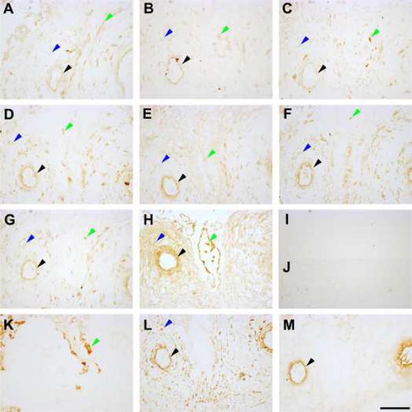
Figure 3. Protein localisation of maternal pre-eclampsia susceptibility genes in the decidua.
Decidual sections shown are from a representative patient sample. A: Activin type I receptor (ACVR1) expression. B: Inhibin α subunit (INHA) expression. C: Inhibin βB subunit (INHBB) expression. D: Endoplasmic reticulum aminopeptidase 1 (ERAP1) expression. E: Endoplasmic reticulum aminopeptidase 2 (ERAP2) expression. F: Placental leucine aminopeptidase (LNPEP) expression. G: Collagen type IV α1 chain (COL4A1) expression. H: Collagen type IV α2 chain (COL4A2) expression. I: Non-immune mouse IgG control. J: Non-immune rabbit IgG control. K: Cytokeratin 7 staining. L: Vimentin staining. M: von Willebrand factor staining. Black arrowheads denote endothelium staining, blue arrowheads denote decidual stromal cell staining and green arrowheads denote cytotrophoblast cell staining. Images were taken under 200X magnification. Scale bar denotes 100 μm.

