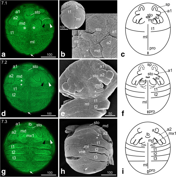
Figure 5.
Stage 7 (Postnaupliar segments, 7.1 (a-c); 7.2 (d-f); 7.3 (g-i)). (a, d, g) Sytox; (b, e, h) SEM; (c, f, i) Schematic drawing. (a) Stage 7.1, ventral view: the tips of the second antennae and the mandibles are arranged in a horizontal line. The asterisks mark the maxillary zone. The arrow marks the area of the proctodeum. (b) The insertions of the first and second antennae have come to lie next to each other. The ‘flap’-like structure posterior of the proctodeum (arrow) disappeared. (c) Schematic drawing of stage 7.1. (d) Stage 7.2, ventral view: The first antennae have moved medially. The second antennae are elongated, and the tips of their exo- and endopodites delimit the maxillary zone laterally. The asterisks mark the segments of the first and second maxillae. The second thoracic segment is separated from the rest of the trunk by an intersegmental furrow except for the area of the neuroectoderm. (e) Ventral view: the SEM of stage 7.2 provides a clear view of the intersegmental furrows between the first and second maxillae (asterisks), between the first and second thoracopod and the rest of the trunk as well as of the longitudinal midline of the neuroectoderm. (f) Schematic drawing of stage 7.2. (g) Stage 7.3. Ventral view: the second antennae have continued elongation and almost extend to the first thoracic segment. The mandibles have increased in size, and the emerging first maxillae become evident. The third thoracic segment has formed. The limb anlagen of the thoracic segments become evident by uneven boarders; in late stage 7.3 embryos slight elevations can be detected. (h) The prolonged second antennae and the enlarged mandibles are evident. Also the undulated boarders of the first, second and third thoracopod due to the developing limb buds are obvious. The lateral ventral neuroectoderm can be distinguished from the midline cells by its uneven surface, which seems to be related to the development of neuroblasts. (i) Schematic drawing of stage 7.3. a1, a2 = first and second antennae; lb = labrum; md = mandible; ml = midline; mx1, mx2 = first and second maxillae; ne = neuroectoderm; pro = proctodeum; sp = ‘Scheitelplatte’; sto = stomodeum; t = thoracic segment; vne = ventral neuroectoderm.

