ABSTRACT
A 6-year-old female rabbit was presented to a veterinary clinic, and the result of ultrasound examination suggested a tumor in the uterine tube. Subsequently, both ovaries and uterus were surgically removed. In gross, a single large cyst in the right ovary and enlargement of the left uterine tube were observed. Histological examination revealed that the cyst had developed in the hilus of the ovary and was lined by single-layered cuboidal cells. In the left uterine tube, a tumor composed of epithelial cells arranged in tubular structures and pleomorphic cells between the tubular structures was observed. Immunohistochemically, the epithelial cells of the cyst were positive for pan-cytokeratin, cytokeratin 18, CD10, E-cadherin, calretinin and estrogen receptor; the tumor cells of the left uterine tube were positive for pan-cytokeratin, cytokeratin 18, E-cadherin, vimentin, calretinin and estrogen receptor. From these results, the cyst was diagnosed as cystic rete ovarii, and the tumor was diagnosed as adenoma of the uterine tube. This case is the first to demonstrate cystic rete ovarii and uterine tube adenoma in rabbits.
Keywords: cystic rete ovarii, rabbit, uterine tube adenoma
Cystic lesions of the ovary, particularly Graafian follicle cysts, luteinized cysts and ovarian epithelial cysts, are commonly seen in domestic animals. However, cystic rete ovarii, which is another cystic lesion of the ovary, is rather rare in animals including the dog, cat, guinea pig and African green monkey [8,9,10, 13]. Moreover, immunohistochemical observations of cystic rete ovarii in animals are very limited, which makes it difficult to determine the origin of the cyst [1]. Recently, the authors have reported a case of cystic rete testis in a male rabbit [6]. Rete testis is a functional tubular structure comprising the excretory system of the testis. On the other hand, rete ovarii, which is the counterpart of the rete testis in males, is considered to be remnant tissue of the embryonic gonad.
Oviduct adenomas and adenocarcinomas are commonly found and well documented in domestic birds and reptiles [3, 4, 16]. However, uterine tube (the term “uterine tube” is preferred in mammalian species) tumors are very rare in mammalian species [14, 18]. Here, we report pathological findings of cystic rete ovarii and uterine tube adenoma in a rabbit.
A 6-year-old female rabbit was presented to a veterinary clinic with anorexia. Results of ultrasound and radiographic examinations suggested a tumor in the uterine tube with calcification. Subsequently, both ovaries and uterus were surgically removed and fixed in 10% neutral buffered formalin. Gross examination revealed cystic enlargement of the right ovary measuring 2 × 2 × 2 cm and a solid tumor in the left uterine tube measuring 5 × 5 × 5 mm. The tissues were routinely embedded in paraffin, sectioned at 4 µm and stained with hematoxylin and eosin (HE). Endometrial hyperplasia was confirmed in the uterus. Immunohistochemistry was performed in order to further characterize the components of the cyst wall and the tumor. The details of the primary antibodies used for immunohistochemistry are listed in Table 1. The labeling was visualized using the anti-mouse EnVision+ System (Dako, Kyoto, Japan). Also, we immunohistochemically analyzed normal (neither cystic nor tumor lesion) ovarian and uterine tube tissues collected from a 7-year-old rabbit with endometrial hyperplasia. The results of immunohistochemistry of the present case and the normal rabbit are summarized in Table 2.
Table 1. Primary antibodies used in the present study.
| Antibody against | Clone (mouse) | Dilution | Antigen retrieval | Source |
|---|---|---|---|---|
| pan-CK | AE1/AE3 | Autoclave | Dako, Kyoto, Japan | |
| CK 18 | Ks18.04 | Ready to use | Proteinase K | Progen, Heidelberg, Germany |
| CD 10 | 56C6 | 1: 100 | Autoclave | Invitrogen, Camarillo, CA |
| E-cadherin | 4A2C7 | 1: 100 | Autoclave (pH 9) | Invitrogen, Camarillo, CA |
| Estrogen receptor | 6F11 | Ready to use | Autoclave | Leica Biosystems, Newcastle, UK |
| Calretinin | MAB1568 | 1: 100 | Autoclave | Millipore, Temecula,CA |
| p63 | BC4A4 | 1: 100 | Autoclave | Biocare Medical, Concord, CA |
| SMA | 1A4 | 1: 100 | Autoclave | Dako, Kyoto, Japan |
| Vimentin | V9 | 1: 100 | Autoclave | Dako, Kyoto, Japan |
| Melan A | A103 | 1: 50 | Autoclave | Dako, Kyoto, Japan |
Table 2. Results of immunohistochemistry.
| Normal tissue |
Present case |
||||||||||
|---|---|---|---|---|---|---|---|---|---|---|---|
| Ovary |
Uterine tube |
Cyst (right ovary) |
Tumor (left uterine tube) |
||||||||
| Surface epithelial cell |
Spindle stromal cell |
Luteinized stromal cell |
Granulosa cell |
Theca cell |
Epithelial cell |
Stromal cell |
Epithelial cell |
Stromal cell |
Epithelial cell |
Pleomorphic cell |
|
| CK (AE1/AE3) | + | − | − | − | − | + | − | + | − | + | + |
| CK18 | +/− | − | − | − | − | + | − | + | − | + | − |
| CD10 | − | − | − | − | − | + | − | + | − | − | − |
| E-cadherin | + | − | − | − | − | +/− | − | + | − | + | − |
| Vimentin | +/− | + | + | + | + | − | NA | − | + | − | + |
| SMA | − | − | − | − | + | − | − | − | − | − | − |
| Calretinin | − | − | +/− | − | − | +/− | − | + | +/− | − | + |
| Melan A | − | − | + | − | − | − | − | − | + | − | − |
| Estrogen receptor | +/− | − | + | − | +/− | + | + | + | +/− | + | + |
+: Positive, −: Negative, +/−: Positive or negative, NA: Not assessed.
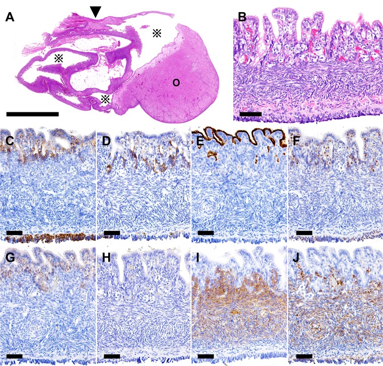
Histological examination revealed a single large cyst in the right ovarian hilus (Fig. 1A). The cyst was lined with monolayer cuboidal epithelial cells and was surrounded by the ovarian tissue (Fig. 1B). The epithelial cells possessed cilia and often invaginated outwards from the cyst forming short tubular structures. Beneath the epithelial layer, luteinized stromal cells and spindle-shaped stromal cells surrounded the cyst. The epithelial cells of the cyst wall in the present case were positive for pan-cytokeratin (CK), CK 18, CD10, estrogen receptor and calretinin (Fig. 1C, 1D, 1E, 1F and 1G) and negative for p63. No muscular layer was observed surrounding the cyst on a smooth muscle actin (SMA)-immunostained section (Fig. 1H). Both luteinized stromal cells and spindle-shaped stromal cells that surrounded the cyst were positive for melan A and vimentin (Fig. 1I and 1 J).
Fig. 1.
Right ovary. (A) A large cyst (※) in the ovarian hilus (arrowhead) is observed. (B) The cyst wall is lined with monolayer cuboidal epithelial cells with cilia. Beneath the epithelial layer, luteinized stromal cells and spindle-shaped stromal cells of the ovary are observed. (C) All the epithelial cells are positive for pan-CK. (D) Epithelial cells invaginating into the stroma are positive for CK18. (E) The apical surface of the epithelial cells is positive for CD10. (F) Nuclei of the epithelial cells are positive for estrogen receptor. (G) Nuclei and the cytoplasms of the epithelial cells are positive for calretinin. (H) No SMA-positive muscular layer is observed beneath the epithelial layer. (I) Luteinized stromal cells and spindle-shaped stromal cells beneath the epithelial layer are positive for vimentin. (J) Luteinized stromal cells and spindle-shaped stromal cells beneath the epithelial layer are positive for melan A. Arrowhead, ovarian hilus; ※, cyst; O, ovary. (A and B) HE; (C–J) immunohistochemistry. (A) Bar=5 mm; (B–J) bar=50 µm.
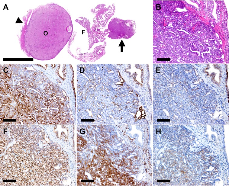
The tumor in the left uterine tube was located adjacent to the fimbria. Histologically, the tumor showed a papillary growth with a distinct margin (Fig. 2A) and was composed of epithelial cells arranged in a tubular pattern and of pleomorphic cells proliferating in a solid pattern between the tubules (Fig. 2B). Calcification was observed at the surface of the tumor tissue. Malignancy, such as atypical mitosis or invasion into the surrounding tissues, was not found. The tumor cells were positive for pan-CK, CK 18, E-cadherin, estrogen receptor, vimentin and calretinin (Fig. 2C, 2D, 2E, 2F, 2G and 2H). However, the expressions of CK18 and E-cadherin were limited to the epithelial cells composing the tubular structure (Fig. 2D and 2E), while those of vimentin and calretinin were limited to the pleomorphic cells between the tubular structures (Fig. 2G and 2H).
Fig. 2.
Left ovary and uterine tube. (A) A nodular tumor (arrow) is observed adjacent to the fimbria (F) of uterine tube. (B) The tumor is composed of epithelial cells arranged in a tubular pattern and pleomorphic cells between the tubular structures. Note the normal uterine tube epithelium in the upper right. (C) All the normal and tumor epithelial cells are positive for pan-CK. (D) The normal and tumor epithelial cells composing the tubular structures are positive for CK18. (E) The normal and tumor epithelial cells composing the tubular structures are positive for E-cadherin. (F) All the normal and tumor epithelial cells are positive for estrogen receptor. (G) The pleomorphic cells between the tubular structures are positive for vimentin. (H) The pleomorphic cells between the tubular structures are positive for calretinin. Arrow, tumor; arrowhead, ovarian hilus; F, fimbria; O, ovary. (A and B) HE; (C–H) immunohistochemistry. (A) Bar=5 mm; (B–H) bar=50 µm.
The cyst in the right ovary was diagnosed as cystic rete ovarii based on the following findings: a single large cyst located in the ovarian hilus and was surrounded by ovarian stromal tissues (Figs. 1A and 2B), differentiating it from ovarian epithelial cyst; epithelial cells lining the cyst wall were negative for vimentin and melan A, differentiating it from Graafian follicle cysts and luteinized cysts (Table 2); the cyst lacked muscular layer (Fig. 1H), differentiating it from cysts arising from other mesonephric tubules [6, 11].
The results of immunohistochemistry in the present study were comparable to those of cystic rete testis in a male rabbit [6], except for additional results of estrogen receptor- and calretinin-positive epithelial cells of cystic rete ovarii (Figs. 1F and 2G). The structure of the cyst is identical between cystic rete ovarii and cystic rete testis in rabbits. However, as it has been demonstrated in the normal ovarian tissue and neoplastic rete ovarii of humans, the epithelial cells of the female counterpart in rabbits expressed estrogen receptor [12, 17]. Though reports on calretinin expression in rete ovarii and rete testis are very limited, Cao et al. reported that 0/2 cases of rete ovaries were calretinin-positive, and 2/8 cases of rete testes were calretinin-positive in humans [5]. CD10 is used as a marker of mesonephric lesions. As the authors have reported in a former study [6], CD10 is likely to be a useful marker to differentiate cystic lesions of the rete testis and also rete ovarii.
The tumor in the left uterine tube was composed of 2 types of cells: epithelial cells arranged in a tubular pattern and pleomorphic cells between the tubular structures (Fig. 2B). Both of the cells were strongly positive for pan-CK and estrogen receptor, suggesting that the tumor was derived from the uterine tube epithelium (Fig. 2C and 2F). The epithelial type cells were also positive for CK18 and E-cadherin, suggesting the characters of the uterine tube epithelium (Fig. 2D and 2E). Interestingly, the pleomorphic cells were negative for CK18 and E-cadherin, but instead were positive for vimentin and calretinin, obtaining features of interstitial cells (Fig. 2G and 2H).
In humans, calretinin is an established marker for mesothelioma, adenomatoid tumor of the female genital system (uterine tube, uterus and ovarian hilus) and synovial sarcoma, all of which are biphasic (epithelioid and/or sarcomatoid) tumor derived from the mesodermal tissue [7, 15]. In addition, it has been reported that calretinin is also expressed in normal functioning endometrial stroma of humans [2]. In the female genital system of the rabbit, calretinin expression was confirmed in luteinized stromal cells of the ovary and in epithelial cells of the uterine tube and uterus.
This is the first case of cystic rete ovarii and uterine tube adenoma in a rabbit. The structure and immunohistochemical characteristics of cystic rete ovarii are comparable to those of rete testis in rabbits [6].
REFERENCES
- 1.Akihara Y., Shimoyama Y., Kawasako K., Komine M., Hirayama K., Kagawa Y., Omachi T., Matsuda K., Okamoto M., Kadosawa T., Taniyama H.2007. Immunohistochemical evaluation of canine ovarian cysts. J. Vet. Med. Sci. 69: 1033–1037. doi: 10.1292/jvms.69.1033 [DOI] [PubMed] [Google Scholar]
- 2.Al Moghrabi H., Elkeilani A., Thomas J. M., Mai K. T.2007. Calretinin: an immunohistochemical marker for the normal functional endometrial stroma and alterations of the immunoreactivity in dysfunctional uterine bleeding. Pathol. Res. Pract. 203: 79–83. doi: 10.1016/j.prp.2006.10.005 [DOI] [PubMed] [Google Scholar]
- 3.Anjum A. D., Payne L. N., Appleby E. C.1989. Oestrogen and progesterone receptors and their relationship to histological grades of epithelial tumours of the magnum region of the oviduct of the domestic fowl. J. Comp. Pathol. 100: 275–286. doi: 10.1016/0021-9975(89)90105-9 [DOI] [PubMed] [Google Scholar]
- 4.Beasley J. N., Klopp S., Terry B.1986. Neoplasms in the oviducts of turkeys. Avian Dis. 30: 433–437. doi: 10.2307/1590553 [DOI] [PubMed] [Google Scholar]
- 5.Cao Q. J., Jones J. G., Li M.2001. Expression of calretinin in human ovary, testis, and ovarian sex cord-stromal tumors. Int. J. Gynecol. Pathol. 20: 346–352. doi: 10.1097/00004347-200110000-00006 [DOI] [PubMed] [Google Scholar]
- 6.Chambers J. K., Uchida K., Murata Y., Watanabe K., Ise K., Miwa Y., Nakayama H.2014. Cystic rete testis with testicular dysplasia in a rabbit. J. Vet. Med. Sci. (in press). [DOI] [PMC free article] [PubMed] [Google Scholar]
- 7.Doglioni C., Dei Tos A. P., Laurino L., Iuzzolino P., Chiarelli C., Celio M. R., Viale G.1996. Calretinin: a novel immunocytochemical marker for mesothelioma. Am. J. Surg. Pathol. 20: 1037–1046. doi: 10.1097/00000478-199609000-00001 [DOI] [PubMed] [Google Scholar]
- 8.Gelberg H. B., McEntee K.1986. Pathology of the canine and feline uterine tube. Vet. Pathol. 23: 770–775 [DOI] [PubMed] [Google Scholar]
- 9.Gelberg H. B., McEntee K., Heath E. H.1984. Feline cystic rete ovarii. Vet. Pathol. 21: 304–307 [DOI] [PubMed] [Google Scholar]
- 10.Kennedy P. C., Cullen J. M., Edwards J. F., Goldschmidt M. H., Larsen L., Munson L., Nielsen S.1998. Histological Classification of Tumors of the Genital System of Domestic Animals. 2nd series, vol IV, WHO, Armed Forces Institute of Pathology, Washington, D.C. [Google Scholar]
- 11.Khan M. S., Dodson A. R., Heatley M. K.1999. Ki-67, oestrogen receptor, and progesterone receptor proteins in the human rete ovarii and in endometriosis. J. Clin. Pathol. 52: 517–520. doi: 10.1136/jcp.52.7.517 [DOI] [PMC free article] [PubMed] [Google Scholar]
- 12.Kim S. W., Lee Y. H., Lee S. R., Kim K. M., Lee Y. J., Jung K. J., Chang K. S., Kim D., Son H. Y., Reu D. S., Chang K. T.2012. Bilateral ovarian cysts originating from rete ovarii in an African green monkey (Cercopithecus aethiops). J. Vet. Med. Sci. 74: 1229–1232. doi: 10.1292/jvms.11-0354 [DOI] [PubMed] [Google Scholar]
- 13.Keller L. S., Griffith J. W., Lang C. M.1987. Reproductive failure associated with cystic rete ovarii in guinea pigs. Vet. Pathol. 24: 335–339 [DOI] [PubMed] [Google Scholar]
- 14.MacLachlan N. J., Kennedy P. C.2002. Tumors of the genital systems. p. 558. In: Tumors in Domestic Animals, 4th ed. (Meuten, D. J. ed.), Iowa State Press, Ames. [Google Scholar]
- 15.Miettinen M., Limon J., Niezabitowski A., Lasota J.2001. Calretinin and other mesothelioma markers in synovial sarcoma: analysis of antigenic similarities and differences with malignant mesothelioma. Am. J. Surg. Pathol. 25: 610–617. doi: 10.1097/00000478-200105000-00007 [DOI] [PubMed] [Google Scholar]
- 16.Pereira M. E., Viner T. C.2008. Oviduct adenocarcinoma in some species of captive snakes. Vet. Pathol. 45: 693–697. doi: 10.1354/vp.45-5-693 [DOI] [PubMed] [Google Scholar]
- 17.Ram M., Abdulla A., Razvi K., Pandeva I., Al-Nafussi A.2009. Cystadenofibroma of the rete ovarii: a case report with review of literature. Rare Tumors 1: e24 [DOI] [PMC free article] [PubMed] [Google Scholar]
- 18.Sailasuta A., Tateyama S., Yamaguchi R., Nosaka D., Otsuka H.1989. Adenomatous papilloma of the uterine tube (oviduct) fimbriae in a dog. Jpn. J. Vet. Sci. 51: 632–633. doi: 10.1292/jvms1939.51.632 [DOI] [PubMed] [Google Scholar]


