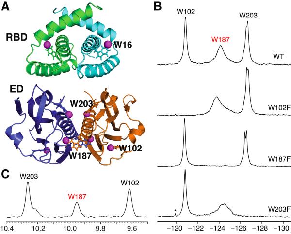
Figure 1.
(A) Locations of the 5-F-Trp residues in the dimer structures of Ud NS1A domains. Top, Ud NS1A RBD (PDB ID, 1NS1) (Chien et al., 1997). Bottom, Ud NS1A ED (PDB ID, 3EE9) (Xia et al., 2009). The fluorine atoms are shown as magenta spheres, and the tryptophan residues within one subunit of each dimer are labeled. Structures were rendered using PyMOL (PyMOL Molecular Graphics System, Version 1.4 Schrödinger, LLC). (B) 19F NMR spectra of 5-F-Trp labeled Ud NS1A ED (530 μM), and its W102F (400 μM), W187F (400 μM), and W203F (200 μM) mutants in high salt pH 8 buffer. Asterisks denote trace amounts of protease inhibitors remaining after purification. Note that the splitting observed for Trp203 under these conditions is more pronounced for the W187F mutant which is a monomer in solution (data not shown). (C) Projection of the indole amide region along the 1H dimension from an 800 MHz 2D 1H-15N TROSY-HSQC spectrum of 400 μM 15N-enriched Ud NS1A ED in pH 6.9 buffer at 27 °C (Aramini et al., 2011).

