Figure 3.

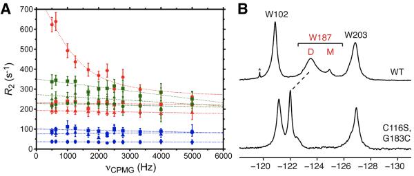
19F NMR CPMG relaxation dispersion data for Trp102 (squares), Trp187 (circles), and Trp203 (triangles) in 5-F-Trp labeled 600 μM Ud NS1A ED (red), 200 μM [K110A] NS1A ED (blue), and 500 μM[C116S,G183C] NS1A ED (disulfide form; no DTT) (green) in low salt pH 8 buffer at 20 °C. (B) 19F NMR spectra of 5-F-Trp labeled Ud NS1A ED (top) and disulfide-bonded 500 μM [C116S,G183C] NS1A ED (bottom), in low salt pH 8 buffer. The downfield shift of the 19F resonance for Trp187 is indicated by a dashed line. The native Cys116 was mutated to a serine in order to prevent a potential mixture of disulfide adducts.
