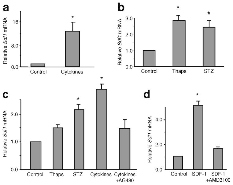
Fig. 3.
Beta cell injury induces expression of Sdf1 in beta cell lines. MIN6 and INS-1 beta cells were treated with stress/apoptosis inducers. MIN6 cells were treated with: (a) mixtures of cytokines (same concentration as indicated in the Fig. 2 legend), or (b) thapsigargin or streptozotocin. INS-1 cells were treated with: (c) thapsigargin (Thaps), streptozotocin (STZ) or cytokines with and without AG490, an inhibitor of JAK/STAT signalling; or (d) 10 nmol/l SDF-1 with and without 25 μmol/l AMD3100, an antagonist of the SDF-1 receptor, CXCR4. In all experiments the amount of Sdf1 mRNA was measured by quantitative RT-PCR. All values are means±SEM (n=8) relative to the value of the control-treated cells. *p<0.01 vs control values

