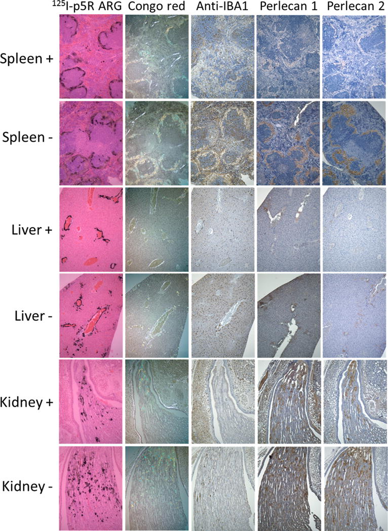
Figure 4.

Stained sections from huIL-6 mice that had been given AEF 4 weeks before sacrifice and were either treated with clodronate (+) or not treated (−) 3 days prior to sacrifice. Sections from liver, spleen and kidney papilla are included. Column 1 images are autoradiography of sections harvested 2 h post iv injection with 125I p5R. Column 2 figures show sections stained with Congo red in birefringence microscopy. Column 3 shows sections stained with iba-1 antibody. Column 4 shows sections stained with rat anti perlecan Mab A7L6 and column 5 shows sections stained with rat Mab HK-102. Original magnification 100×.
