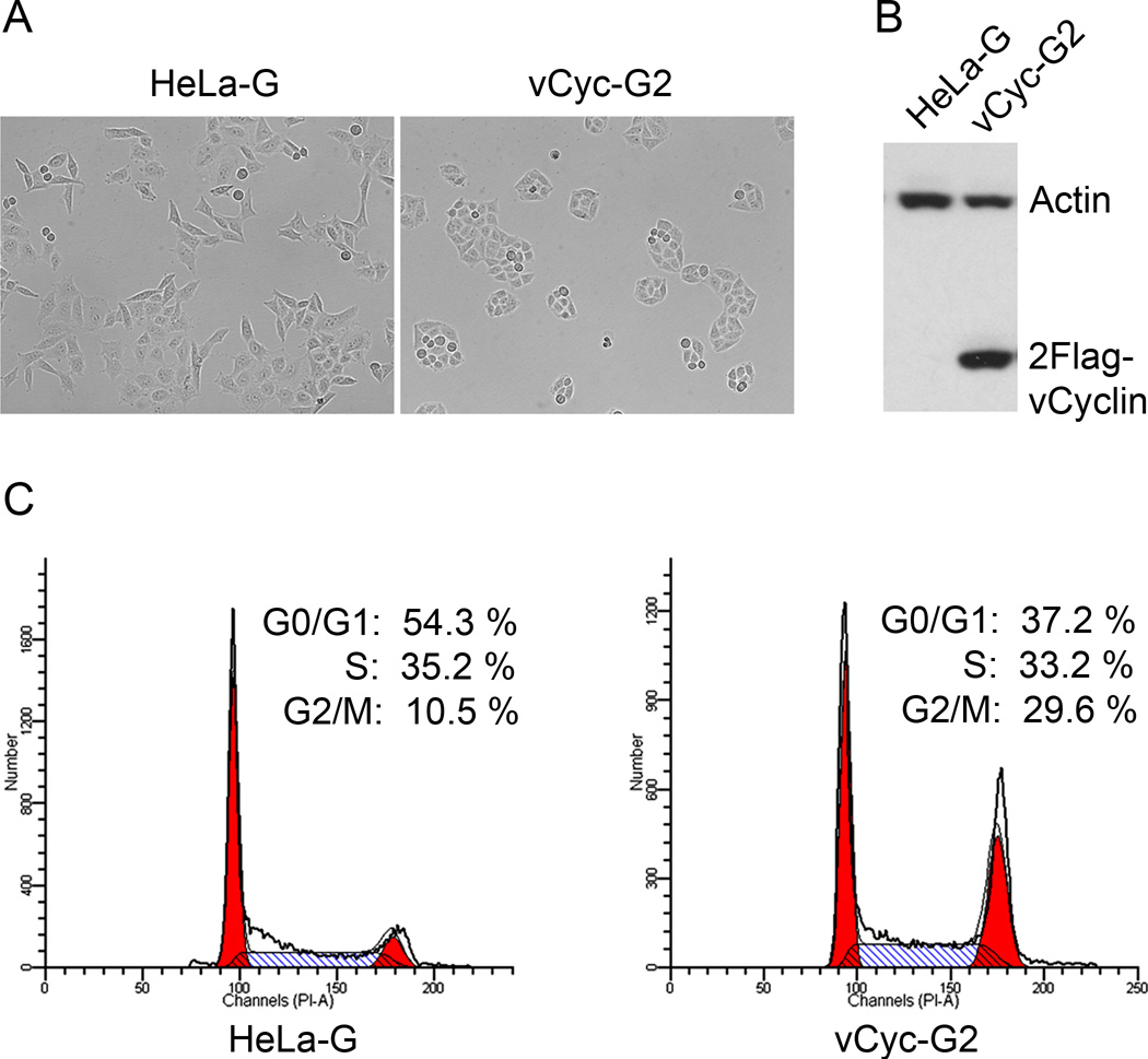
Figure 4. Characterizations of HeLa cells stably expressing KSHV vCyclin.
(A) Phase contrast photographs of HeLa–G and its derivative, vCyc-G2, that expresses dual Flag-tagged- (2Flag-) vCyclin,. (B) Immunoblot analysis of HeLa–G and vCyc-G2 using a Flag-epitope antibody. (C) Flow cytometry histograms of HeLa-G and vCyc-G2. Exponentially growing HeLa-G and vCyc-G2 cells were subjected to flow cytometry as described in Materials and Methods. The percentage of cells in G1, S, and G2/M respectively was determined as in Fig. 1C.

